

AAPM Task Group Report 238: 3D C-arms with volumetric imaging capability
- PMID: 36710257
- PMCID: PMC11584023
- DOI: 10.1002/mp.16245
AAPM Task Group Report 238: 3D C-arms with volumetric imaging capability
Abstract
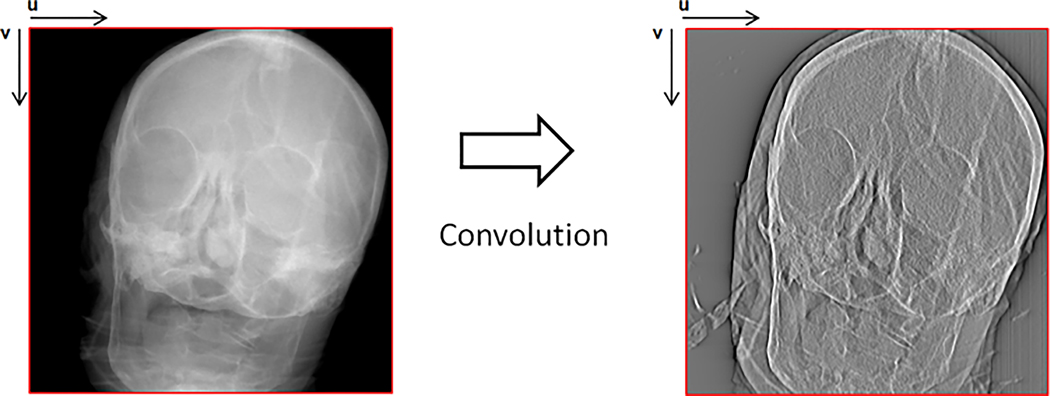
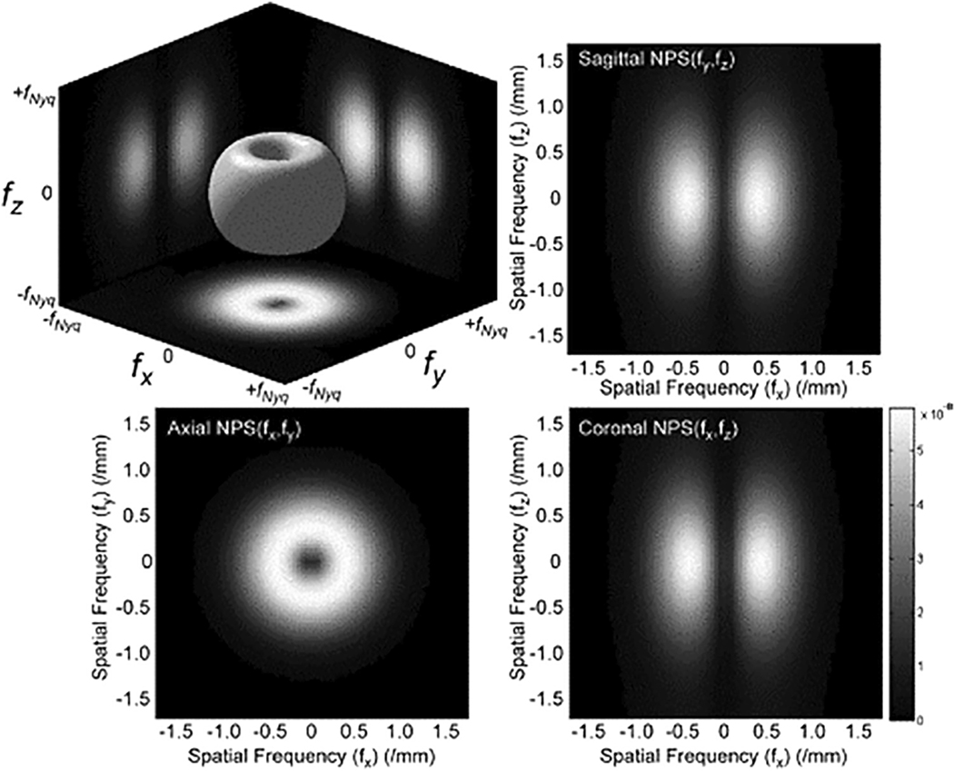
This report reviews the image acquisition and reconstruction characteristics of C-arm Cone Beam Computed Tomography (C-arm CBCT) systems and provides guidance on quality control of C-arm systems with this volumetric imaging capability. The concepts of 3D image reconstruction, geometric calibration, image quality, and dosimetry covered in this report are also pertinent to CBCT for Image-Guided Radiation Therapy (IGRT). However, IGRT systems introduce a number of additional considerations, such as geometric alignment of the imaging at treatment isocenter, which are beyond the scope of the charge to the task group and the report. Section 1 provides an introduction to C-arm CBCT systems and reviews a variety of clinical applications. Section 2 briefly presents nomenclature specific or unique to these systems. A short review of C-arm fluoroscopy quality control (QC) in relation to 3D C-arm imaging is given in Section 3. Section 4 discusses system calibration, including geometric calibration and uniformity calibration. A review of the unique approaches and challenges to 3D reconstruction of data sets acquired by C-arm CBCT systems is give in Section 5. Sections 6 and 7 go in greater depth to address the performance assessment of C-arm CBCT units. First, Section 6 describes testing approaches and phantoms that may be used to evaluate image quality (spatial resolution and image noise and artifacts) and identifies several factors that affect image quality. Section 7 describes both free-in-air and in-phantom approaches to evaluating radiation dose indices. The methodologies described for assessing image quality and radiation dose may be used for annual constancy assessment and comparisons among different systems to help medical physicists determine when a system is not operating as expected. Baseline measurements taken either at installation or after a full preventative maintenance service call can also provide valuable data to help determine whether the performance of the system is acceptable. Collecting image quality and radiation dose data on existing phantoms used for CT image quality and radiation dose assessment, or on newly developed phantoms, will inform the development of performance criteria and standards. Phantom images are also useful for identifying and evaluating artifacts. In particular, comparing baseline data with those from current phantom images can reveal the need for system calibration before image artifacts are detected in clinical practice. Examples of artifacts are provided in Sections 4, 5, and 6.
© 2023 American Association of Physicists in Medicine.
Conflict of interest statement
CONFLICTS OF INTEREST
1. The members of TG 238 3D C-Arms with Volumetric Imaging Capability listed below attest that they have no potential Conflicts of Interest related to the subject matter or materials presented in this document: Keyvan Farahani, Grace Jianan Gang, Beth Schueler, Jie Zhang
2. The member of TG 238 3D C-Arms with Volumetric Imaging Capability listed below disclose the following potential Conflict(s) of Interest related to the subject matter or materials presented in this document: Rebecca Fahrig (founder, Tibaray Inc. employee, Siemens Healthineers GmbH), Jan Jans (employee, Philips Healthcare), A. Kyle Jones (President, Fluoroscopic Safety, LLC), Thomas Koenig (employee, Ziehm Imaging), Andrew Kuhls-Gilcrist (employee Canon Medical Systems USA, Inc; Vice Chair, Medical Imaging and Technology Alliance (MITA) US NC Expert, International Electrotechnical Commission (IEC)), MingDe Lin (employee, Visage Imaging, Inc. and former employee Philips Research North America), Cyril Ridell (employee GE Healthcare), Ludwig Ritschl (employee, Siemens Healthineers and former employee, Ziehm Imaging), Sebastian Schafer (employee, Siemens Healthineers), Jeffrey Siewerdsen (Academic-Industry Research Grant, Siemens Healthineers. AcademicIndustry Research Grant, Carestream Health. Research Grant, Medtronicc. Advisory Board, Siemens Healthineers. Advisory Board, Carestream Health. Licensing Agreement, Elekta Oncology. Licensing Agreement, Carestream Health. Licensing Agreement, The Phantom Lab), Michael Silver (former employee, Canon Medical Systems USA, inc), Mark Supanich (Co-Technical Advisor for US Technical Advisory Group 62B (ANSI), US NC Expert, International Electrotechnical Commission (IEC)).
Figures


References
-
- Edward Rainier GS, Sembrano JN, Castro C, Ledonio C, Truong W, Polly DW. Accuracy of intra-operative O-arm images in determining pedicle screw position. Spine J. 2010;10(9):S50.
-
- Michael F, Powell DD, Arra S. Reddy C-arm fluoroscopic cone beam CT for guidance of minimally invasive spine interventions. Pain Physician. 2010;13(1):51–59. - PubMed
Publication types
MeSH terms
Grants and funding
LinkOut - more resources
Full Text Sources
Miscellaneous
