Review
. 2023 Jan 9:15:1068185.
doi: 10.3389/fnmol.2022.1068185. eCollection 2022.

Gene-agnostic therapeutic approaches for inherited retinal degenerations

Affiliations
Review

Gene-agnostic therapeutic approaches for inherited retinal degenerations

Molly C John et al. Front Mol Neurosci. .

Abstract

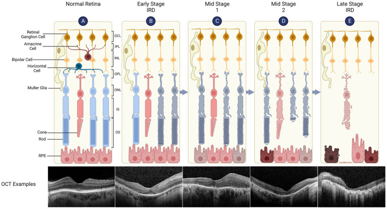
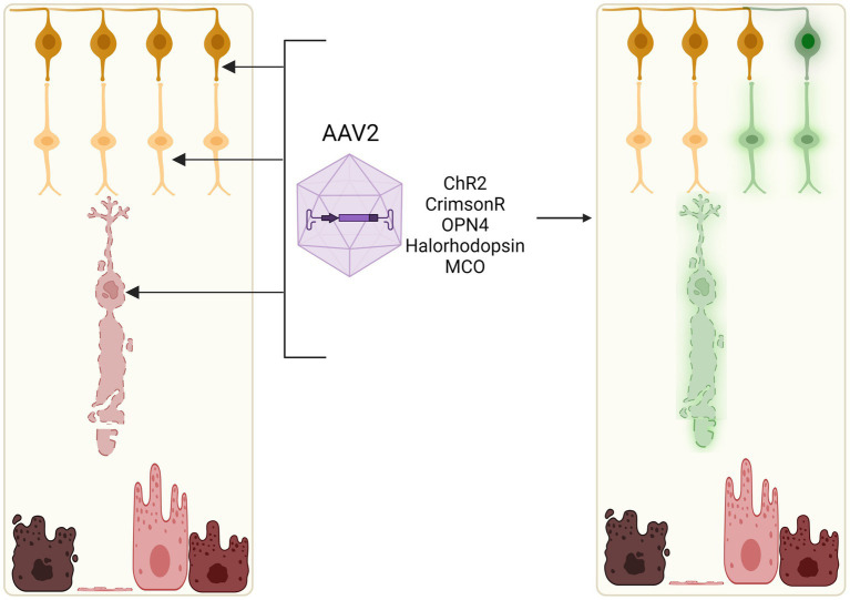
Inherited retinal diseases (IRDs) are associated with mutations in over 250 genes and represent a major cause of irreversible blindness worldwide. While gene augmentation or gene editing therapies could address the underlying genetic mutations in a small subset of patients, their utility remains limited by the great genetic heterogeneity of IRDs and the costs of developing individualised therapies. Gene-agnostic therapeutic approaches target common pathogenic pathways that drive retinal degeneration or provide functional rescue of vision independent of the genetic cause, thus offering potential clinical benefits to all IRD patients. Here, we review the key gene-agnostic approaches, including retinal cell reprogramming and replacement, neurotrophic support, immune modulation and optogenetics. The relative benefits and limitations of these strategies and the timing of clinical interventions are discussed.

Keywords: cellular reprogramming; gene-independent; immune modulation; inherited retinal degeneration; optogenetics; retina - medical therapies; stem cells.


Conflict of interest statement

The authors declare that the research was conducted in the absence of any commercial or financial relationships that could be construed as a potential conflict of interest.

Figures

Figure 1
Figure 1
Retinal structure and degeneration in inherited retinal diseases (IRDs). The retina has a laminar structure consisting of distinct cell types (A). The neural retina consists of the ganglion cell layer (GCL, containing the cell bodies of retinal ganglion cells), inner plexiform layer (IPL), inner nuclear layer (INL - bipolar cell bodies), outer plexiform layer (OPL), outer nuclear layer (ONL - photoreceptor cell bodies), inner segments (IS) and outer segments (OS). The retinal pigment epithelium (RPE) supports the metabolism of overlying photoreceptors, is attached to the Bruch’s membrane/choroid and forms outer blood-retinal barrier. A representative spectral domain optical coherence tomography (OCT) of the retina is shown which demonstrates normal retinal layers. (B) Early Stage IRD, such as retinitis pigmentosa, is typically characterised by dysfunction and degeneration of rod, which can be seen as peripheral outer retinal thinning on the OCT (note that parafoveal architecture is relatively preserved). (C) Retinal degeneration progresses to Mid Stage 1 where cone function (day light vision) remains relatively intact while rod function (night vision) is severely impaired. The OCT shows widespread disruption of the ellipsoid line which represents IS/OS junctions. RPE thinning can also be seen. (D) Mid Stage 2 sees cone degeneration with shortened OS and loss of rods. (E) In Late Stage (or end stage) IRD, there is complete loss of photoreceptors while inner retinal layers remain relatively preserved. OCT shows complete outer retinal atrophy with areas of RPE hypertrophy which correspond to bone spicules seen clinically.
Figure 2
Figure 2
In situ reprogramming of rod photoreceptors. Rods may be reprogrammed in early stage IRDs to generate ‘pseudocones’ through manipulation of the NRL and NR2E3 transcription factors which normally determine rod fate (e.g., by CRISPR-mediated gene knockout or molecular inhibitors, PR1 and PR3). Pseudocones confer resistance against rod-specific gene mutations, thus slowing the rate of retinal degeneration.
Figure 3
Figure 3
Reprogramming Müller glia to retinal cells. Manipulation of key signalling factors (ASCL-1, PTB, or miRNAs) could induce expansion and differentiation of resident retinal Müller glia into a number of retinal cell types. These include photoreceptors, amacrine cells and ganglion cells, which could replace lost and degenerating cells in the retina.
Figure 4
Figure 4
Cell therapies for retinal degenerations. Allogeneic stem cell-based cell replacement strategies rely on induced pluripotent stem cells (iPSCs) or embryonic stem cells (ESCs) which can be differentiated into photoreceptor or RPE cell suspensions, sheets, or retinal organoids for transplantation. The donor cells or tissue replace lost photoreceptors and RPE in late stage IRDs. Subretinal injection of cell suspensions allows direct contact between donor cells and the surviving neuronal cells in the retina but can result in disorganised engraftment. Subretinal implantation of structured retinal sheet helps to retain anatomical organisation, which may facilitate appropriate cellular differentiation but is surgically challenging.
Figure 5
Figure 5
Optogenetic induction of light sensitivity in inner retinal cells. As bipolar and ganglion cell layers often remain relatively intact in retinal degenerations, light sensitivity may be induced in these neuronal cell types through viral vector-mediated delivery of opsins, e.g., channelrhodopsin-2 (ChR2), channelrhodopsin-CrimsonR, halorhodopsin, or multicharacteristic-opsin (MCO). Current clinical trials of optogenetic therapies utilise adeno-associated viral vectors which can efficiently transduce retinal ganglion cells, bipolar cells, and some remaining cone cell bodies in vivo.

References

    1. Aires I. D., Madeira M. H., Boia R., Rodrigues-Neves A. C., Martins J. M., Ambrósio A. F., et al. (2019). Intravitreal injection of adenosine A(2A) receptor antagonist reduces neuroinflammation, vascular leakage and cell death in the retina of diabetic mice. Sci. Rep. 9, 1–14. doi: 10.1038/s41598-019-53627-y, PMID: - DOI - PMC - PubMed
    1. Aït-Ali N., Fridlich R., Millet-Puel G., Clérin E., Delalande F., Jaillard C., et al. (2015). Rod-derived cone viability factor promotes cone survival by stimulating aerobic glycolysis. Cells 161, 817–832. doi: 10.1016/j.cell.2015.03.023, PMID: - DOI - PubMed
    1. Akhtar-Schäfer I., Wang L., Krohne T. U., Xu H., Langmann T. (2018). Modulation of three key innate immune pathways for the most common retinal degenerative diseases. EMBO Mol. Med. 10:e8259. doi: 10.15252/emmm.201708259, PMID: - DOI - PMC - PubMed
    1. Algvere P. V., Gouras P., Kopp E. D. (1999). Long-term outcome of RPE allografts in non-immunosuppressed patients with AMD. Eur. J. Ophthalmol. 9, 217–230. doi: 10.1177/112067219900900310, PMID: - DOI - PubMed
    1. Angbohang A., Wu N., Charalambous T., Eastlake K., Lei Y., Kim Y. S., et al. (2016). Downregulation of the canonical WNT signaling pathway by TGFb1 inhibits photoreceptor differentiation of adult human Müller glia with stem cell characteristics. Stem Cells Dev. 25, 1–12. doi: 10.1089/scd.2015.0262, PMID: - DOI - PMC - PubMed

LinkOut - more resources