. 2023 Jan 19:2023:3335959.
doi: 10.1155/2023/3335959. eCollection 2023.

LncRNA SNHG5 Suppresses Cell Migration and Invasion of Human Lung Adenocarcinoma via Regulation of Epithelial-Mesenchymal Transition

Affiliations

LncRNA SNHG5 Suppresses Cell Migration and Invasion of Human Lung Adenocarcinoma via Regulation of Epithelial-Mesenchymal Transition

Zhirong Li et al. J Oncol. .

Abstract

Long noncoding RNAs (lncRNAs) are gradually being annotated as important regulators of multiple cellular processes. The goal of our study was to investigate the effects of the lncRNA small nucleolar RNA host gene 5 (SNHG5) in lung adenocarcinoma (LAD) and its underlying mechanisms. The findings revealed a substantial drop in SNHG5 expression in LAD tissues, which correlated with clinical-pathological parameters. Transcriptome sequencing analysis demonstrated that the inhibitory effect of SNHG5 was associated with cell adhesion molecules. Moreover, the expression of SNHG5 was shown to be correlated with epithelial-mesenchymal transition (EMT) markers in western blots and immunofluorescence. SNHG5 also had significant effects of antimigration and anti-invasion on LAD cells in vitro. Furthermore, the migration and invasion of A549 cells were suppressed by overexpressed SNHG5 in the EMT progress induced by transforming growth factor β1 (TGF-β1), and this might be due to the inhibition of the expression of EMT-associated transcription factors involving Snail, SLUG, and ZEB1. In LAD tissues, the expression of SNHG5 exhibited a positive association with E-cadherin protein expression but a negative correlation with N-cadherin and vimentin, according to the results of quantitative real-time PCR (qRT-PCR). In summary, the current work demonstrated that the lncRNA SNHG5 might limit cell migration and invasion of LAD cancer via decreasing the EMT process, indicating that SNHG5 might be used as a target for LAD therapeutic methods.


Conflict of interest statement

The authors declare that they have no conflicts of interest.

Figures

Figure 1
Figure 1
The LncRNA SNHG5 was found to be downregulated in LAD tissues and was associated with the aggressiveness of the disease. (a) The qRT-PCR was used to examine the expression of SNHG5 in 61 samples of LAD tissues and their corresponding neighboring nontumor tissues. The internal control was set to GAPDH. (b) The correlation between SNHG5 expression and size of the tumor. According to the most recent Union for International Cancer Control (UICC) and AJCC (American Joint Committee on Cancer) guidelines, there are four grades of tumor (T) that reflect the size of the tumor (area of cancer). (c) The correlation between the expression of SNHG5 and the TNM stages. a: ∗∗∗p < 0.001; b: p < 0.05; c: ∗∗p < 0.01. (d) The association of lymph node metastasis and SNHG5 expression. Negative results indicate no lymph node metastasis, whereas positive results show lymph node metastasis. ∗∗p < 0.01. (e, f) Kaplan-Meier survival curves for progression-free interval, and for disease-specific survival, based on SNHG5 expression data from TCGA. p < 0.05.
Figure 2
Figure 2
In LAD A549 cells, overexpression of SNHG5 decreases the expression of EMT-associated genes. (a) To evaluate the effect of lncRNA SNHG5 on lung A549 cells, transcriptome sequencing analysis (GSE143766) was performed. According to the color bar in the logarithmic scale given above the heatmap, the red and green colors in the heatmap respectively reflect high and low expression of target genes. (b) Volcano plot analysis of the differential gene expression after SNHG5 overexpression. (c) KEGG analysis of downregulated genes in SNHG5-overexpressing A549 cells. (d) The qRT-PCR analyzed the expression of target genes associated with migration and invasion, which were downregulated in the results of the transcriptome sequencing after SNHG5-overexpression. p < 0.05; (e, f) Western blot and immunofluorescence analysis evaluated the expression of EMT markers in control and A549 cancer cells after transfection with SNHG5 or siRNAs. p < 0.05.
Figure 3
Figure 3
In vitro, SNHG5 inhibits LAD cell migration and invasion. (a) In the controls and LAD cell lines transfected with SNHG5, qRT-PCR analysis of SNHG5 was performed. Statistically, groups identified with various letters differ from one another. p < 0.01. (b) The MTS assay was performed in the control and LAD cell lines after transfection with SNHG5, (c, d) Transwell analyses were carried out in normal and SNHG5-overexpressing LAD cell lines; the original magnification was set at ×200. a, c: p < 0.05; b, d: ∗∗p < 0.01. (e, f) Wound healing assay was conducted in the control and SNHG5-overexpressed LAD cell lines. Original magnification, ×100. The groups marked with “a” are statistically distinct. p < 0.05. (g) In the normal and A549 cells transfected with different siRNAs, qRT-PCR analysis of SNHG5 was performed. (h–j) MTS, Transwell, and wound healing analyses were conducted in controls and A549 tumor cells transfected with various siRNAs. a,b: p < 0.05.
Figure 4
Figure 4
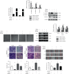
In the EMT of LAD A549 cells caused by TGF1, the LncRNA SNHG5 was downregulated. (a) An inverted microscope was used to examine morphological alterations in A549 cells generated by vehicle control and 10 ng/mL TGF-β1. (b, c) The levels of expression of EMT markers in A549 cells after TGF-β1 exposure were investigated using western blot and immunofluorescence. (d) The expression of SNHG5 in A549 cells was investigated using qRT-PCR at various time periods subsequent to TGF-β1 exposure.
Figure 5
Figure 5
Overexpression of SNHG5 inhibits EMT, preventing LAD cells from migrating and invading. (a) After TGF-β1 or vehicle control treatment, qRT-PCR was used to examine the relative fold change of SNHG5 expression in control and SNHG5-overexpressing A549 cells. ∗∗∗p < 0.001. (b) After TGF-β1 exposure, qRT–PCR was used to examine the relative fold change of EMT markers in normal and SNHG5-overexpressing A549 cancerous cells. (c) Western blot analysis was used to assess the level of expression of markers in control and SNHG5-overexpressing A549 cancer cells after TGF-β1 treatment. (d) The inverted microscope determined the morphological changes in control and SNHG5-overexpressing A549 cells after TGF-β1 or vehicle control treatment. (e, f) Western blot and qRT–PCR analysis of EMT-related transcription factors in normal and SNHG5-overexpressing A549 cancer cells following TGF-β1 or vehicle control treatment. (g–i) Transwell and wound-healing experiments were conducted on control and SNHG5-overexpressing A549 cancer tissues after TGF-β1 treatment. Wound-healing distance was measured and expressed as a percentage of the distance at zero hours. a: ∗∗p < 0.01; b: ∗∗p < 0.01; c: ∗∗∗p < 0.001; d: ∗∗p < 0.01; e: ∗∗∗p < 0.001; f: ∗∗∗p < 0.001.
Figure 6
Figure 6
In lung adenocarcinoma tissues, the expression of EMT markers was negatively linked with SNHG5. (a) The expression of CDH1, N-cadherin, and vimentin was examined by qRT-PCR in 34 samples of LAD tissues and their neighboring nontumor tissues. The internal control was set to GAPDH. (b) Pearson's correlation analysis indicated that levels of SNHG5 RNA expression in LAD tissues were positively related to CDH1, but negatively related to both N-cadherin and vimentin ∗∗∗p < 0.001.

Similar articles

Cited by

References

    1. Sardenberg R. A. S., Mello E. S., Younes R. N. The lung adenocarcinoma guidelines: what to be considered by surgeons. Journal of Thoracic Disease . 2014;6(Suppl 5):S561–S567. doi: 10.3978/j.issn.2072-1439.2014.08.25. - DOI - PMC - PubMed
    1. Chaudhary K. R., Yan S. X., Heilbroner S. P., et al. Effects of beta-adrenergic antagonists on chemoradiation therapy for locally advanced non-small cell lung cancer. Journal of Clinical Medicine . 2019;8(5):p. 575. doi: 10.3390/jcm8050575. - DOI - PMC - PubMed
    1. Han B., Jin B., Chu T., et al. Combination of chemotherapy and gefitinib as first-line treatment for patients with advanced lung adenocarcinoma and sensitive EGFR mutations: a randomized controlled trial. International Journal of Cancer . 2017;141(6):1249–1256. doi: 10.1002/ijc.30806. - DOI - PubMed
    1. Jin Z., Guan L., Xiang G. m., Gao B. a. Radiation resistance of the lung adenocarcinoma is related to the AKT-Onzin-POU5F1 axis. Biochemical and Biophysical Research Communications . 2018;499(3):538–543. doi: 10.1016/j.bbrc.2018.03.185. - DOI - PubMed
    1. Wang Z., Pan L., Yu H., Wang Y. The long non-coding RNA SNHG5 regulates gefitinib resistance in lung adenocarcinoma cells by targetting miR-377/CASP1 axis. Bioscience Reports . 2018;38(4):p. BSR20180400. doi: 10.1042/bsr20180400. - DOI - PMC - PubMed