[Preprint]. 2023 Jan 4:2023.01.04.522773.
doi: 10.1101/2023.01.04.522773.

MNK1 and MNK2 expression in the human dorsal root and trigeminal ganglion

Affiliations

MNK1 and MNK2 expression in the human dorsal root and trigeminal ganglion

Stephanie Shiers et al. bioRxiv. .

Update in

Abstract

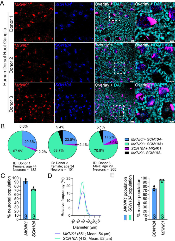
Mitogen activated protein kinase interacting kinases (MNK) 1 and 2 are serine/threonine protein kinases that play an important role in translation of mRNAs through their phosphorylation of the RNA 5’-cap binding protein, eukaryotic translation initiation factor (eIF) 4E. These kinases are downstream targets for mitogen activated protein kinases (MAPKs), extracellular activity regulated protein kinase (ERK) and p38. MNKs have been implicated in the sensitization of peripheral nociceptors of the dorsal root and trigeminal ganglion (DRG and TG) using transgenic mouse lines and through the use of specific inhibitors of MNK1 and MNK2. While specific knockout of the Mknk1 gene suggests that it is the key isoform for regulation of nociceptor excitability and nociceptive behaviors in mice, both MKNK1 and MKNK2 genes are expressed in the DRG and TG of mice and humans based on RNA sequencing experiments. Single cell sequencing in mice suggests that Mknk1 and Mknk2 may be expressed in different populations of nociceptors. We sought to characterize mRNA expression in human DRG and TG for both MNK1 and MNK2. Our results show that both genes are expressed by nearly all neurons in both human ganglia with expression in other cell types as well. Our findings provide evidence that MNK1 and MNK2 are expressed by human nociceptors and suggest that efforts to pharmacologically target MNKs for pain would likely be translatable due its conserved expression in both species.


Conflict of interest statement

Conflict of Interest Statement

JJS is an employee of 4E Therapeutics and TJP is a founder of 4E Therapeutics, a company developing MNK inhibitors for pain treatment. SS, JJS and TJP are inventors on patents related to MNK inhibition for pain treatment

Figures

Figure 1.
Figure 1.. Distribution of MKNK1 (MNK1) mRNA in human dorsal root ganglia.
A) Representative 20X images of human lumbar DRGs labeled with RNAscope in situ hybridization for MKNK1 (red) and SCN10A (blue) mRNAs and co-stained with DAPI (cyan). The fourth panel for each donor is a zoomed-in region demarcated by white boundaries in the 20X overlay image. Lipofuscin (globular structures) that autofluoresced in both channels and appear magenta in the overlay image were not analyzed as this is background signal that is present in all human nervous tissue. MKNK1 mRNA was expressed in neurons and non-neuronal cells. B) Pie-charts showing the distribution of MKNK1 neuronal subpopulations in human DRG for each donor. C) 92.4% of human DRG sensory neurons were positive for MKNK1. D) Histogram with Gaussian distribution displaying the size profile of all MKNK1+ and SCN10A+ neurons in human DRG. E) Percentage of MKNK1+ neurons that coexpressed SCN10A (blue bar), and the percentage of SCN10A+ neurons that coexpressed MKNK1 (green bar). Scale bars: 20X = 50μm. Zoomed in panel = 20μm.
Figure 2.
Figure 2.. Distribution of MKNK2 (MNK2) mRNA in human dorsal root ganglia.
A) Representative 20X images of human lumbar DRGs labeled with RNAscope in situ hybridization for MKNK2 (red) and SCN10A (blue) mRNAs and co-stained with DAPI (cyan). The fourth panel for each donor is a zoomed-in region demarcated by white boundaries in the 20X overlay image. Lipofuscin (globular structures) that autofluoresced in both channels and appear magenta in the overlay image were not analyzed as this is background signal that is present in all human nervous tissue. MKNK2 mRNA was expressed in neurons and non-neuronal cells. B) Pie-charts showing the distribution of MKNK2 neuronal subpopulations in human DRG for each donor. C) 92.5% of human DRG sensory neurons were positive for MKNK2. D) Histogram with Gaussian distribution displaying the size profile of all MKNK2+ and SCN10A+ neurons in human DRG. E) Percentage of MKNK2+ neurons that coexpressed SCN10A (blue bar), and the percentage of SCN10A+ neurons that coexpressed MKNK2 (green bar). Scale bars: 20X = 50μm. Zoomed in panel = 20μm.
Figure 3.
Figure 3.. Distribution of MKNK1 (MNK1) mRNA in human trigeminal ganglia.
A) Representative 20X images of human TGs labeled with RNAscope in situ hybridization for MKNK1 (red) and SCN10A (blue) mRNAs and co-stained with DAPI (cyan). The fourth panel for each donor is a zoomed-in region demarcated by white boundaries in the 20X overlay image. Lipofuscin (globular structures) were not analyzed as this is background signal that is present in all human nervous tissue. MKNK1 mRNA was expressed in neurons and non-neuronal cells. B) Pie-charts showing the distribution of MKNK1 neuronal subpopulations in human TG for each donor. C) 100% of human TG sensory neurons were positive for MKNK1. D) Histogram with Gaussian distribution displaying the size profile of all MKNK1+ and SCN10A+ neurons in human TG. E) Percentage of MKNK1+ neurons that coexpressed SCN10A (blue bar), and the percentage of SCN10A+ neurons that coexpressed MKNK1 (green bar). Scale bars: 20X = 50μm. Zoomed in panel = 20μm.
Figure 4.
Figure 4.. Distribution of MKNK2 (MNK2) mRNA in human trigeminal ganglia.
A) Representative 20X images of human TGs labeled with RNAscope in situ hybridization for MKNK2 (red) and SCN10A (blue) mRNAs and co-stained with DAPI (cyan). The fourth panel for each donor is a zoomed-in region demarcated by white boundaries in the 20X overlay image. Lipofuscin (globular structures) were not analyzed as this is background signal that is present in all human nervous tissue. MKNK2 mRNA was expressed in neurons and non-neuronal cells. B) Pie-charts showing the distribution of MKNK2 neuronal subpopulations in human TG for each donor. C) 100% of human TG sensory neurons were positive for MKNK2. D) Histogram with Gaussian distribution displaying the size profile of all MKNK2+ and SCN10A+ neurons in human TG. E) Percentage of MKNK2+ neurons that coexpressed SCN10A (blue bar), and the percentage of SCN10A+ neurons that coexpressed MKNK2 (green bar). Scale bars: 20X = 50μm. Zoomed in panel = 20μm.
Figure 5.
Figure 5.. Axonal MKNK1 (MNK1) and MKNK2 (MNK2) mRNAs in the human trigeminal ganglia.
A) Representative 20X image of the axonal-rich area of the human TG labeled with RNAscope in situ hybridization for MKNK1 (red) mRNA and co-stained with DAPI (blue). Background autofluorescence was brightened to visualize the axons (green). B) Zoomed-in region of the nerve demarcated by white boundaries in panel A. MKNK1 mRNA can be seen in or adjacent to non-neuronal nuclei. White arrows point towards MKNK1 mRNA puncta that are localized within the axon. C) Representative 20X image of the axonal-rich area of the human TG labeled with RNAscope in situ hybridization for MKNK2 (red) mRNA and co-stained with DAPI (blue). Background autofluorescence was brightened to visualize the axons (green). D) Zoomed-in region of the nerve demarcated by white boundaries in panel C. MKNK2 mRNA can be seen in or adjacent to non-neuronal nuclei. White arrows point towards MKNK2 mRNA puncta that are localized within the axon. Scale bars: 20X = 50μm. Zoomed in panel = 10μm.

Similar articles

References

    1. Aguilar-Valles A, Haji N, De Gregorio D, Matta-Camacho E, Eslamizade MJ, Popic J, Sharma V, Cao R, Rummel C, Tanti A, Wiebe S, Nunez N, Comai S, Nadon R, Luheshi G, Mechawar N, Turecki G, Lacaille JC, Gobbi G, Sonenberg N (2018) Translational control of depression-like behavior via phosphorylation of eukaryotic translation initiation factor 4E. Nat Commun 9:2459. - PMC - PubMed
    1. Akopian AN, Sivilotti L, Wood JN (1996) A tetrodotoxin-resistant voltage-gated sodium channel expressed by sensory neurons. Nature 379:257–262. - PubMed
    1. Barragan-Iglesias P, Franco-Enzastiga U, Jeevakumar V, Shiers S, Wangzhou A, Granados-Soto V, Campbell ZT, Dussor G, Price TJ (2020) Type I Interferons Act Directly on Nociceptors to Produce Pain Sensitization: Implications for Viral Infection-Induced Pain. J Neurosci 40:3517–3532. - PMC - PubMed
    1. Brosius Lutz A, Lucas TA, Carson GA, Caneda C, Zhou L, Barres BA, Buckwalter MS, Sloan SA (2022) An RNA-sequencing transcriptome of the rodent Schwann cell response to peripheral nerve injury. J Neuroinflammation 19:105. - PMC - PubMed
    1. Buxade M, Parra-Palau JL, Proud CG (2008) The Mnks: MAP kinase-interacting kinases (MAP kinase signal-integrating kinases). Front Biosci 13:5359–5373. - PubMed

Publication types