[Preprint]. 2023 Jan 19:rs.3.rs-2458794.
doi: 10.21203/rs.3.rs-2458794/v1.

Paneth cells as the origin of intestinal cancer in the context of inflammation

Affiliations

Paneth cells as the origin of intestinal cancer in the context of inflammation

Mathijs P Verhagen et al. Res Sq. .

Update in

Abstract

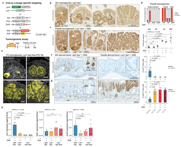
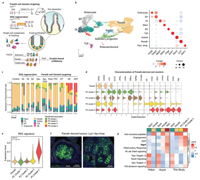
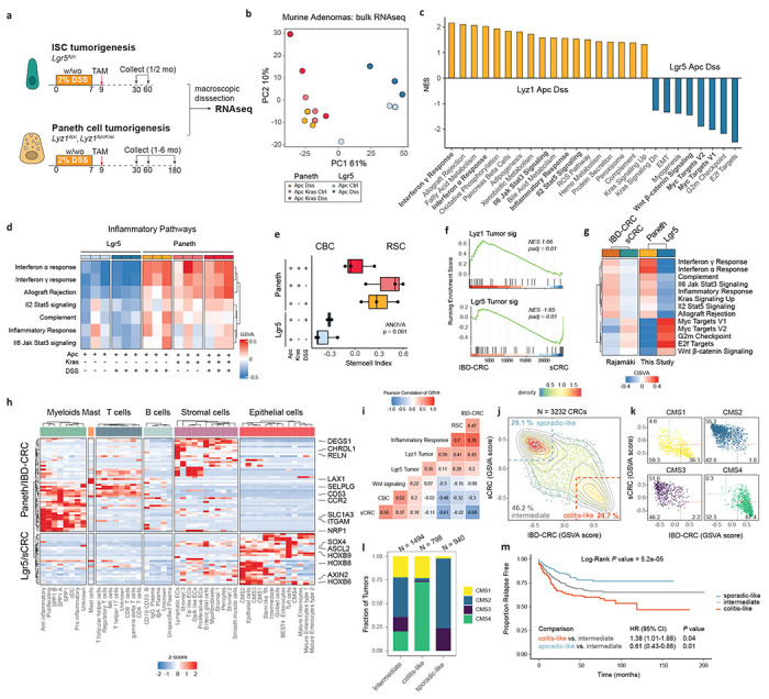
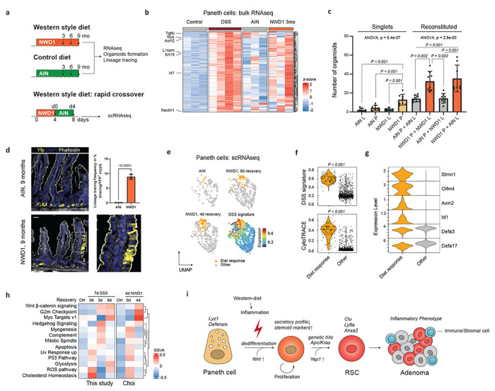
Paneth cells (PCs), responsible for the secretion of antimicrobial peptides in the small intestine and for niche support to Lgr5+ crypt-base columnar stem cells (CBCs), have been shown to respond to inflammation by dedifferentiating into stem-like cells in order to sustain a regenerative response1,2. Therefore, PCs may represent the cells-of-origin of intestinal cancer in the context of inflammation. To test this hypothesis, we targeted Apc, Kras, and Tp53 mutations in Paneth cells by Cre-Lox technology and modelled inflammation by dextran sodium sulfate (DSS) administration. PC-specific loss of Apc resulted in multiple small intestinal tumors, whereas Kras or Tp53 mutations did not. Compound Apc and Kras mutations in PCs resulted in a striking increase in tumor multiplicity even in the absence of the inflammatory insult. By combining scRNAseq with lineage tracing to capture the conversion of PCs into bona fide tumor cells, we show that they progress through a "revival stem cell" (RSC) state characterized by high Clusterin (Clu) expression and Yap1 signaling, reminiscent of what has been previously observed upon irradiation of the mouse digestive tract3. Accordingly, comparison of PC- and Lgr5-derived murine intestinal tumors revealed differences related to Wnt signaling and inflammatory pathways which match the dichotomy of CBCs and injury-induced RSCs4 between human sporadic colon cancers and those arising in the context of inflammatory bowel diseases. Last, we show that western-style dietary habits, known to trigger a low-grade inflammation throughout the intestinal tract, underlie the analogous dedifferentiation of Paneth cells and their acquisition of stem-like features. Taken together, our results show that intestinal cancer arises in the context of inflammation through the dedifferentiation of committed secretory lineages such as Paneth cells and the activation of the revival stem cell state. As such, a true quiescent stem cell identity may be hidden in fully committed and postmitotic lineages which, upon inflammation, support the regenerative response by re-entering the cell cycle and dedifferentiating into RSCs. The chronic nature of the tissue insult in inflammatory bowel diseases and even in the context of western-style dietary habits is likely to result in the expansion of cell targets for tumor initiation and progression.


Conflict of interest statement

Competing interests The authors declare no competing interests.

Figures

Fig. 1 |
Fig. 1 |. Paneth cells as the cell-of-origin of intestinal cancer.
a. Cre-Lox strategy to target Apc, Kras, and Tp53 mutations in intestinal stem cells (Lgr5+ ISCs) and Paneth cells (Lyz1+ PCs). b. and c. β-catenin IHC analysis of intestinal tumors initiated from Lgr5+ ISCs (b) and PCs (c). The asterisks indicate Lgr5+ ISCs and PCs with enhanced cytoplasmic and nuclear β-catenin accumulation; tumor foci and adenomas are indicated by dashed lines. d. Tumor multiplicity was calculated according to tumor-bearing animals (top panel) and by tumor number per genotype (lower panel) in the presence/absence of DSS based on Swiss roll counts. Error bars denote standard deviations of the mean. P values denote one-way ANOVA and Tukey post-hoc tests for group comparisons. e. Lineage tracing analysis of Paneth cells (labelled by YFP) at different stages of tumor-initiation and progression. f. and g. Left panels: Lyz1 (f) and Dclk1 (g) IHC analysis Lgr5+ ISCs and PCs-derived adenomas. Right panels: quantification of number of Lyz1- and Dclk1-positive tumor cells. P values depict one-way ANOVA and Tukey post-hoc tests for group comparisons. h. Lyz1, Dclk1, and Axin2 qPCR expression analysis across different adenoma genotypes. P values represent one-way ANOVA and Tukey post-hoc tests for group comparisons.
Fig. 2 |
Fig. 2 |. Paneth cells dedifferentiate into a revival stem cell identity.
a. Schematics of the experimental approach. After genetic targeting of Paneth cells, intestinal crypts were extracted, and the isolated cells labeled with hashing antibodies and sorted according to three different sorting strategies: epithelium, PC-enriched and -traced cells. b. UMAP embedding of the different cell clusters/lineages (left panel), annotated according to the expression of canonical marker genes (right panel). c. Bar plot of the distribution of traced cells across the different mouse genotypes and experimental conditions. d. Violin plots representing marker genes of the newly identified Paneth-derived cell clusters (PC Cluster 1-4). e. Association analysis of the revival stem cell (RSC) signature with PC Cluster 1-4. f. RNA in situ hybridizations of the Clu gene in small tumors derived from Paneth cells upon compound targeting of Apc and Kras mutations. g. Gene sets variation analysis among the Haber et al., Ayyaz et al., and the present studies.
Fig. 3 |
Fig. 3 |. Paneth-derived adenomas have an inflammatory phenotype mimicking colitis-associated cancer
a. Schematics of the experimental approach to compare PC- and Lgr5-derived adenomas. b. Principal Component Analysis plot showing that the cell-of-origin is the dominant discriminator of variance. c. Bar plot summarizing the gene set enrichment analysis between Paneth- (Lyz1 Apc DSS) and ISC- (Lgr5 Apc DSS) derived tumors. Pathways were filtered based on Pval < 0.05 and abs NES > 0.5. d. Subset of inflammatory pathways, visualized as heat map based on values from the gene set variation analysis. e. Box plots showing results of the stem cell index. P value depicts result of one-way ANOVA. f. Gene set enrichment analysis showing significant but opposite associations between the Lyz1 tumor signature and IBD-CRCs, and between the Lgr5 tumor signature and sCRC. g. Heatmap showing GSVA scores, averaged per tumor group, of pathways with similar patterns between the murine and human tumor groups. h. Heatmap highlighting the differentially expressed genes (log2FC >1.5, Padj < 0.01) shared between the Paneth/IBD-tumors and the Lgr5/sCRC-tumors. Values denote z-scores of average expression per cell type. i-m. Two distinct sporadic colon cancer identities become apparent upon analysis of a large cohort of CRC tumors (N = 3232 samples). i. Heatmap showing Pearson Correlations of the GSVA scores. j-k. Scatter plot showing the two distinct clusters of sporadic- and colit6is-like in all (j) colon cancers and (k) per molecular subtype. Grey lines indicate contours lines, dashed lines show thresholds to classify tumors in colitis-like, sporadic-like and intermediate groups. I. Stacked bar plot analysis showing the distribution of consensus molecular subtypes (CMS1 to 4) across the colitis-like and sporadic-like colon cancers. m. Kaplan-Meier survival analysis for relapse free survival. Pvalues denote result of log-rank test and cox regression models for univeriate analyses. Hazard ratios (HR) and confidence intervals (CI) are displayed for pairwise comparisons.
Fig. 4 |
Fig. 4 |. Western-style diet triggers an inflammatory response leading to dedifferentiation of Paneth cells.
a. Schematics of the experimental approach to investigate the consequences of short- and long-term exposure to western-style diet (NWD1) vs. control (AIN76A) diets. b. Heatmap showing z-scored DSS signature (DSS vs. Control; Padj < 0.05, Log2FC > 0.25) in Paneth cells exposed to DSS or NWD1. c. Organoid multiplicities derived either from single ISCs and PCs and reconstituted doublets (L: Lgr5+ ISCs, P: Paneth cells). Pooled data from N = 4 independent experiments. P values were calculated using one-way ANOVA and Tukey tests for group comparisons. Error bars depict standard deviations from the mean. d. Representative image of lineage tracings from a NWD1-fed Lyz1-Yfp mouse. Scale bar: 50 μm. P value depicts result of the student t-test and error bar represents the standard deviation. Data from N = 3 mice. e. UMAP showing Paneth cells from AIN76A- and NWD1-fed mice (N = 3 mice per condition). The DSS signature portrayed on UMAP embedding, highlights a subcluster of Paneth cells responsive to the NWD1 diet. f. Violin plots showing different levels of the DSS signature (top) and CytoTRACE score (bottom) between the PCs responsive to the NWD1 diet and other Paneth cells. P values depict significance values of Wilcoxon test. g. Violin plots representing marker genes of PCs responsive to the NWD1 diet, showing co-expression of stem and secretory markers. h. Heatmap visualization of gene set variation analysis, indicating pathways that are activated in Paneth cells after exposure to DSS or NWD1. i. Model: Paneth cells react to dietary and inflammatory cues by dedifferentiation to aid epithelial regeneration. During this process, Paneth cells become potential cells-of-origin of cancer, leading to an alternative bottom-up route to intestinal tumorigenesis.

References

    1. Schmitt M. et al. Paneth Cells Respond to Inflammation and Contribute to Tissue Regeneration by Acquiring Stem-like Features through SCF/c-Kit Signaling. Cell Rep 24, 2312–2328 e2317, doi:S2211-1247(18)31203-8 [pii]10.1016/j.celrep.2018.07.085 (2018). - DOI - PubMed
    1. Yu S. et al. Paneth Cell Multipotency Induced by Notch Activation following Injury. Cell Stem Cell 23, 46–59 e45, doi:10.1016/j.stem.2018.05.002 (2018). - DOI - PMC - PubMed
    1. Ayyaz A. et al. Single-cell transcriptomes of the regenerating intestine reveal a revival stem cell. Nature 569, 121–125, doi:10.1038/s41586-019-1154-y10.1038/s41586-019-1154-y [pii] (2019). - DOI - PubMed
    1. Vasquez E. G. et al. Dynamic and adaptive cancer stem cell population admixture in colorectal neoplasia. Cell Stem Cell 29, 1213–1228 e1218, doi:S1934-5909(22)00303-4 [pii]10.1016/j.stem.2022.07.008 (2022). - DOI - PMC - PubMed
    1. Barker N. et al. Crypt stem cells as the cells-of-origin of intestinal cancer. Nature 457, 608–611, doi:nature07602 [pii]10.1038/nature07602 (2009). - DOI - PubMed

Publication types