[Preprint]. 2023 Jan 2:2023.01.02.521806.
doi: 10.1101/2023.01.02.521806.

Arginine limitation causes a directed DNA sequence evolution response in colorectal cancer cells

Affiliations

Arginine limitation causes a directed DNA sequence evolution response in colorectal cancer cells

Dennis J Hsu et al. bioRxiv. .

Update in

Abstract

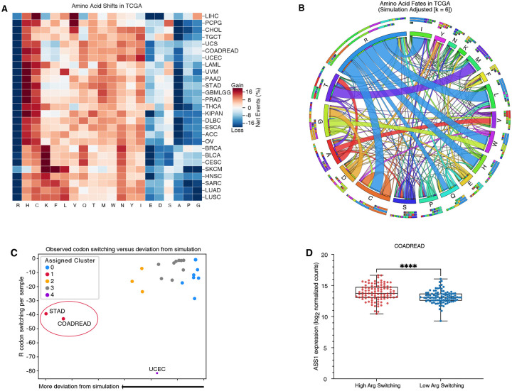
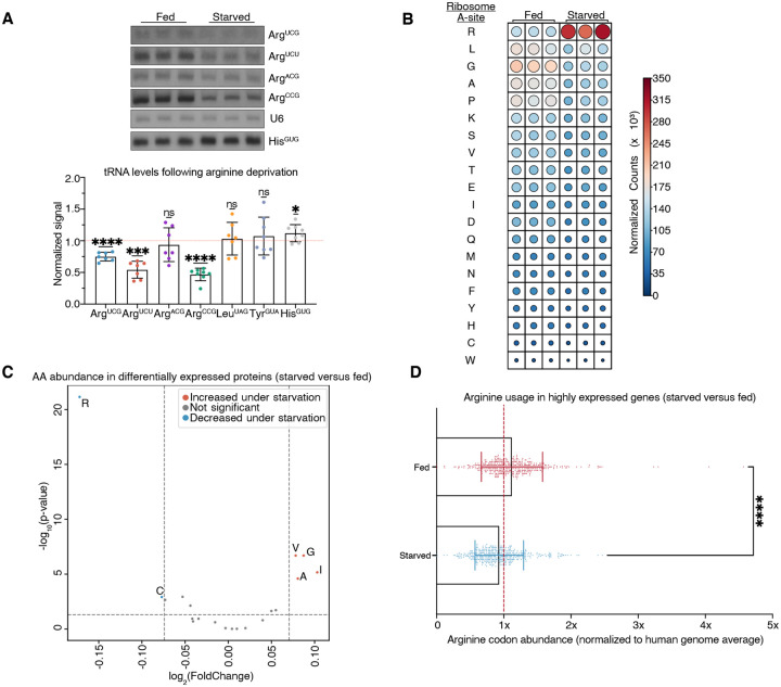
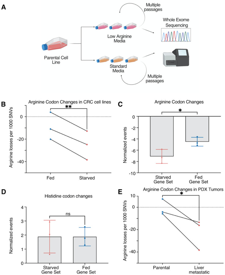
Utilization of specific codons varies significantly across organisms. Cancer represents a model for understanding DNA sequence evolution and could reveal causal factors underlying codon evolution. We found that across human cancer, arginine codons are frequently mutated to other codons. Moreover, arginine restriction-a feature of tumor microenvironments-is sufficient to induce arginine codon-switching mutations in human colon cancer cells. Such DNA codon switching events encode mutant proteins with arginine residue substitutions. Mechanistically, arginine limitation caused rapid reduction of arginine transfer RNAs and the stalling of ribosomes over arginine codons. Such selective pressure against arginine codon translation induced a proteomic shift towards low arginine codon containing genes, including specific amino acid transporters, and caused mutational evolution away from arginine codons-reducing translational bottlenecks that occurred during arginine starvation. Thus, environmental availability of a specific amino acid can influence DNA sequence evolution away from its cognate codons and generate altered proteins.


Conflict of interest statement

Competing interests: Authors declare that they have no competing interests.

Figures

Fig. 1.
Fig. 1.. Arginine codons and residues are frequently lost and are associated with an increase in ASS1 expression.
(A) Heatmap depicting codons gained (red) and lost (blue) across the TCGA. Gains and losses are normalized to the total number of missense and silent mutation events for each cancer type (B) Qualitative chord diagram showing amino acid switching events in cancer after adjustment from simulations. Ribbons which directly touch a column segment indicate loss of that specific amino acid codon during a mutational event and gain of the corresponding amino acid codon in which the ribbon terminates. Ribbons that begin and end at the same amino acid represent synonymous mutations (C) Arginine codon switching events observed versus predicted. Clusters were assigned with Affinity Propagation (D) ASS1 expression in colorectal cancer samples with either a high-degree or low-degree of arginine codon switching events (n=96 per group) (DESeq2 ****Padjusted < 0.0001).
Fig. 2.
Fig. 2.. Arginine codon losses are associated with increased dependence on extracellular arginine and nucleotide pool instability during starvation.
(A) Cell line viability under low arginine (12.5 uM) conditions. (n = 3 per group) (B) Effect of nucleotide supplementation on colon cancer cell viability with arginine deprivation (n = 6 per group, two-tailed t-test) (C) Metabolite profiling differences after exposure to low arginine concentrations for 24 hours. Each point represents a purine/pyrimidine pathway metabolite and is the average log2FC difference between high arginine codon-mutated lines and low-codon mutated lines (one-sample t-test with μ0 = 0). (D) Volcano plot of metabolite changes following arginine deprivation. Only detected citric acid cycle, urea cycle, amino acids, and nucleotide intermediates are labeled. (*P < 0.05, **P < 0.01, ***P<0.001)
Fig. 3.
Fig. 3.. Arginine deprivation reduces arginine tRNA availability, increases arginine ribosome localization, and reduces arginine usage in the tumor proteome.
(A) tRNA quantification as assessed via northern blot. Each dot represents the average abundance in an independent colon cancer or gastric cancer cell line (n = 7 per group, one-sample t-test with μ0 = 0) (B) Ribosome A-site localization counts from ribosomal profiling experiments under starved or fed conditions. Circle size is scaled to counts (C) Amino acid usage in genes that are highly expressed under fed or starved states (D) Arginine codon abundance in genes expressed in fed or starved states (n ≥ 450 per group, two-tailed Mann-Whitney test). Proteins are stratified based on the top 10% most changed in either fed or starved states. (*P<0.05, **P<0.01, ***P<0.001, ****P<0.0001)
Fig. 4.
Fig. 4.. Arginine deprivation promotes arginine-losing mutations.
(A) Schematic of arginine deprivation experiments. (B) Arginine codon changes in cells serially passaged in either full media or low arginine media (n=3 per group, two-tailed paired t-test). (C) Arginine codon changes in proteins that are increased during the fed or arginine-starved states (n = 3 per group, two-tailed t-test). (D) Histidine codon changes in proteins that are increased during the fed or arginine-starved states (n = 3 per group, two-tailed t-test) (E) Arginine codon changes in PDX tumors that underwent multiple rounds of in vivo liver metastatic selection (n = 3 per group, one-tailed paired t-test). (*P<0.05, **P<0.01)

References

    1. Hershberg R., Petrov D. A., Selection on Codon Bias. Annual Review of Genetics. 42, 287–299 (2008). - PubMed
    1. The Cancer Genome Atlas Research Network, Weinstein J. N., Collisson E. A., Mills G. B., Shaw K. R. M., Ozenberger B. A., Ellrott K., Shmulevich I., Sander C., Stuart J. M., The Cancer Genome Atlas Pan-Cancer analysis project. Nat Genet. 45, 1113–1120 (2013). - PMC - PubMed
    1. Alexandrov L. B., Australian Pancreatic Cancer Genome Initiative, ICGC Breast Cancer Consortium, ICGC MMML-Seq Consortium, ICGC PedBrain, Nik-Zainal S., Wedge D. C., Aparicio S. A. J. R., Behjati S., Biankin A. V., Bignell G. R., Bolli N., Borg A., Børresen-Dale A.-L., Boyault S., Burkhardt B., Butler A. P., Caldas C., Davies H. R., Desmedt C., Eils R., Eyfjörd J. E., Foekens J. A., Greaves M., Hosoda F., Hutter B., Ilicic T., Imbeaud S., Imielinski M., Jäger N., Jones D. T. W., Jones D., Knappskog S., Kool M., Lakhani S. R., López-Otín C., Martin S., Munshi N. C., Nakamura H., Northcott P. A., Pajic M., Papaemmanuil E., Paradiso A., Pearson J. V., Puente X. S., Raine K., Ramakrishna M., Richardson A. L., Richter J., Rosenstiel P., Schlesner M., Schumacher T. N., Span P. N., Teague J. W., Totoki Y., Tutt A. N. J., Valdés-Mas R., van Buuren M. M., van ‘t Veer L., Vincent-Salomon A., Waddell N., Yates L. R., Zucman-Rossi J., Andrew Futreal P., McDermott U., Lichter P., Meyerson M., Grimmond S. M., Siebert R., Campo E., Shibata T., Pfister S. M., Campbell P. J., Stratton M. R., Signatures of mutational processes in human cancer. Nature. 500, 415–421 (2013). - PMC - PubMed
    1. Alexandrov L. B., Stratton M. R., Mutational signatures: the patterns of somatic mutations hidden in cancer genomes. Current Opinion in Genetics & Development. 24, 52–60 (2014). - PMC - PubMed
    1. Pan M., Reid M. A., Lowman X. H., Kulkarni R. P., Tran T. Q., Liu X., Yang Y., Hernandez-Davies J. E., Rosales K. K., Li H., Hugo W., Song C., Xu X., Schones D. E., Ann D. K., Gradinaru V., Lo R. S., Locasale J. W., Kong M., Regional glutamine deficiency in tumours promotes dedifferentiation through inhibition of histone demethylation. Nat. Cell Biol. 18, 1090–1101 (2016). - PMC - PubMed

Publication types