[Preprint]. 2023 Jan 19:2023.01.19.524776.
doi: 10.1101/2023.01.19.524776.

Maturation of glutamatergic transmission onto dorsal raphe serotonergic neurons

Affiliations

Maturation of glutamatergic transmission onto dorsal raphe serotonergic neurons

Alexandre Kisner et al. bioRxiv. .

Update in

Abstract

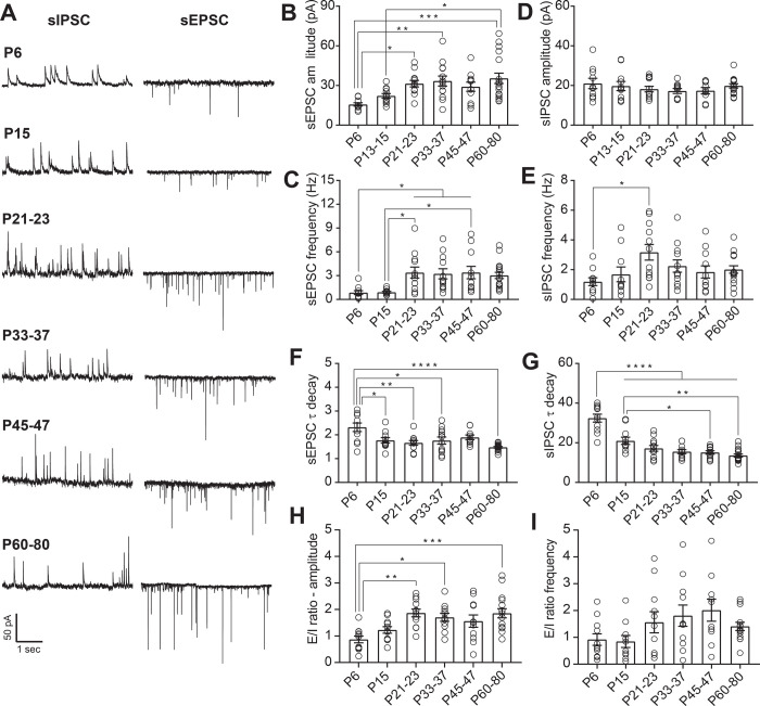
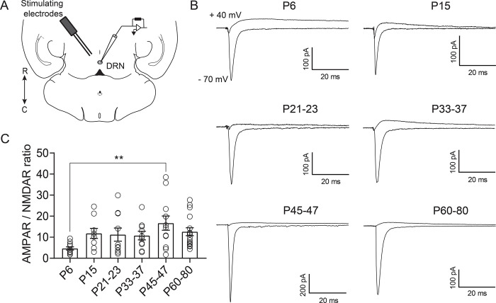
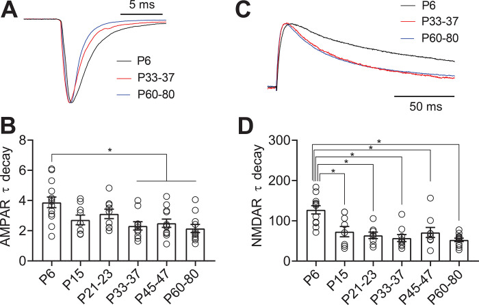
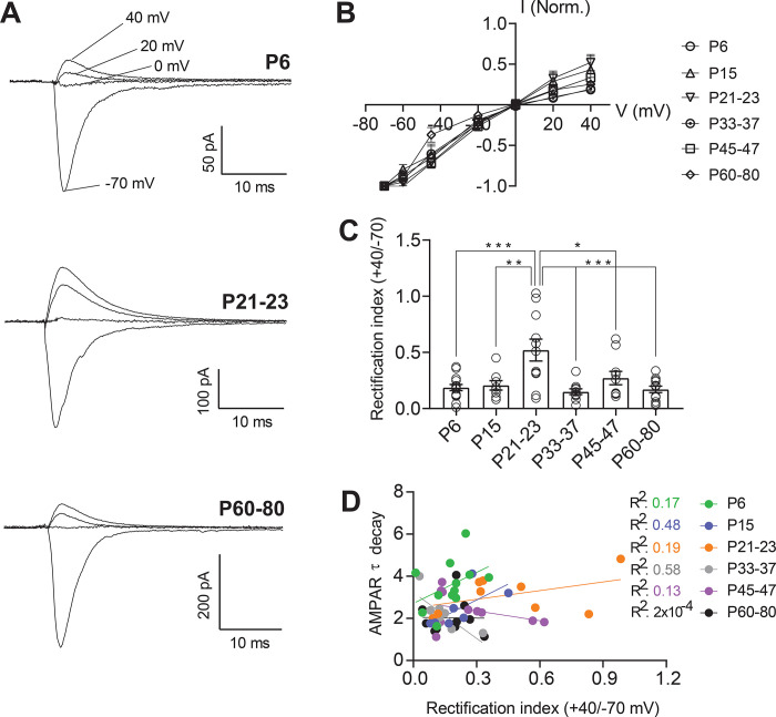
Serotonergic neurons in the dorsal raphe nucleus (DRN) play important roles early in postnatal development in the maturation and modulation of higher order emotional, sensory, and cognitive circuitry. This unique position makes these cells a substrate by which early experience can be wired into brain. In this study, we have investigated the maturation of synapses onto dorsal raphe serotonergic neurons in typically developing male and female mice using whole-cell patch-clamp recordings in ex vivo brain slices. We show that while inhibition of these neurons is relatively stable across development, glutamatergic synapses greatly increase in strength between P6 and P21-23. In contrast to forebrain regions, where the components making up glutamatergic synapses are dynamic across early life, we find that the makeup of these synapses onto DRN serotonergic neurons is largely stable after P15. DRN excitatory synapses maintain a very high ratio of AMPA to NMDA receptors and a rectifying component of the AMPA response throughout the lifespan. Overall, these findings reveal that the development of serotonergic neurons is marked by a significant refinement of glutamatergic synapses during the first 3 postnatal weeks. This suggests this time as a sensitive period of heightened plasticity for integration of information from upstream brain areas and that genetic and environmental insults during this period could lead to alterations in serotonergic output, impacting both the development of forebrain circuits and lifelong neuromodulatory actions.


Figures

Figure 1.
Figure 1.
Maturation of spontaneous synaptic transmission onto DRN serotonergic neurons. A: Representative traces of spontaneous synaptic transmission at developmental timepoint. For each timepoint, traces were recorded from the same 5-HT neuron at holding potentials of −70 mV (sEPSC) and 0 mV (sIPSC). B, C: Developmental changes in the mean amplitude (B) and frequency (C) of sEPSCs. D, E: Developmental changes in the mean amplitude (D) and frequency (E) of sIPSCs. F: Time constant of decay (τdecay) for sEPSCs. G: τdecay for sIPSCs. H: Excitation/Inhibition (E/I) ratio for amplitude of sPSCs. I: E/I ratio for frequency of sPSCs. PND 6, n = 10 cells (5 mice); PND 15, n = 11 cells (7 mice); PND 21–23, n = 12 cells (8 mice); PND 33–37, n = 12 cells (8 mice); PND 45–47, n = 11 cells (8 mice); PND 60–80, n = 17 cells (10 mice). Bar graphs represent mean ± SEM. *p < 0.05, one-way ANOVA followed by Tukey’s multiple comparison test.
Figure 2.
Figure 2.
Excitatory currents onto DRN serotonergic neurons are primarily mediated by AMPAR. A: Schematic of the experiment illustrating the placement of the bipolar stimulating electrode in the DRN and recording of evoked excitatory currents from 5-HT neurons. B: Representative traces of electrically evoked excitatory synaptic currents recorded from 5-HT neurons at holding potentials of −70 mV and +40 mV. Recordings at +40 mV were performed in the presence of the AMPA antagonist NBQX (10 μM). C: Quantification of AMPA/NMDA ratios across maturation. PND 6, n = 11 cells (6 mice); PND 15, n = 9 cells (6 mice); PND 21–23, n = 10 cells (9 mice); PND 33–37, n = 10 cells (7 mice); PND 45–47, n = 12 cells (8 mice); PND 60–80, n = 16 cells (11 mice). Bar graphs represent mean ± SEM. ** p < 0.01, one-way ANOVA followed by Tukey’s multiple comparison test.
Figure 3.
Figure 3.
AMPA and NMDA receptor subunits are developmentally regulated in 5-HT neurons. A: Scaled representative traces of evoked AMPAR currents. B: Time constant of decay (τdecay) for evoked AMPAR currents recorded at −70 mV. C: Scaled representative traces of evoked NMDAR currents. D: Time constant of decay (τdecay) for evoked NMDAR currents recorded at +40 mV in the presence of the AMPA antagonist NBQX (10 μM). Bar diagrams represent mean ± SEM. * p < 0.05, one-way ANOVA followed by Tukey’s multiple comparison test.
Figure 4.
Figure 4.
AMPAR-mediated currents are inwardly rectifying throughout development in a subset of 5-HT neurons A: Representative traces of evoked excitatory synaptic currents recorded from 5-HT neurons at holding potentials of −70 mV to +40 mV. B: Average peak current – voltage curves with slight deviation from a linear relationship. C: Rectification index of AMPAR currents, calculated as the ratio of peak amplitude at +40 mV divided by the peak amplitude at −70 mV for each cell. D: Correlation between AMPAR EPSC τ decay and rectification index for each time point. PND 6, n = 14 cells (7 mice); PND 15, n = 8 cells (7 mice); PND 21–23, n = 11 cells (6 mice); PND 33–37, n = 10 cells (5 mice); PND 45–47, n = 10 cells (7 mice); PND 60–80, n = 12 cells (7 mice). Bar graphs represent mean ± SEM. *p < 0.05, one-way ANOVA followed by Tukey’s multiple comparison test.
Figure 5.
Figure 5.
Schematic summarizing the maturation of synaptic inputs onto 5-HT neurons. A: The overall synaptic drive is led by glutamatergic transmission, which undergoes an abrupt enhancement between juvenile and the pre-adolescent period and then remained stable throughout adulthood. B: Glutamatergic transmission is mainly carried by AMPA receptors and in a subset of 5-HT neurons these receptors have a high permeability to calcium and are inwardly rectifying across the whole developmental period.

References

    1. Tau GZ, Peterson BS. Normal development of brain circuits. Neuropsychopharmacol Off Publ Am Coll Neuropsychopharmacol 35: 147–168, 2010. doi: 10.1038/npp.2009.115. - DOI - PMC - PubMed
    1. Suri D, Teixeira CM, Cagliostro MKC, Mahadevia D, Ansorge MS. Monoamine-sensitive developmental periods impacting adult emotional and cognitive behaviors. Neuropsychopharmacol Off Publ Am Coll Neuropsychopharmacol 40: 88–112, 2015. doi: 10.1038/npp.2014.231. - DOI - PMC - PubMed
    1. Hartley CA, Lee FS. Sensitive periods in affective development: nonlinear maturation of fear learning. Neuropsychopharmacol Off Publ Am Coll Neuropsychopharmacol 40: 50–60, 2015. doi: 10.1038/npp.2014.179. - DOI - PMC - PubMed
    1. Schoenfeld TJ, Cameron HA. Adult neurogenesis and mental illness. Neuropsychopharmacol Off Publ Am Coll Neuropsychopharmacol 40: 113–128, 2015. doi: 10.1038/npp.2014.230. - DOI - PMC - PubMed
    1. Guang S, Pang N, Deng X, Yang L, He F, Wu L, Chen C, Yin F, Peng J. Synaptopathology Involved in Autism Spectrum Disorder. Front Cell Neurosci 12: 470, 2018. doi: 10.3389/fncel.2018.00470. - DOI - PMC - PubMed

Publication types