[Preprint]. 2023 Jan 18:2023.01.17.524254.
doi: 10.1101/2023.01.17.524254.

Higher Angiotensin I Converting Enzyme 2 (ACE2) levels in the brain of individuals with Alzheimer's disease

Affiliations

Higher Angiotensin I Converting Enzyme 2 (ACE2) levels in the brain of individuals with Alzheimer's disease

Reveret Louise et al. bioRxiv. .

Update in

Abstract

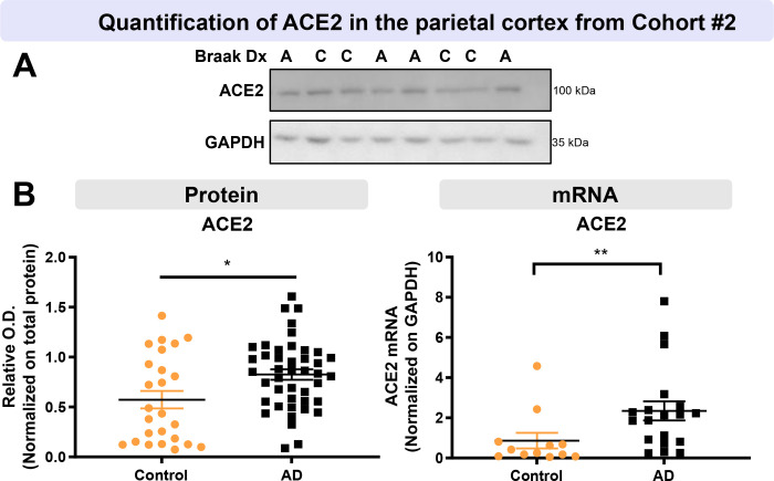
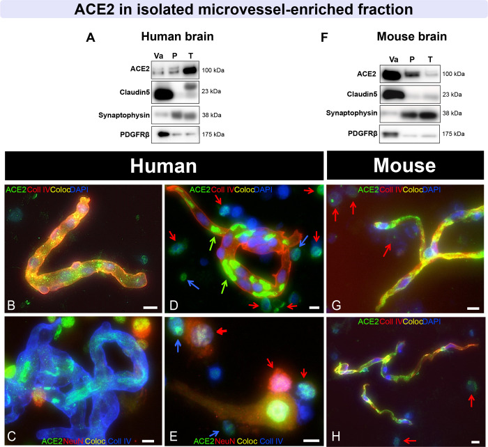
The severe acute respiratory syndrome coronavirus 2 (SARS-CoV-2) is a major cause of death in the elderly. Cognitive decline due to Alzheimer's disease (AD) is frequent in the geriatric population disproportionately affected by the COVID-19 pandemic. Interestingly, central nervous system (CNS) manifestations have been reported in SARS-CoV-2-infected patients. In this study, we investigated the levels of Angiotensin I Converting Enzyme 2 (ACE2), the main entry receptor of SARS-COV-2 in cells, in postmortem parietal cortex samples from two independent AD cohorts, totalling 142 persons. Higher concentrations of ACE2 protein and mRNA were found in individuals with a neuropathological diagnosis of AD compared to age-matched healthy control subjects. Brain levels of soluble ACE2 were inversely associated with cognitive scores (p = 0.02), markers of pericytes (PDGFRβ, p=0.02 and ANPEP, p = 0.007) and caveolin1 (p = 0.03), but positively correlated with soluble amyloid-β peptides (Aβ) concentrations (p = 0.01) and insoluble phospho- tau (S396/404, p = 0.002). No significant differences in ACE2 were observed in the 3xTgAD mouse model of tau and Aβ neuropathology. Results from immunofluorescence and Western blots showed that ACE2 protein is mainly localized in neurons in the human brain but predominantly in microvessels in the mouse brain. The present data show that an AD diagnosis is associated with higher levels of soluble ACE2 in the human brain, which might contribute to a higher risk of CNS SARS-CoV-2 infection.


Conflict of interest statement

Competing interests

The authors report no competing interests.

Figures

Figure 1:
Figure 1:. Levels of TBS-Soluble ACE2 protein are higher in AD individuals and are negatively correlated with global cognitive score.
Parietal cortex levels of ACE2 protein from Cohort #1 were determined by Western Blot in three fractions: a TBS-soluble fraction (A-D), a detergent-soluble fraction (E-H) and a microvessel-enriched fraction (I-L). No statistical difference was detected for ACE2 in the three fractions when subjects were classified according to antemortem clinical diagnosis (B, F, J). Levels of the ACE2 protein were higher in the TBS-soluble fraction in individuals with a neuropathological diagnosis of AD based on ABC scoring (C, G, K). TBS-soluble ACE2 and microvascular ACE2 levels were negatively correlated with the global cognitive score (D, L). An equal amount (12 μg) of proteins per sample for both TBS-soluble and detergent soluble fractions was loaded and 8 μg of proteins per sample was loaded for microvessel-enriched fractions. All samples, loaded in a random order, were run on the same gel and transferred on the same membrane before immunoblotting for quantification. Examples were taken from the same experiment, and consecutive bands loaded in random order are shown. Actin and cyclophilin B are shown as loading controls. Data are represented as a scatterplot. Horizontal lines indicate mean ± SEM. Statistical analysis: Mann-Whitney test **p < 0.01, Coefficient of determination &p < 0.05. Abbreviations: ACE2, Angiotensin-Converting Enzyme 2; A/AD, Alzheimer’s disease; C, control; Clin Dx, clinical diagnosis; ABC Dx, ABC neuropathological diagnosis; CyB, cyclophilin B; M/MCI, mild cognitive impairment; N/NCI, healthy controls with no cognitive impairment; O.D., optical density; TBS, Tris-Buffered Saline.
Figure 2:
Figure 2:. Higher ACE2 protein and mRNA levels in parietal cortex of AD participants from the second cohort.
AD subjects from Cohort #2 had higher levels of ACE2 protein and mRNA compared to controls. Diagnosis was determined using Braak staging. ACE2 levels were determined by Western-Blot and qPCR analysis (A, B). Statistical analysis: Mann-Whitney test *p < 0.05, Unpaired t-test **p < 0.01. All samples, loaded in a random order, were run on the same immunoblot experiment for quantification. Examples were taken from the same immunoblot experiment, and consecutive bands loaded in random order are shown. GAPDH is shown as a loading control. Data are represented as a scatterplot. Horizontal lines indicate mean ± SEM. Abbreviations: A, Alzheimer’s disease (Braak scores III-VI); ACE2, Angiotensin-Converting Enzyme 2; C, control (Braak scores I or II); Dx, diagnostic; O.D., optical density.
Figure 3:
Figure 3:. ACE2 protein in TBS-soluble/microvascular protein fractions show opposite relationships with detergent-soluble ACE2 when correlating with AD markers.
Heat-map of hierarchical clustering analysis of correlation coefficients from partial correlation analyses with antemortem evaluation, neuropathological markers (soluble and insoluble proteins of the parietal cortex) and BBB markers (microvascular fractions of the parietal cortex). The significance of the correlation (inverse in blue, positive in red) between two elements is entered in the associated box. Statistical analysis: Coefficient of determination *p < 0.05, **p < 0.01 and ***p < 0.001. All proteins presented in these heat-maps were determined by Western-blot analysis except Aβ peptides which were quantified using ELISA. Total soluble and insoluble Tau was detected using Tau (640–680) antibody. AD2 antibody recognized Tau phosphorylated at S396. Phosphorylated TDP43 antibody recognized at pSer409/410. Abbreviations: ABCB1, ATP Binding Cassette Subfamily B Member 1; ACE2, Angiotensin-Converting Enzyme 2; ANPEP, Aminopeptidase N; BBB, blood-brain barrier; BACE1, Beta-Secretase 1; CD31 or PECAM1, Platelet endothelial cell adhesion molecule; LRP1, Low density lipoprotein receptor-related protein 1; NS, non significative; PDGFRß, Platelet Derived Growth Factor Receptor Beta; RAGE, Receptor for Advanced Glycation Endproducts; αSMA, alpha smooth muscle actin; TDP-43, TAR DNA binding protein 43; TBS, Tris-Buffered Saline.
Figure 4:
Figure 4:. In fractionated brain homogenates, ACE2 immunosignal is predominantly observed in neurons in human samples and in the vasculature in mice.
(A, F) Immunoblotting detection of ACE2 in human (parietal cortex) (A) and mouse (whole brain)(F) vascular fractions “Va”, compared to postvascular parenchymal samples depleted in vascular cells “P” and total unfractionated homogenates “T”. For comparison purposes, synaptophysin (a synaptic/neuronal marker), Claudin5 (endothelial marker), and PDGFRß (pericyte marker), are also shown. In the mouse brain, ACE2 is highly enriched in microvessels compared to the postvascular fraction, different to what is observed in the human (A, F). (B-E, G, H) Representative immunostaining of ACE2 (green) in human (B-E) and murine cerebrovascular fractions (G, H), with collagen IV (endothelial marker) in red (B, D, G, H) or blue (C, E), as well as NeuN (neuronal marker) in red (C, E) and DAPI (nuclei) in blue (B-H). In human samples, moderate ACE2 staining is observed in neurons, whereas vascular ACE2 staining is strong in mice. Red arrows point to ACE2+/NeuN+ cells, blue arrows to ACE2+/NeuN− cells, and green arrows to erythrocytes. ACE2 antibodies: rb mAb #ab108252 (A, F), rb pAb #HPA000288 (B, D, G) and #35–1875 (C, E, H). Scale bar: 10 μm. Abbreviations: ACE2, Angiotensin-Converting Enzyme 2; Coll IV, Collagen IV; Coloc, Colocalization; PDGFRß, Platelet Derived Growth Factor Receptor Beta. mAb, monoclonal antibody; pAb, polyclonal antibody.
Figure 5:
Figure 5:. In tissue sections, ACE2 immunostaining is predominantly observed in neurons in the human brain and in the cerebrovasculature in mice.
(A-E) Representative immunostaining of ACE2 (green or red) in fresh frozen human hippocampus (A, B), formalin-fixed paraffin-embedded parietal cortex (C-E), and in murine fresh frozen hippocampal and cerebellar sections (F-H). For immunofluorescence, NeuN (neuronal marker)(A, B), collagen IV (endothelial marker)(F, G) or PDGFRß (pericyte marker) (H) are in red, and DAPI (nuclei) is in blue (A-C, F-H). For immunohistochemistry, sections were counterstained with hematoxylin (nuclei)(D, E), and negative control is inserted in (E). In human samples, strong ACE2 staining is observed in large and small neurons, whereas in mice, neuronal signal is moderate and vascular ACE2 staining is strong and colocalizes well with PDGFRß. Red arrows point to ACE2+/NeuN+ cells and green arrows to erythrocytes; dashed white line highlights small ACE2+/NeuN+ cells that stain strongly for DAPI. ACE2 antibodies: rabbit mAb #ab108252 (C, G), rabbit pAb #HPA000288 (A, B, E, F) and #35–1875 (D) or goat pAb #AF3437. Scale bars: 200 μm (A, F, G, H), 20 μm (B, D, E) and 10 μm (C). Abbreviations: ACE2, Angiotensin-Converting Enzyme 2; Coll IV, Collagen IV; Coloc, Colocalization; PDGFRβ, Platelet Derived Growth Factor Receptor Beta; mAb, monoclonal antibody; pAb, polyclonal antibody.
Figure 6:
Figure 6:. ACE2 levels are not altered in a model of AD, the 3xTg-AD mouse.
(A) Determination of ACE2 levels by Western immunoblotting in brain homogenates from NonTg and 3xTg-AD mice aged 4, 12, and 18 months. No difference was observed in TBS-soluble and detergent-soluble ACE2. (B) In brain microvessel-enriched fractions from NonTg and 3xTg-AD mice aged 6, 12, 18 months, and in 18-month-old animals fed either a control diet or a high fat diet, no difference was observed in microvascular ACE2 levels. ACE2 levels in the mouse brain are not influenced by age or a diet that exacerbates AD-like neuropathology. Examples were taken from the same immunoblot experiment, and consecutive bands loaded in random order are shown. Actin and cyclophilin B are shown as loading controls. Data are represented as mean ± SEM. Statistical analysis: Kruskal-Wallis, ns, non-significant. Abbreviations: Angiotensin-Converting Enzyme 2; Non-Tg,/NT, non-transgenic mice; 3x, 3xTg-AD mice; CD/C, control diet; CyB, cyclophilin B; HFD/H, high-fat diet; O.D., optical density.

References

    1. Abdi A, Jalilian M, Sarbarzeh PA, Vlaisavljevic Z (2020) Diabetes and COVID-19: A systematic review on the current evidences. Diabetes Research and Clinical Practice. Elsevier Ireland Ltd, City - PMC - PubMed
    1. Abiodun OA, Ola MS (2020) Role of brain renin angiotensin system in neurodegeneration: An update. Saudi Journal of Biological Sciences. Elsevier B.V., City, pp 905–912 - PMC - PubMed
    1. Alnefeesi Y, Siegel A, Lui LMW, Teopiz KM, Ho RCM, Lee Y, Nasri F, Gill H, Lin K, Cao B et al. (2020) Impact of SARS-CoV-2 Infection on Cognitive Function: A Systematic Review. Front Psychiatry 11: 621773 Doi 10.3389/fpsyt.2020.621773 - DOI - PMC - PubMed
    1. Barron AM, Rosario ER, Elteriefi R, Pike CJ (2013) Sex-Specific Effects of High Fat Diet on Indices of Metabolic Syndrome in 3xTg-AD Mice: Implications for Alzheimer’s Disease. PLoS ONE. PLoS One, City - PMC - PubMed
    1. Bennett DA, Schneider J A., Arvanitakis Z, Wilson R S. (2012) Overview and Findings from the Religious Orders Study. Current Alzheimer Research. Bentham Science Publishers Ltd., City, pp 628–645 - PMC - PubMed

Publication types