[Preprint]. 2023 Jan 9:rs.3.rs-2382345.
doi: 10.21203/rs.3.rs-2382345/v1.

Spatiotemporal immune atlas of the first clinical-grade, gene-edited pig-to-human kidney xenotransplant

Affiliations

Spatiotemporal immune atlas of the first clinical-grade, gene-edited pig-to-human kidney xenotransplant

Matthew D Cheung et al. Res Sq. .

Update in

  • Spatiotemporal immune atlas of a clinical-grade gene-edited pig-to-human kidney xenotransplant.
    Cheung MD, Asiimwe R, Erman EN, Fucile CF, Liu S, Sun CW, Hanumanthu VS, Pal HC, Wright ED, Ghajar-Rahimi G, Epstein D, Orandi BJ, Kumar V, Anderson DJ, Greene ME, Bell M, Yates S, Moore KH, LaFontaine J, Killian JT Jr, Baker G, Perry J, Khan Z, Reed R, Little SC, Rosenberg AF, George JF, Locke JE, Porrett PM. Cheung MD, et al. Nat Commun. 2024 Apr 11;15(1):3140. doi: 10.1038/s41467-024-47454-7. Nat Commun. 2024. PMID: 38605083 Free PMC article.

Abstract

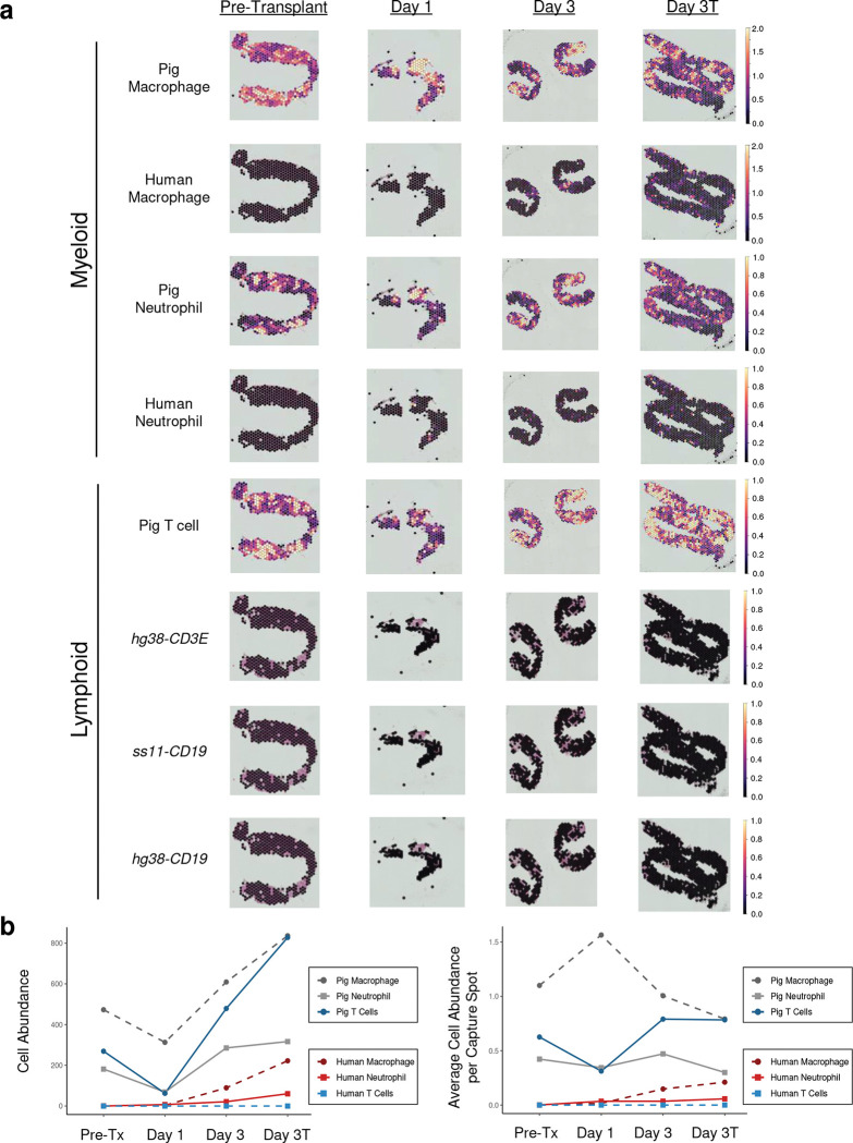
Pig-to-human xenotransplantation is rapidly approaching the clinical arena; however, it is unclear which immunomodulatory regimens will effectively control human immune responses to pig xenografts. We transplanted a gene-edited pig kidney into a brain-dead human recipient on pharmacologic immunosuppression and studied the human immune response to the xenograft using spatial transcriptomics and single-cell RNA sequencing. Human immune cells were uncommon in the porcine kidney cortex early after xenotransplantation and consisted of primarily myeloid cells. Both the porcine resident macrophages and human infiltrating macrophages expressed genes consistent with an alternatively activated, anti-inflammatory phenotype. No significant infiltration of human B or T cells into the porcine kidney xenograft was detected. Altogether, these findings provide proof of concept that conventional pharmacologic immunosuppression is sufficient to restrict infiltration of human immune cells into the xenograft early after compatible pig-to-human kidney xenotransplantation.


Conflict of interest statement

Competing Interests Statement The following authors receive or have received salary support from a research grant from United Therapeutics: RA, EDW, CFF, DE, BJO, MB, GB, JP, RR, SCL, AFR, JEL, and PMP.

Figures

Figure 1.
Figure 1.. Limited infiltration of the porcine kidney xenograft by human immune cells.
Spatial transcriptomics was performed on serial needle core biopsies of 10-GE porcine kidneys before and after transplantation into a brain-dead human recipient. Biopsies were obtained from either the right (pre-transplant and Day 3 samples) or left (Day 1 and Day 3T) xenografts. Cell type signatures were identified from reference transcriptomes using Cell2location or expression of individual marker genes (CD3E and CD19). a) Top: Human myeloid cells are detected in biopsies of the porcine kidney xenograft three days after transplantation. Capture spot color corresponds to cell abundance, and color scales to the right of each spatial plot indicate cell abundance. Note that scaling is conserved across time for a given cell type but differs between macrophages (cell abundance range: 0–2) and neutrophils (cell abundance range: 0–1). Visualization of cell abundance in a given capture spot is thus capped at 1 or 2 cells. Bottom: No detection of human T or B cells in xenograft biopsies at any time point. b) Calculated total (left) and normalized (right) cell abundance for various immune cell types in the indicated biopsies. For clarity, quantification of B cells is not shown. Note that cell abundance for human T cells and all B cells was imputed from expression of hg38-CD3E, ss11-CD19, and hg38-CD19 genes as shown in (a). “Pre-tx” = pre-transplant. Day 3T biopsy was taken on post-transplant day 3 at study termination.
Figure 2.
Figure 2.. Predominance of M2-like macrophages in the porcine kidney xenograft.
scRNA-seq was performed on CD45+ immune cells sorted from the right porcine kidney xenograft at explant (see Extended Fig. 3), and macrophage clusters were selected for analysis. a) Expression of M1 (red) and M2 (blue) genes in human and pig macrophages (see Extended Data Table 1 and ref for full gene list). b) Expanded view of top 50 most highly expressed M1 and M2 genes in each species. Note M2 > M1 genes for both species. c) Composite gene expression score of pig and human macrophages of M1-like pro-inflammatory (red) and M2-like anti-inflammatory (blue) gene signatures (ref 16). UMAPs were generated from re-clustering of macrophage clusters selected from Extended Fig. 3. d) Expression of select anti- and pro-inflammatory cytokine genes in pig and human macrophages. Average gene expression is visualized such that the mean of the scaled expression dataset is set at 0 with a standard deviation of 1. ss11-IL6 was not detected.

References

    1. Wolfe R.A. et al. New Engl J Med 341, 1725–1730 (1999). - PubMed
    1. United States Renal Data System. 2020 USRDS Annual Data Report.
    1. Sykes M. & Sachs D.H. Sci Immunol 4, eaau6298 (2019). - PMC - PubMed
    1. Adams A.B. et al. Ann Surg 268, 264–573 (2018).
    1. Yamamoto T. et al. Sci Rep 10, 9771 (2020). - PMC - PubMed

Supplemental References:

    1. Satija R. et al. Nat Biotechnol 33, 495–502 (2015). - PMC - PubMed
    1. Young M.D. & Behjati S. Gigascience 9, giaa151 (2020). - PMC - PubMed
    1. Korsunsky I. et al. Nat Methods 16, 1289–1296 (2019). - PMC - PubMed
    1. Wolf F. et al. Genome Biol 19, 15 (2018). - PMC - PubMed

Publication types

LinkOut - more resources