[Preprint]. 2023 Jan 9:2023.01.09.522931.
doi: 10.1101/2023.01.09.522931.

Neutrophil extracellular trap stabilization by platelet factor 4 reduces thrombogenicity and endothelial cell injury

Affiliations

Neutrophil extracellular trap stabilization by platelet factor 4 reduces thrombogenicity and endothelial cell injury

Anh T P Ngo et al. bioRxiv. .

Update in

Abstract

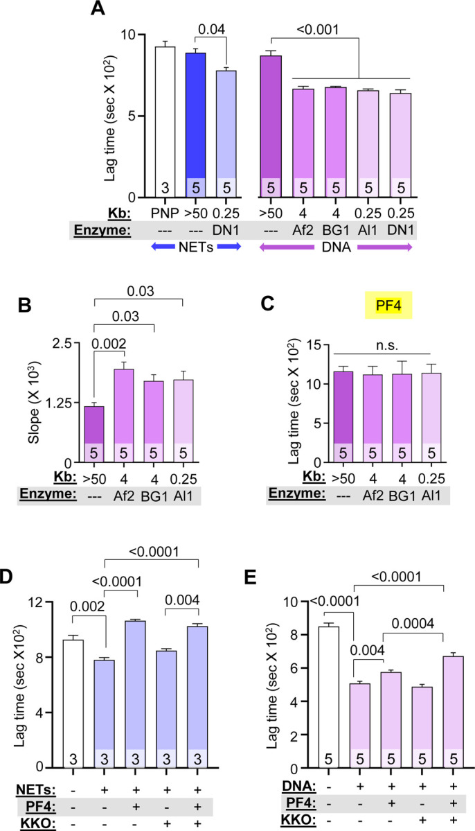
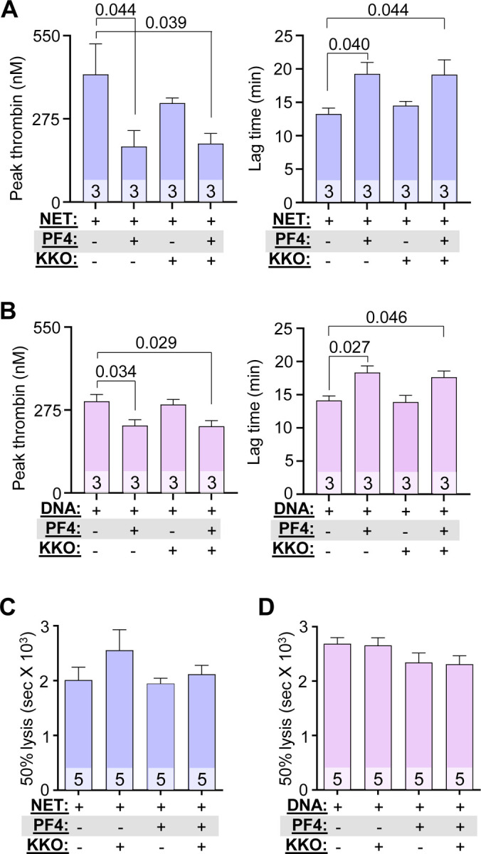
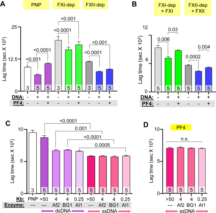
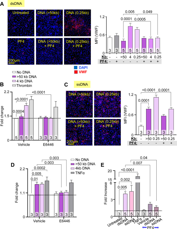
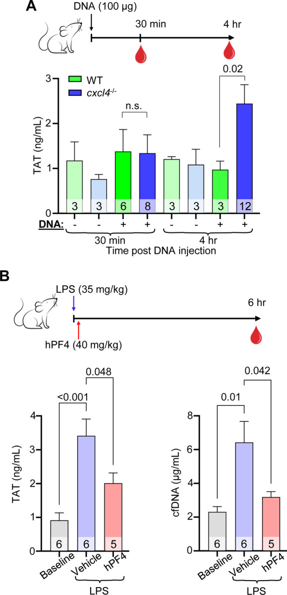
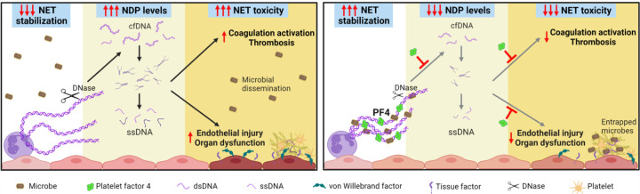
Neutrophil extracellular traps (NETs) are abundant in sepsis, and proposed NET-directed therapies in sepsis prevent their formation or accelerate degradation. Yet NETs are important for microbial entrapment, as NET digestion liberates pathogens and NET degradation products (NDPs) that deleteriously promote thrombosis and endothelial cell injury. We proposed an alternative strategy of NET-stabilization with the chemokine, platelet factor 4 (PF4, CXCL4), which we have shown enhances NET-mediated microbial entrapment. We now show that NET compaction by PF4 reduces their thrombogenicity. In vitro, we quantified plasma thrombin and fibrin generation by intact or degraded NETs and cell-free (cf) DNA fragments, and found that digested NETs and short DNA fragments were more thrombogenic than intact NETs and high molecular weight genomic DNA, respectively. PF4 reduced the thrombogenicity of digested NETs and DNA by interfering, in part, with contact pathway activation. In endothelial cell culture studies, short DNA fragments promoted von Willebrand factor release and tissue factor expression via a toll-like receptor 9-dependent mechanism. PF4 blocked these effects. Cxcl4-/- mice infused with cfDNA exhibited higher plasma thrombin anti-thrombin (TAT) levels compared to wild-type controls. Following challenge with bacterial lipopolysaccharide, Cxcl4-/- mice had similar elevations in plasma TAT and cfDNA, effects prevented by PF4 infusion. Thus, NET-stabilization by PF4 prevents the release of short fragments of cfDNA, limiting the activation of the contact coagulation pathway and reducing endothelial injury. These results support our hypothesis that NET-stabilization reduces pathologic sequelae in sepsis, an observation of potential clinical benefit.

Keywords: cell-free DNA; neutrophil extracellular trap; platelet factor 4; sepsis; thrombosis.


Conflict of interest statement

Conflict-of-interest disclosure The authors declare no conflict-of-interests.

Figures

Figure 1.
Figure 1.. PF4 and KKO inhibits fibrin generation initiated by NET and DNA fragments.
Fibrin generation in pooled normal plasma (PNP) with or without added HMW, digested NETs (blue), or DNA (purple). DN1 = DNase I, Af2 = AflII, BsGrI-HF = BG1 and Al1 = AluI. Data are mean ± SEM) of at least 3 independent experiments as indicated in each bar. (A) Lag time determined from kinetic curves of fibrin generation with NETs (blue) and DNA (purple). (B) Slope (rate) of fibrin generation was determined from the same kinetic curves as in (A). (C) Lag time of DNA-induced fibrin generation was determined from kinetic curves similarly to (A), in the presence of PF4 (20 μg/mL). (D-E) Lag times are shown for fibrin generation studies of DNase I-digested NETs (D) and DNA (E) in the absence or presence of 1 μg/mL low PF4 and/or 10 μg/mL KKO. Data are mean ± SEM of at least 3 independent experiments as indicated in each bar.
Figure 2.
Figure 2.. PF4 and KKO inhibit thrombin generation but not fibrinolysis.
(A and B) Thrombin generation studies of DNase-digested NETs (A) and DNA (B) in the absence or presence of PF4 (20 μg/mL) and/or KKO (10 μg/mL). (C and D) Time to 50% clot lysis of NETs- (C) and DNA- (D) induced coagulation, as determined based on kinetic curves. Data are mean ± SEM of at least 3 independent experiments as indicated in each bar.
Figure 3.
Figure 3.. Effect of PF4 on fibrin generation in FXI- and FXII-depleted plasma and on thrombogenicity of ssDNA.
(A) Lag time, determined from kinetic curves, of fibrin generation in PNP or FXI- or FXII-depleted (dep) plasma, induced by digested DNA in the absence or presence of PF4. (B) Lag time of fibrin generation induced by digested DNA in depleted plasma supplemented with missing coagulation factors. (C) Lag time of ssDNA-induced (pink) fibrin generation compared to dsDNA (purple). (D) Lag time of ssDNA-induced fibrin generation with added PF4 (20 μg/mL). Data are mean ± SEM of at least 3 independent experiments as indicated in each bar.
Figure 4.
Figure 4.. PF4 protects against dsDNA and ssDNA-induced procoagulant responses by endothelium.
Mean fluorescence intensity (MFI) of released VWF from HUVECs exposed to fragments of dsDNA (A and B) or ssDNA (C and D), with or without PF4 or TLR9 inhibitor (E6446). Data were normalized to MFI of untreated cells without exposure to dsDNA or inhibitor (as indicated by dotted lines). Exposure to α-thrombin (B) or TNFα (D) serves as positive controls. (E) TF expression by HUVEC (shown as fold increase from untreated cells without PF4 exposure) induced by dsDNA or ssDNA in the absence or presence of PF4. TNFα exposure was used as a positive control. Data are mean ± SEM of at least 3 independent experiments as indicated in each bar.
Figure 5.
Figure 5.. PF4 attenuates cfDNA-induced coagulability in vivo.
(A) Top: schematic of study in which WT mice or cxcl4−/− littermates were given normal saline vehicle or normal saline containing digested DNA prior to blood collection at 30 minutes or 4 hours. Bar graph: TAT levels using a commercial ELISA kit in platelet-poor plasma. Data are mean ± SEM of at least 3 independent experiments as indicated in each bar. (B) Top: schematic of study in which WT mice received normal saline vehicle or LPS, and a subset of animals was given hPF4 by tail-vein injection immediately following LPS injection. TAT levels were measured in blood was drawn at baseline and 6-hours post LPS ± PF4 infusion. Bar graphs: TAT levels (left) and cfDNA levels (right) at 6-hours post LPS challenge. Data are mean ± SEM of at least 5 independent experiments as indicated in each bar.
Figure 6.
Figure 6.. Proposed protective mechanisms of NET-stabilization
Left: NETs are subjected to digestion by DNase I, reducing microbial capture and liberating toxic NDPs, including cfDNA. These cfDNA fragments expose ssDNA at termini that trigger coagulation activation and induce endothelial injury, leading to thrombosis and end-organ dysfunction in sepsis. Right: NET stabilization by PF4 enhances DNase I resistance, enhancing NET microbial capture, reducing circulating cfDNA levels, and attenuating cfDNA-induced thrombogenicity and toxicity to endothelial cells.

References

    1. Rudd KE, Johnson SC, Agesa KM, et al. Global, regional, and national sepsis incidence and mortality, 1990–2017: analysis for the Global Burden of Disease Study. Lancet. 2020;395(10219):200–211. - PMC - PubMed
    1. Mayr FB, Yende S, Angus DC. Epidemiology of severe sepsis. Virulence. 2014;5(1):4–11. - PMC - PubMed
    1. Dwivedi DJ, Toltl LJ, Swystun LL, et al. Prognostic utility and characterization of cell-free DNA in patients with severe sepsis. Crit Care. 2012;16(4):R151. - PMC - PubMed
    1. Avriel A, Paryente Wiessman M, Almog Y, et al. Admission cell free DNA levels predict 28-day mortality in patients with severe sepsis in intensive care. PLoS One. 2014;9(6):e100514. - PMC - PubMed
    1. Jing Q, Leung CHC, Wu AR. Cell-Free DNA as Biomarker for Sepsis by Integration of Microbial and Host Information. Clin Chem. 2022;68(9):1184–1195. - PubMed

Publication types