[Preprint]. 2023 Jan 10:rs.3.rs-2423179.
doi: 10.21203/rs.3.rs-2423179/v1.

The DACH1 gene is frequently deleted in prostate cancer, restrains prostatic intraepithelial neoplasia, decreases DNA damage repair, and predicts therapy responses

Affiliations

The DACH1 gene is frequently deleted in prostate cancer, restrains prostatic intraepithelial neoplasia, decreases DNA damage repair, and predicts therapy responses

Zhiping Li et al. Res Sq. .

Update in

Abstract

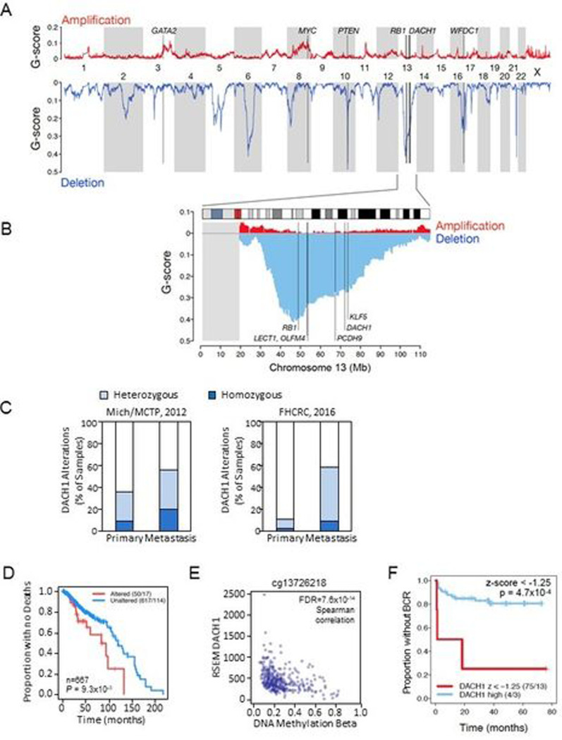
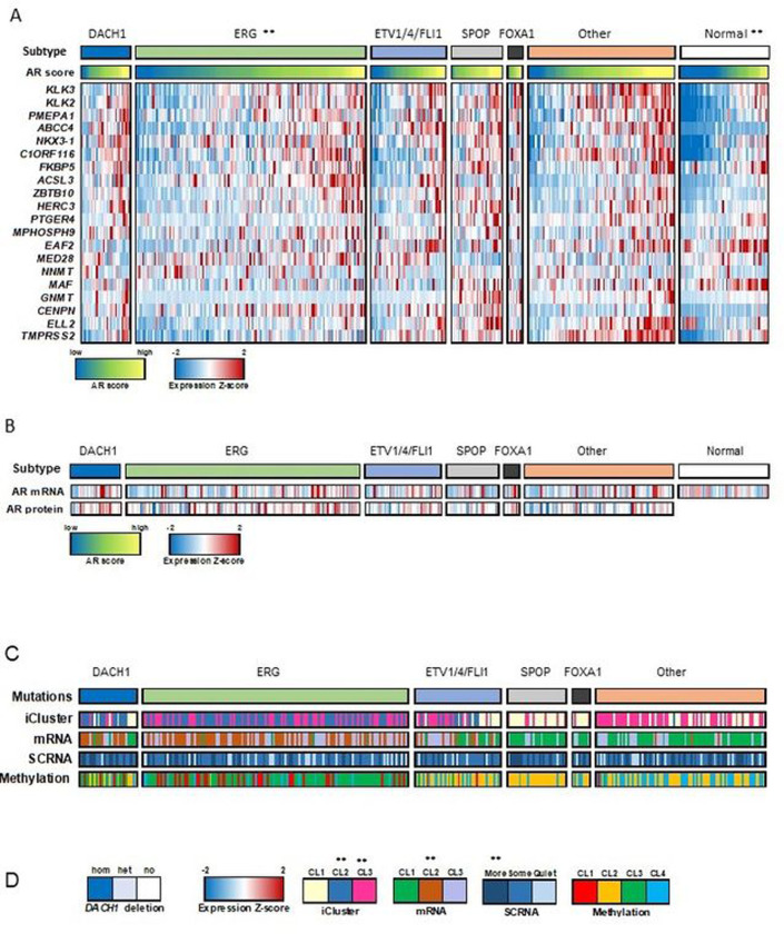
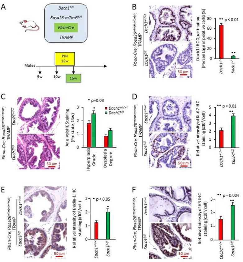
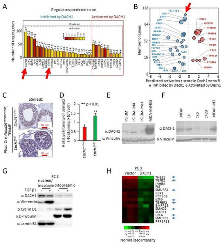
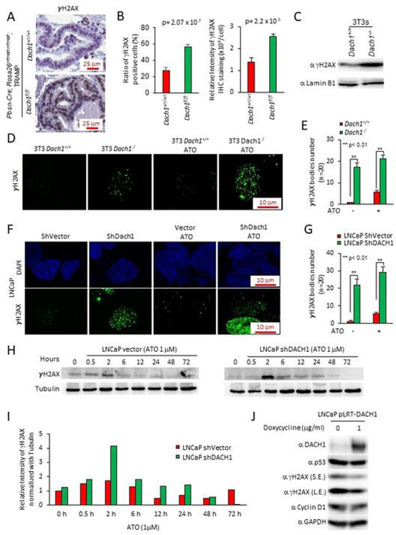
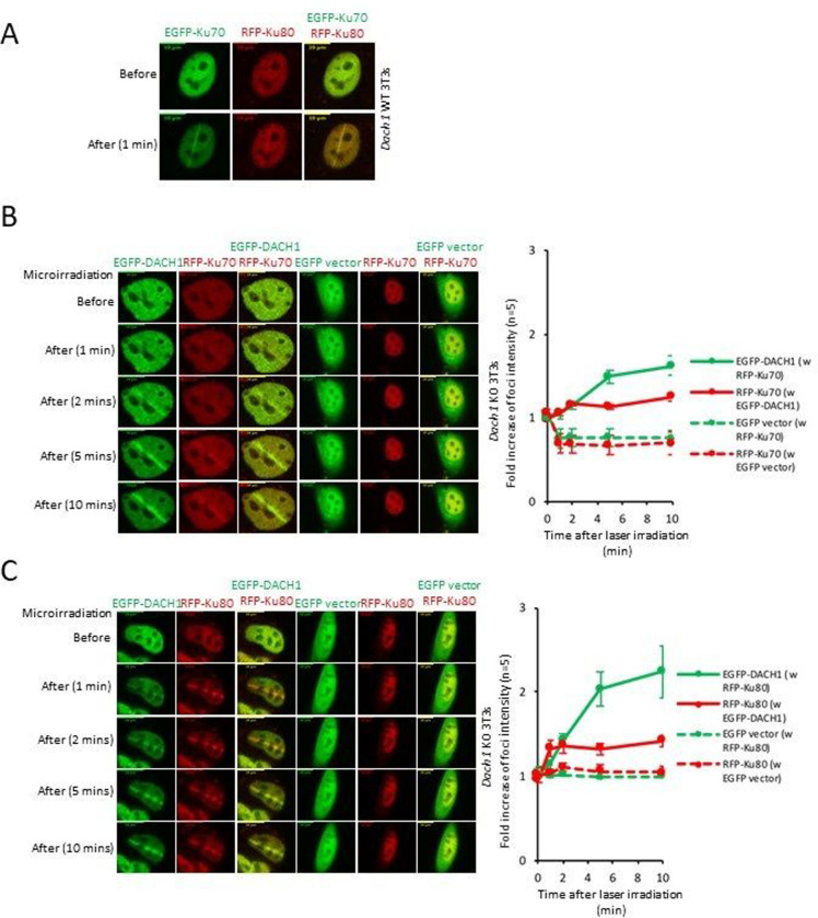
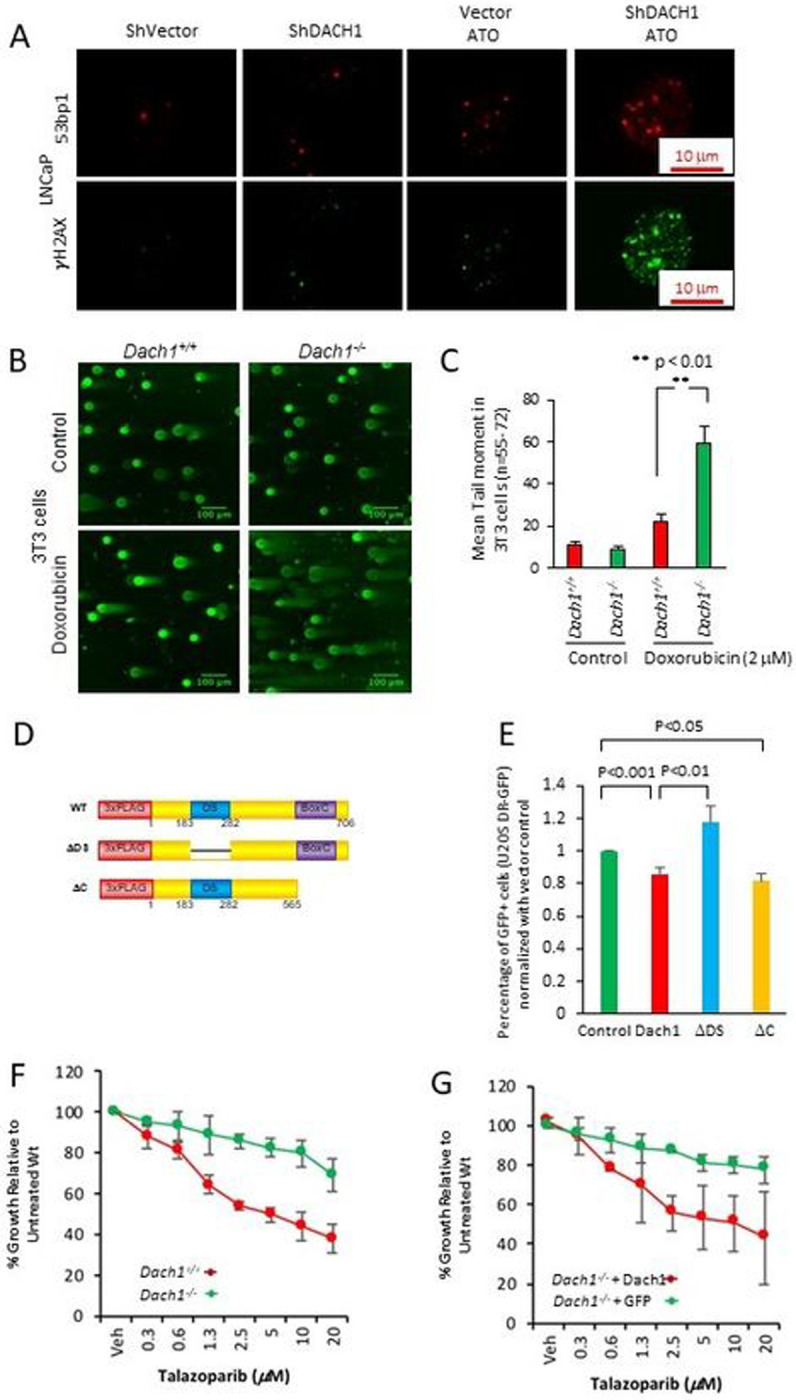
Prostate cancer (PCa), the second leading cause of death in American men, includes distinct genetic subtypes with distinct therapeutic vulnerabilities. The DACH1 gene encodes a winged helix/Forkhead DNA-binding protein that competes for binding to FOXM1 sites. Herein, DACH1 gene deletion within the 13q21.31-q21.33 region occurs in up to 18% of human PCa and was associated with increased AR activity and poor prognosis. In prostate OncoMice, prostate-specific deletion of the Dach1 gene enhanced prostatic intraepithelial neoplasia (PIN), and was associated with increased TGFb activity and DNA damage. Reduced Dach1 increased DNA damage in response to genotoxic stresses. DACH1 was recruited to sites of DNA damage, augmenting recruitment of Ku70/Ku80. Reduced Dach1 expression was associated with increased homology directed repair and resistance to PARP inhibitors and TGFb kinase inhibitors. Reduced Dach1 expression may define a subclass of PCa that warrants specific therapies.


Conflict of interest statement

CONFLICT OF INTEREST:

The authors declare that they have no relevant financial conflicts of interest. R.G.P. holds ownership interests in CytoDyn, EcoGenome, StromaGenesis, and LightSeed, Inc. R.G.P. additionally holds ownership interests (value unknown) for several patents and submitted patent applications.

Figures

Figure 1.
Figure 1.. The DACH1 gene is deleted in human prostate cancer.
(A). Landscape of somatic copy number alteration (SCNA) [26] in the TCGA PCa cohort (firebrowse.org) shown as profiles of GISTIC2 G-scores [76] for GRCh37/hg19 SNP6-based data for n=492 primary tumors. (B) as from (A), for chromosome 13, vertical lines indicate positions of candidate tumor suppressor genes (TSGs) PCDH9-DACH1-KLF5. The grey rectangle at the left indicates that TCGA/firebrowse.org reported no GISTIC2 data for the 13p arm. (C). Analysis of DACH1 gene status in human prostate cancer (PCa) from two cBioPortal cohorts (for which copy number data was available for primary and the metstatic sites) shows DACH1 homozygous deletions (dark blue) in 2.3% to 20% of patients and a higher frequency of heterozygous deletions (light blue). (D). DACH1 copy number and overall survival data was determined by combining three cBioPortal cohorts (TCGA PanCancer Atlas 2018, SU2C 2019, and MCTP) (N=667 tumor samples). Kaplan-Meier plot for overall survival is shown using a copy number threshold of −2 to segregate the data into samples with altered vs. unaltered DACH1 [26]. Patients with homozygous DACH1 deletions (“Altered” in the figure caption) showed reduced overall survival (log-rank P<9.3 ×10⁻³) (“Altered” 50/17 vs. “Unaltered” 617/114). The numbers (50/17, 617/114) indicate the number of samples in the group (e.g., Altered = 50, Unaltered = 617) and the number of events, i.e., for overall survival, deaths (e.g., Altered = 17, Unaltered = 114). (E). The range of DNA methylation beta values for probe cg13726218 (reported for DACH1 at cBioPortal) and RNA-Seq by Expectation Maximization (RSEM) DACH1 normalized expression (n=333 cohort) [26]. The (negative) Spearman correlation between beta and RSEM DACH1 normalized expression was rho=−0.41, FDR=7.6×10⁻¹⁴. (F). Kaplan-Meier plot data from Gerhausen et al.[27] showing low DACH1 gene expression (expressed as a z-score with a z-score threshold of −1.25 ) is significantly correlated with earlier biochemical recurrence (BCR). (log rank p value = 4.7×10⁻⁴ (n=79).
Figure 2.
Figure 2.. DACH1 deletion PCa enhances AR signaling.
(A). Interrogation of human PCa gene expression data [26], showing candidate genetic drivers ERG, ETV1/ETV4/FLI1, SPOP, FOXA1, and unknown. Samples with DACH1 homozygous (deep) genetic deletions (29/333) are shown as an additional subtype. The AR score (the average of the AR target gene expression) refers to a group of AR-responsive genes[26], and together with the expression Z-score of the AR target genes, are shown as colorimetric scales. The AR score-based gene names are shown. The androgen receptor (AR) activity, inferred by the induction of AR target genes, was increased in DACH1 homozygous (‘deep’) deletion PCa compared with normal (P=2×10⁻⁵ by t-test) and ERG mutation groups (P=0.003 by t-test). (B). AR mRNA and AR protein levels, shown for each DACH1 deletion sample, were not significantly different. (C). The iCluster[29], mRNA cluster, and SCNA (somatic copy-number alteration), and DNA methylation status are shown for the PCa classified by the corresponding gene deletion subtypes. (D). DACH1 homozygous deletions were enriched for iCluster 2 and 3[29], mRNA cluster 2 (P=0.0003 by Fisher exact test, SCNA (“more” somatic copy-number alteration, P=0.0004 by Fisher exact test), but not for DNA methylation.
Figure 3.
Figure 3.. Prostate-specific Dach1 gene deletion promotes prostate hyperplasia and dysplasia in OncoMice (15 weeks).
(A). Schematic representation of transgenes integrated into mice. (B). Representative immunohistochemistry for Dach1, with data quantitated as mean ± standard error of the mean (SEM) for N=20 (4 separate mice, with 5 views per mouse, in each group). (C). Blinded quantitative histology grading of prostate of multigenic mice at 15 weeks. Data are shown as mean ± SEM for N=15 (5 separate mice, with 3 prostate areas [anterior, ventral, lateral] per mouse) in each group). H&E staining demonstrates the presence of a focal atypical intraductal proliferation in Dach1−/− prostate, compatible with prostatic intraepithelial neoplasia (PIN). Representative immunohistochemistry with results shown as mean ± SEM for Ki-67 (n=20, 4 separate mice for each genotype, 5 views per mouse) (D), Beclin 1 (n=9, 3 separate mice for each genotype, 3 views per mouse) (E); and AR (n=15 for Dach1wt/wt mice, 3 separate mice, 5 views per mouse) (n=12 for Dach1fl/fl mice, 3 separate mice, 2 views for one mouse and 5 views for other two mice) (F). Scale bars, 50 μm. A Student’s t-test was performed for all comparisons.
Figure 4.
Figure 4.. Prostate-specific Dach1 gene deletion in TRAMP mice induces PIN lesions with increased TGFβ activity.
(A-B). Genome-wide expression analysis of TRAMP Dach1+/+ vs. Dach1−/− PIN lesions was analyzed for enrichment of known targets of upstream regulators using Ingenuity Pathway Analysis (IPA) and represented as (A) barplot with calculated by IPA activation Z-score labeled and as (B) bubble plot with size of the bubbles proportional to –log10 p values. (C). IHC was conducted for SMAD activation using SMAD2P, quantitated and shown as (D) mean ± SEM (n=15 for Dach1wt/wt mice, 3 separate mice, 5 views per mouse) (n=10 for Dach1fl/fl mice, 2 separate mice, 5 views per mouse). (E, F). Western blot of either PCa cell lines for the presence of DACH1 or (G) TGFβ-treated (10 ng/ml for 24 hrs) PC3 cells illustrating induction of nuclear vimentin and cytoplasmic cyclin D1. Protein loading controls are β-tubulin (a marker of cytoplasmic proteins) and Lamin B1 (a marker for nuclear protein enrichment). (H). Microarray-based gene expression analysis of PC3 cells stably expressing DACH1, showing restraint of genes mediating TGFβ signaling (shown with blue arrows), including reduction of TGFB2 and TGFBR2 [30].
Figure 5.
Figure 5.. Prostatic Dach1 governs the DNA damage response in TRAMP mice.
(A). Immunohistochemical staining for markers of DNA damage (γH2AX) in TRAMP mice prostate, with (B). Quantitation data shown as mean ± SEM for percentage of γH2AX positive cells (P=2.07×10⁻⁷ by Student’s t-test) (n=19 for Dach1wt/wt mice, 3 separate mice, 6 views for two mice, 7 views for one mouse) (n=23 for Dach1fl/fl mice, 4 separate mice, 7 views for two mice, 6 views for one mouse, 3 views for one mouse) (left panel). Data shown as mean ± SEM for relative intensity of γH2AX (n=16 for Dach1wt/wt mice, 3 separate mice, 5 views for two mice, 6 views for one mouse) (n=20 for Dach1fl/fl mice, 4 separate mice, 6 views for two mice, 5 views for one mouse, 3 views for one mouse) (P=2.2×10⁻⁵ by Student’s t-test) (right panel). (C). Western blot of Dach1+/+ or Dach1−/− 3T3 cells. (D). γH2AX immunofluorescent staining of Dach1+/+ or Dach1−/− 3T3 cells, with (E). quantitation shown as mean ± SEM, (n=20 separate cells). (F). LNCaP cells transduced with shDACH1, treated with arsenic trioxide (ATO 1 hrs) (10 μM) and (G). quantitation shown as mean ± SEM (n=20 cells). (H). LNCaP cells stably transduced with control vector or shDACH1 were treated with ATO (1 μM) for the time points indicated. Western blotting was conducted for γH2AX, with quantitation of a representative experiment shown in (I). (J). LNCaP cell line stably expressing doxycycline-inducible DACH1 were analyzed for the abundance of γH2AX and other proteins as indicated. GAPDH was used as a protein loading control. S.E., short exposure; L.E., long exposure.
Figure 6.
Figure 6.. DACH1 facilitates the recruitment of, and co-accumulates with, Ku70/Ku80 proteins at sites of DNA damage.
(A). Co-accumulation of Ku-70/Ku-80 at laser micro irradiation-induced DSBs sites in Dach1+/+ 3T3 cells. (B,C). 24 h after transfection, the accumulation of DACH1 and Ku70/Ku80 in Dach1−/− 3T3 cells transfected with EGFP or EGFP-tagged DACH1 and red fluorescent protein (RFP)-tagged Ku70 or RFP-tagged Ku80 expression vectors were treated with laser micro-irradiation (403 nm) to induce DSBs. Time is shown after micro-irradiation. Accumulation of the transfected proteins was indicated by EGFP (green) or RFP (red) fluorescence at laser-irradiated sites. Co-accumulation was visualized in yellow merged images. Time is shown in minutes and -fold increase in foci intensity is shown as mean ± SEM for N=5 separate cells.
Figure 7.
Figure 7.. DACH1 enhances DNA repair.
(A). LNCaP cells stably transduced with control vector or shDACH1 were treated with ATO (1μM), and immunofluorescence for 53BP1 or γH2AX was conducted. (B). The neutral pH comet assay, which mainly detects DNA double-strand breaks (DSBs), was conducted as a single-cell DNA damage assay. Dach1+/+ and Dach1−/− 3T3 cells were treated with 2 μM doxorubicin for 18 hrs. Scale bar, 100 μm with (C). data shown as mean ± SEM. (D). Schematic representation of DACH1 expression vectors, which were introduced into (E) U2OS cells expressing I-SceI based reporter assays for homologous repair (DR-GFP). Cells were analyzed after 48 hrs with data shown as mean ± SEM for N=12 (DACH1), N=7 (DACH1DDS), and N=5 (DACH1DC). (F). Dach1+/+ and Dach1−/− 3T3 cells or (G). Dach1−/− 3T3 transduced with a MSCV/DACH1-IRES-GFP expression vector or GFP vector control, were treated for 3 days with increasing doses of Talazoparib, a PARP inhibitor. Data are shown as mean ± SEM for N=3 separate experiments in triplicate.

References

    1. Wang G, Zhao D, Spring DJ, DePinho RA. Genetics and biology of prostate cancer. Genes & Development 2018; 32: 1105–1140. - PMC - PubMed
    1. Dimakakos A, Armakolas A, Koutsilieris M. Novel tools for prostate cancer prognosis, diagnosis, and follow-up. Biomed Res Int 2014; 2014: 890697. - PMC - PubMed
    1. Draisma G, Etzioni R, Tsodikov A, Mariotto A, Wever E, Gulati R et al. Lead time and overdiagnosis in prostate-specific antigen screening: importance of methods and context. J Natl Cancer Inst 2009; 101: 374–383. - PMC - PubMed
    1. Li S, Silvestri V, Leslie G, Rebbeck TR, Neuhausen SL, Hopper JL et al. Cancer Risks Associated With BRCA1 and BRCA2 Pathogenic Variants. J Clin Oncol 2022; 40: 1529–1541. - PMC - PubMed
    1. Teyssonneau D, Margot H, Cabart M, Anonnay M, Sargos P, Vuong NS et al. Prostate cancer and PARP inhibitors: progress and challenges. J Hematol Oncol 2021; 14: 51. - PMC - PubMed

Publication types