[Preprint]. 2023 Jan 12:2023.01.10.523481.
doi: 10.1101/2023.01.10.523481.

Spatial transcriptomics analysis of neoadjuvant cabozantinib and nivolumab in advanced hepatocellular carcinoma identifies independent mechanisms of resistance and recurrence

Affiliations

Spatial transcriptomics analysis of neoadjuvant cabozantinib and nivolumab in advanced hepatocellular carcinoma identifies independent mechanisms of resistance and recurrence

Shuming Zhang et al. bioRxiv. .

Update in

Abstract

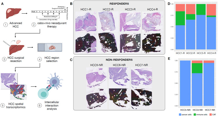
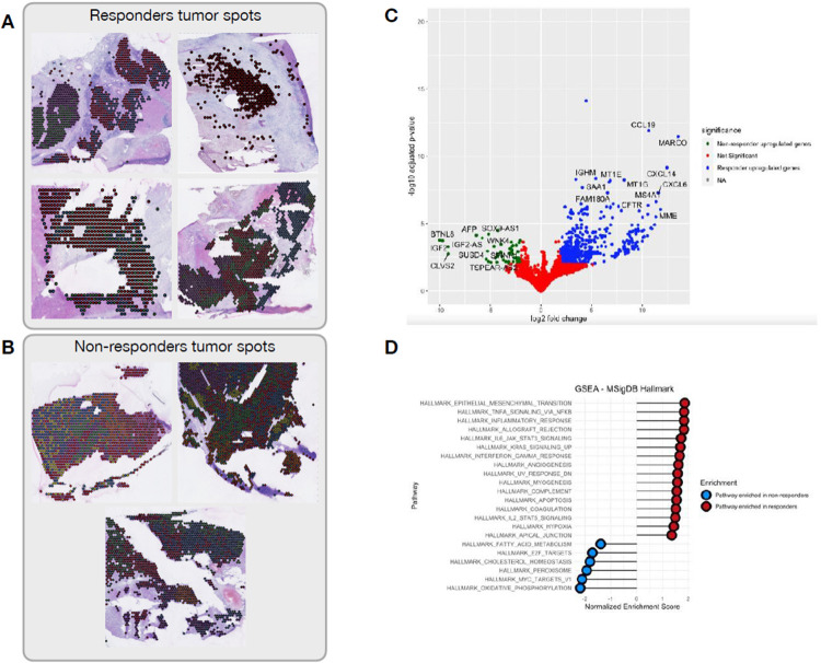
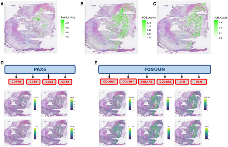
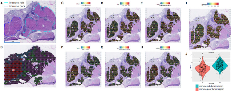
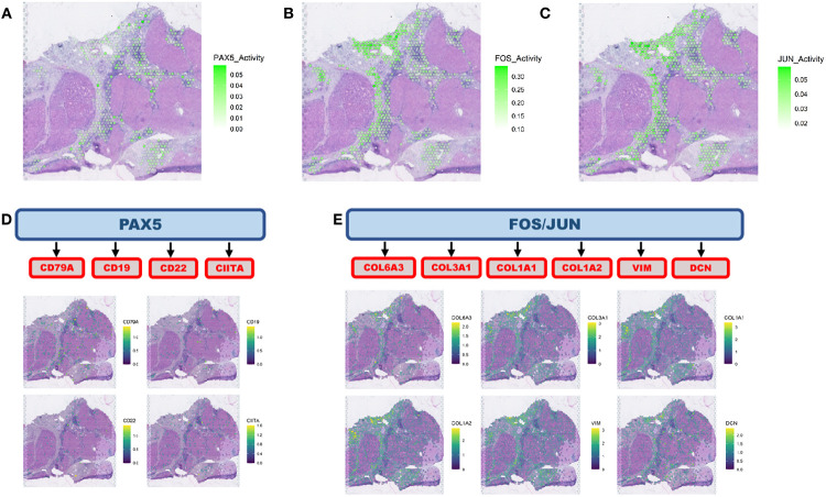
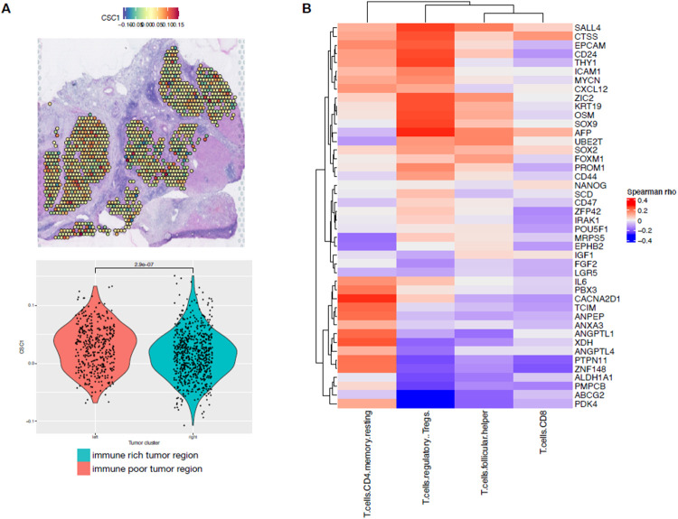
Novel immunotherapy combination therapies have improved outcomes for patients with hepatocellular carcinoma (HCC), but responses are limited to a subset of patients and recurrence can also occur. Little is known about the inter- and intra-tumor heterogeneity in cellular signaling networks within the HCC tumor microenvironment (TME) that underlie responses to modern systemic therapy. We applied spatial transcriptomics (ST) profiling to characterize the tumor microenvironment in HCC resection specimens from a clinical trial of neoadjuvant cabozantinib, a multi-tyrosine kinase inhibitor that primarily blocks VEGF, and nivolumab, a PD-1 inhibitor in which 5 out of 15 patients were found to have a pathologic response. ST profiling demonstrated that the TME of responding tumors was enriched for immune cells and cancer associated fibroblasts (CAF) with pro-inflammatory signaling relative to the non-responders. The enriched cancer-immune interactions in responding tumors are characterized by activation of the PAX5 module, a known regulator of B cell maturation, which colocalized with spots with increased B cell markers expression suggesting strong activity of these cells. Cancer-CAF interactions were also enriched in the responding tumors and were associated with extracellular matrix (ECM) remodeling as there was high activation of FOS and JUN in CAFs adjacent to tumor. The ECM remodeling is consistent with proliferative fibrosis in association with immune-mediated tumor regression. Among the patients with major pathologic response, a single patient experienced early HCC recurrence. ST analysis of this clinical outlier demonstrated marked tumor heterogeneity, with a distinctive immune-poor tumor region that resembles the non-responding TME across patients and was characterized by cancer-CAF interactions and expression of cancer stem cell markers, potentially mediating early tumor immune escape and recurrence in this patient. These data show that responses to modern systemic therapy in HCC are associated with distinctive molecular and cellular landscapes and provide new targets to enhance and prolong responses to systemic therapy in HCC.


Figures

FIGURE 1
FIGURE 1
Spatial transcriptomics analysis of HCC samples treated with cabozantinib and nivolumab. A. Experimental workflow. B. Hematoxylin and eosin (H&E) stained images of the samples profiled and the spatial clusters for responders. C. H&E and spatial clusters for non-responders. D. Tumor, immune and cancer associated fibroblasts composition in each responder samples as determined by spatial transcriptomics. E. Tumor, immune and cancer associated fibroblasts proportions in non-responders samples.
FIGURE 2
FIGURE 2
Differential expression analysis of (A) tumor clusters from responders versus (B) tumor clusters from non-responders across all patients. C. Volcano plot of the differential expression analysis showing the most differentially expressed genes in responders (blue dots) and non-responders (green dots), showing up-regulation of immune genes in responders relative to the up-regulation of hepatocellular markers among non-responders. D. Pathway enrichment analysis between responders (red dots) and non-responders (blue dots) reveal activation of immune related pathways in responders tumors, while non-responders tumors have activation of proliferation and metabolic pathways.
FIGURE 3
FIGURE 3
Intercellular interaction analysis. A. Cancer-immune interaction analysis identified the activation of PAX5 network. B. and C. Cancer-CAF interaction analysis pointed to the activation of FOS and JUN networks. D. PAX5 is a transcription factor that regulate B cell activity and the PAX5 network identified co-localizes with the distribution of B cells as determined by the spatial distribution of B cell markers. E. FOS and JUN are transcription factors that can regulate genes involved in extracellular matrix remodeling and the networks regulated by these genes colocalize with CAF marker genes.
FIGURE 4
FIGURE 4
Intra-sample heterogeneity analysis with spatial transcriptomics. A. Responder sample with remarkable heterogeneity with two tumor regions, one immune-rich (cyan) and one immune-poor (dark blue). B. Each of these regions present distinct transcriptional profiles as determined by clustering analysis. C, D and E. The immune rich tumor region highly expresses immune related pathways that were initially observed to be enriched across responders’ samples. F, G and H. Proliferation and metabolic pathways, that are enriched across non-responders’ tumors, are expressed in high levels at the immune poor region. I. The immune rich tumor region expresses high levels of the antigen processing and presentation machinery genes, which is associated with more efficient attraction of immune cells.
FIGURE 5
FIGURE 5
Intercellular interaction analysis in the context of intra-sample heterogeneity. A. Intercellular interaction analysis in immune rich tumor region (cancer-immune) reveals activation of PAX5. B and C. FOS and JUN are the active networks from the interaction analysis at the immune poor tumor region (cancer-CAF). D. PAX5 activation co-localizes with the expression of B cell markers concentrated at the borders of the immune rich tumor region. E. FOS and JUN networks are co-expressed with CAF markers adjacent to the immune poor tumor region.
FIGURE 6
FIGURE 6
Cancer stem cells detection in hepatocellular carcinoma heterogeneous sample. A. Cancer stem cell markers are highly expressed at the immune poor tumor region of the responder sample with intrasample heterogeneity as noted by the spatial expression and the levels of expression are significantly different between the distinct tumor regions. B. Hepatocellular carcinoma samples from TCGA that expresses cancer stem cell markers are highly infiltrated by T CD4+ regulatory cells while T CD8+ cells are rare.

References

    1. Sung H, Ferlay J, Siegel RL, Laversanne M, Soerjomataram I, Jemal A, et al. Global cancer statistics 2020: GLOBOCAN estimates of incidence and mortality worldwide for 36 cancers in 185 countries. CA Cancer J Clin. 2021;71:209–49. doi: 10.3322/caac.21660. - DOI - PubMed
    1. Llovet JM, Zucman-Rossi J, Pikarsky E, Sangro B, Schwartz M, Sherman M, et al. Hepatocellular carcinoma. Nat Rev Dis Primers. 2016;2:16018. doi: 10.1038/nrdp.2016.18. - DOI - PubMed
    1. Villanueva A. Hepatocellular Carcinoma. N Engl J Med. 2019;380:1450–62. doi: 10.1056/NEJMra1713263. - DOI - PubMed
    1. Gordan JD, Kennedy EB, Abou-Alfa GK, Beg MS, Brower ST, Gade TP, et al. Systemic therapy for advanced hepatocellular carcinoma: ASCO guideline. J Clin Oncol. 2020;38:4317–45. doi: 10.1200/JCO.20.02672. - DOI - PubMed
    1. Finn RS, Qin S, Ikeda M, Galle PR, Ducreux M, Kim T-Y, et al. Atezolizumab plus Bevacizumab in Unresectable Hepatocellular Carcinoma. N Engl J Med. 2020;382:1894–905. doi: 10.1056/NEJMoa1915745. - DOI - PubMed

Publication types