[Preprint]. 2024 Sep 23:2023.01.19.524721.
doi: 10.1101/2023.01.19.524721.

Ferroptosis Integrates Mitochondrial Derangements and Pathological Inflammation to Promote Pulmonary Hypertension

Affiliations

Ferroptosis Integrates Mitochondrial Derangements and Pathological Inflammation to Promote Pulmonary Hypertension

Felipe Kazmirczak et al. bioRxiv. .

Abstract

Background: Mitochondrial dysfunction, characterized by impaired lipid metabolism and heightened reactive oxygen species (ROS) generation, results in lipid peroxidation and ferroptosis. Ferroptosis is an inflammatory mode of cell death that promotes complement activation and macrophage recruitment. In pulmonary arterial hypertension (PAH), pulmonary arterial endothelial cells (PAEC) exhibit cellular phenotypes that promote ferroptosis. Moreover, there is ectopic complement deposition and inflammatory macrophage accumulation in the pulmonary vasculature. However, the effects of ferroptosis inhibition on these pathogenic mechanisms and the cellular landscape of the pulmonary vasculature are incompletely defined.

Methods: Multi-omics and physiological analyses evaluated how ferroptosis inhibition modulated preclinical PAH. The impact of AAV1-mediated expression of the pro-ferroptotic protein ACSL4 on PAH was determined, and a genetic association study in humans further probed the relationship between ferroptosis and pulmonary hypertension (PH).

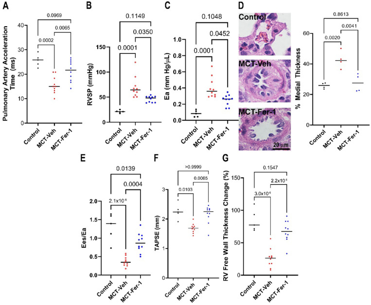
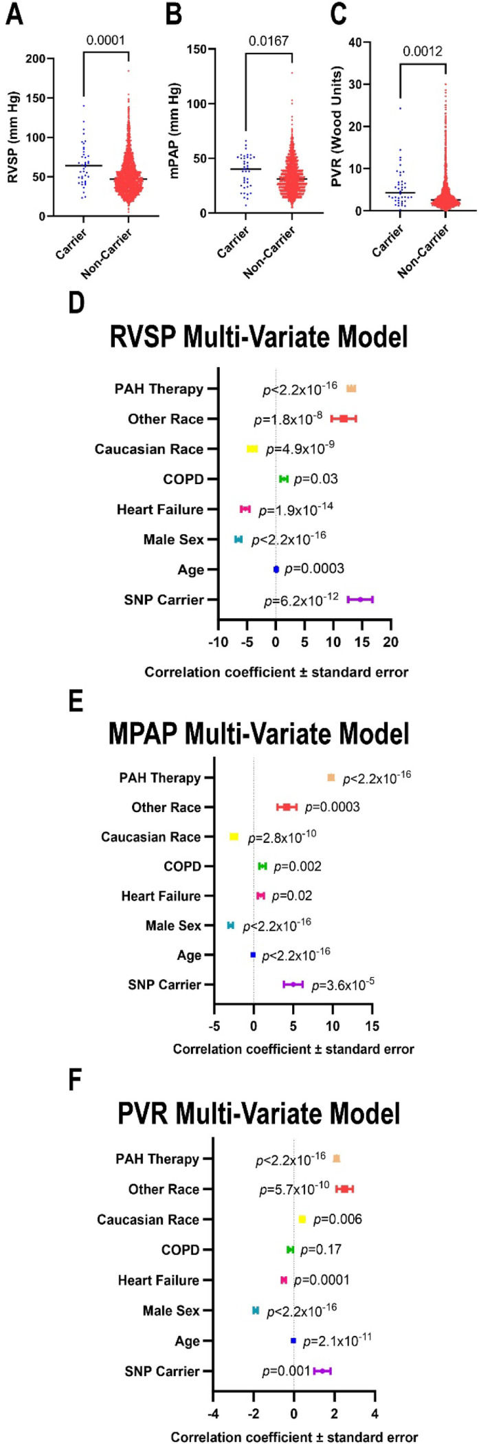
Results: Ferrostatin-1, a small-molecule ferroptosis inhibitor, mitigated PAH severity in monocrotaline rats. RNA-seq and proteomics analyses demonstrated ferroptosis was associated with PAH severity. RNA-seq, proteomics, and confocal microscopy revealed complement activation and pro-inflammatory cytokines/chemokines were suppressed by ferrostatin-1. Additionally, ferrostatin-1 combatted changes in endothelial, smooth muscle, and interstitial macrophage abundance and gene activation patterns as revealed by deconvolution RNA-seq. Ferroptotic PAEC damage associated molecular patterns restructured the transcriptomic signature, mitochondrial morphology, and promoted proliferation of pulmonary artery smooth muscle cells, and created a pro-inflammatory phenotype in monocytes in vitro. AAV1-Acsl4 induced an inflammatory PAH phenotype in rats. Finally, single-nucleotide polymorphisms in six ferroptosis genes identified a potential link between ferroptosis and PH severity in the Vanderbilt BioVU repository.

Conclusions: Ferroptosis promotes PAH through metabolic and inflammatory mechanisms in the pulmonary vasculature.


Figures

Figure 1:
Figure 1:. Ferrostatin-1 Treatment Combatted Pulmonary Hypertension Severity and Improved Right Ventricular Function in Monocrotaline Rats.
(A) Ferrostatin-1 (1 mg/kg) treatment increased pulmonary artery acceleration time in MCT rats [Con: 26±1 (n=5), MCT-Veh: 16±1 (n=10), MCT-Fer-1: 21±1 ms (n=10)]. (B) Ferroptosis inhibition with ferrostatin-1 reduced right ventricular systolic pressure (RVSP) (Con: 21±1, MCT-Veh: 69±7, MCT-Fer-1: 46±2 mmHg), p-values determined by Kruskal-Wallis test. (C) Effective arterial elastance (Ea) was lowered by ferroptosis inhibition (Con: 0.08±0.02, MCT-Veh: 0.40±0.04, MCT-Fer-1: 0.25±0.02 mmHg/μL), p-values determined by Kruskal-Wallis test. (D) Small vessel pulmonary arterial remodeling was blunted by ferrostatin-1 (Median values averaged from 4 animals per group: Con: 26±1, MCT-Veh: 43±3 MCT-Fer-1: 28±3, p-values determined by ANOVA with Tukey’s multiple comparisons test]. (E) Ferrostatin-1 treatment improved right ventricular-pulmonary artery coupling (Ees/Ea) (Con: 1.3±0.2, MCT-Veh: 0.37±0.04, MCT-Fer-1: 0.88±0.08), p-values determined by Brown-Forsythe and Welch ANOVA test), TAPSE (Con: 2.3±0.1, MCT-Veh: 1.7±0.05, MCT-Fer-1: 2.2±0.1, p-values determined by Kruskal-Wallis test) (F) and RV free wall thickening (Con: 84±8, MCT-Veh: 26±5, MCT-Fer-1: 67±5, p-values determined by ANOVA with Tukey’s multiple comparisons test) (G).
Figure 2:
Figure 2:. Lung RNA-sequencing and Proteomics Analyses Identified Associations Between Ferroptosis and Pulmonary Hypertension Severity.
(A) Pathway analysis of transcripts isolated from whole lung specimens from (n=4 control, n=4 MCT-Veh, and n=3 MCT-Fer-1) significantly associated with Ea. (B) Correlational heat mapping of ferroptosis pathway transcripts and Ea. (C) Ten transcripts most strongly associated with Ea. (D) Pathway analysis of proteins (n=5 control, n=5 MCT-Veh, and n=6 MCT-Fer-1 animals) significantly associated with Ea. (E) Correlational heat map of ferroptosis proteins with Ea. (F) Ferroptosis proteins most strongly associated with Ea. (G) Pathway analysis of proteins significantly associated with right ventricular systolic pressure (RVSP). (H) Correlational heat map of ferroptosis proteins and RVSP. (I) Ten most strongly associated ferroptosis proteins with RVSP.
Figure 3:
Figure 3:. Ferroptosis Inhibition Mitigated Ectopic Complement Activation in the Lungs of MCT Rats
(A) Hierarchical cluster analysis of transcripts in the complement coagulation pathway. (A) Ferrostatin-1 reduced levels of many transcripts as compared to MCT-Veh. (B) Correlational heat map of complement/coagulation transcripts with Ea. (C) Identification of 10 transcripts most strongly associated with Ea. Classical complement components C1Qa and C1Qb were higher as Ea increased. (D) Correlational heat map of complement/coagulation transcripts with RVSP. (E) Ten transcripts most strongly associated with RVSP. (F) Hierarchical cluster analysis of proteins in the complement/coagulation cascade. (G) Correlational heat map of complement/coagulation proteins with Ea. (H) Identification of 10 proteins most strongly associated with Ea. (I) Correlational heat map of complement/coagulation proteins with RVSP. (J) Identification of 10 proteins most strongly associated with RVSP. (K) Representative confocal micrographs showed increased complement deposition around the pulmonary vasculature in MCT-Vehicle, which was mitigated by ferrostatin-1. (L) Quantification of perivascular C3 intensity. p-values determined by Kruskal-Wallis test and Dunn’s multiple comparisons test.
Figure 4:
Figure 4:. Deconvolution RNA-sequencing Demonstrated Ferroptosis Inhibition Modulated Endothelial, Smooth Muscle, and Interstitial Macrophage Abundances and Gene Activation Patterns.
(A) Map of cell type abundances in control, MCT-Veh, and MCT-Fer-1 animals as determined by deconvolution RNA-seq (n=4 control, n=4 MCT-Veh, and n=4 MCT-Fer-1). Correlational heat mapping identified relationships between cell type abundances and Ea (B) and RVSP (C). (D) Relationships between smooth muscle cell abundance and Ea and RVSP. (E) Relationships between endothelial cell population 1 (EA1) and Ea and RVSP. (F) Relationships between interstitial macrophage population and Ea and RVSP. (G) Pathway analysis of transcriptomic changes in endothelial cells, macrophages, and total cells with ferrostatin-1 treatment. (H) Representative images of pulmonary arteriole demonstrating perivascular CD11b+ positive cells (arrow) and quantification of peri-vascular CD11b+ cells. p-values determined by Kruskal-Wallis test and Dunn’s multiple comparisons test.
Figure 5:
Figure 5:. DAMPs from Ferroptotic Human Pulmonary Arterial Endothelial Cells Recapitulated Pathogenic Changes in Human Pulmonary Artery Smooth Muscle Cells and Promoted a Pro-Inflammatory Phenotype in Human Monocytes In Vitro.
(A) Diagram of experimental approach to evaluate the effects of ferroptotic pulmonary artery endothelial cells on pulmonary artery smooth muscle cell phenotypes in vitro. (B) Hierarchical cluster analysis of top 699 transcripts from RNA isolated from n=4 replicates of PASMC treated with control media and n=4 replicates of PASMC treated with ferroptotic media. (C) KEGG pathway analysis of upregulated and downregulated transcripts comparing control and ferroptotic media treated PASMC. (D) Super resolution confocal micrographs of PASMC incubated with control and ferroptotic PAEC spent media stained with MitoTracker Orange and DAPI and quantification of the mitochondrial footprint. p-value determined by Mann Whitney test. (E) Confocal micrographs of PASMC stained with DAPI and Ki67 to identify replicating cells. Red arrows indicate Ki67+ cells, white arrows indicate Ki67- cells. Quantification of the proportion of Ki67+ cells, p-value determined by Mann Whitney test. (F) Diagram of experimental conditions employed to evaluate in vitro effects of ferroptotic PAECs on isolated human monocytes. (G) Quantification of the number of viable monocytes following media exposure. p-value determined by unpaired t-test. Flow cytometry analysis of monocyte expression of CD86 (H), CD209 (H), MerTK (J), and CD80 (K). p-values determined by unpaired t-test in H, I, and J and unpaired t-test with Welch correction in K.
Figure 6:
Figure 6:. AAV1-Mediated Endothelial ACSL4 Overexpression Induced Perivascular Complement Deposition, Pulmonary Hypertension, and Right Ventricular Dysfunction in Rats.
(A) Schematic presentation of experimental approach of rats treated with 0.3×10¹¹ vector genomes of AAV1-Acsl4 or 0.3×10¹¹ vector genomes of AAV1-GFP with concurrent exposure to low dose (30 mg/kg) monocrotaline with confocal micrographs demonstrating heightened ACSL4 immunoreactivity in the endothelial cell layer (arrows). AAV1-Acsl4 treatment induced pulmonary hypertension as pulmonary artery acceleration time was reduced [AAV1-GFP: 19±1 ms (n=5) AAV1-Acsl4: 15±0.7 ms (n=5)] (B) and RVSP [AAV1-GFP: 33±2 mm Hg (n=5), AAV1-Acsl4: 55±3 mm Hg (n=4)] (C), Ea [AAV1-GFP: 0.12±0.2 (n=5), AAV1-Acsl4: 0.29±0.05 (n=4)] (D) were increased and small vessel remodeling was heightened (E). (F) Representative confocal micrographs and quantification of C3 immunofluorescence intensity demonstrated AAV1-Acsl4 promoted perivascular complement deposition. AAV1-Acsl4 induced RV cardiomyocyte hypertrophy (AAV1-GFP: 364±5 μm2, AAV1-Acsl4: 407±5 μm2) (G) and RV dysfunction as RV-PA coupling was compromised [AAV1-GFP: 1.4±0.1 (n=5), AAV1-Acsl4: 0.6±0.1 (n=4)] (H) and cardiac output [AAV1-GFP: 140±12 mL/min (n=5) (I), AAV1-Acsl4: 99±11 mL/min (n=5)] and RV free wall thickening [AAV1-GFP: 75±9% (n=5), AAV1-Acsl4: 38±8% (n=5)] were impaired (J). p-values as determined by unpaired t-test (B, C, H, and I) or Mann-Whitney test (D-G, and J)
Figure 7:
Figure 7:. Presence of Single Nucleotide Polymorphisms in Ferroptosis Regulating Genes Was Associated With More Severe Pulmonary Hypertension.
(A) Patients harboring SNPs in ferroptosis genes had higher right ventricular systolic pressures [Carriers: 63.9 (45.0–80.6), Noncarriers 46.8 (35.0–62.0), median (25th-75th percentiles)] as determined by echocardiography, (B) invasively measured mean pulmonary arterial pressure [Carriers: 40.0 (25.0–51.0), Noncarriers 31.0 (22.0–41.0), median (25th-75th percentiles)], and pulmonary vascular resistance [Carriers: 4.3 (2.4–6.9), Noncarriers 2.6 (1.7–4.6), median (25th-75th percentiles)] as determined by invasive hemodynamic studies (C). p-values as determined by Mann-Whitney U-test. Forest plots depicted correlational co-efficient with standard errors and calculated p-values when RVSP (D), mPAP (E), and PVR (F) were modeled in multivariate regression analysis.
Central Figure:
Central Figure:
Proposed Model for the Pathogenic Role of Endothelial Cell Ferroptosis in PAH Pathobiology

Similar articles

References

    1. Stockwell BR. Ferroptosis turns 10: Emerging mechanisms, physiological functions, and therapeutic applications. Cell. 2022;185:2401–2421. doi: 10.1016/j.cell.2022.06.003 - DOI - PMC - PubMed
    1. Dixon SJ, Lemberg KM, Lamprecht MR, Skouta R, Zaitsev EM, Gleason CE, Patel DN, Bauer AJ, Cantley AM, Yang WS, et al. Ferroptosis: an iron-dependent form of nonapoptotic cell death. Cell. 2012;149:1060–1072. doi: 10.1016/j.cell.2012.03.042 - DOI - PMC - PubMed
    1. Friedmann Angeli JP, Schneider M, Proneth B, Tyurina YY, Tyurin VA, Hammond VJ, Herbach N, Aichler M, Walch A, Eggenhofer E, et al. Inactivation of the ferroptosis regulator Gpx4 triggers acute renal failure in mice. Nat Cell Biol. 2014;16:1180–1191. doi: 10.1038/ncb3064 - DOI - PMC - PubMed
    1. Bai T, Li M, Liu Y, Qiao Z, Wang Z. Inhibition of ferroptosis alleviates atherosclerosis through attenuating lipid peroxidation and endothelial dysfunction in mouse aortic endothelial cell. Free Radic Biol Med. 2020;160:92–102. doi: 10.1016/j.freeradbiomed.2020.07.026 - DOI - PubMed
    1. Culley MK, Chan SY. Mitochondrial metabolism in pulmonary hypertension: beyond mountains there are mountains. J Clin Invest. 2018;128:3704–3715. doi: 10.1172/JCI120847 - DOI - PMC - PubMed

Publication types