

This is a preprint.
It has not yet been peer reviewed by a journal.
The National Library of Medicine is
running a pilot
to include preprints that result from research funded by NIH in PMC and PubMed.
[Preprint]. 2023 Jan 20:rs.3.rs-2365576.
doi: 10.21203/rs.3.rs-2365576/v1.
Focused ultrasound-mediated brain genome editing
Affiliations
- PMID: 36712096
- PMCID: PMC9882596
- DOI: 10.21203/rs.3.rs-2365576/v1
Item in Clipboard
Focused ultrasound-mediated brain genome editing
Res Sq.
.
Update in
-
Focused ultrasound-mediated brain genome editing.Proc Natl Acad Sci U S A. 2023 Aug 22;120(34):e2302910120. doi: 10.1073/pnas.2302910120. Epub 2023 Aug 14. Proc Natl Acad Sci U S A. 2023. PMID: 37579143 Free PMC article.
Abstract
Gene editing in the mammalian brain has been challenging because of the restricted transport imposed by the blood-brain barrier (BBB). Current approaches rely on local injection to bypass the BBB. However, such administration is highly invasive and not amenable to treating certain delicate regions of the brain. We demonstrate a safe and effective gene editing technique by using focused ultrasound (FUS) to transiently open the BBB for the transport of intravenously delivered CRISPR/Cas9 machinery to the brain.
Figures
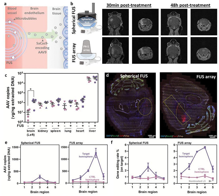
a, Schematic overview of FUS-mediated BBB opening. b, Two types of FUS systems used in this study and the representative MRI images showing the transient opening induced by FUS. c, Biodistribution of SaCas9/AAV9 vector at week 2 post-administration. Both FUS and control groups received an AAV dose of 2×10¹¹ GC/mouse (N=5 for the FUS group and N=4 for the control group, adult male C57BL/6). d, Representative RNAscope images to confirm the SaCas9 expression in the FUS-targeted region in adult C57BL/6 mice e, Deposition of SaCas9/AAV9 vector in different brain regions (Two biological repeats for each group). f, Gene editing efficiency in different brain regions (determined by amplicon sequencing; two biological repeats for each group). For Figs. 1d, e and f, adult C57BL/6 mice (aged between 9 to 10 weeks old) were given intravenously with SaCas9/AAV9 vectors in a dose of 1012 GC/mouse, and the brain tissues were dissected at week 3 post-administration.
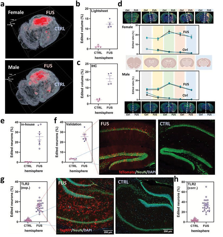
a, Representative reconstructed 3D Lightsheet images showing the Cas9-activated TdTomato signals (red) in the FUS-targeted regions. b, Gene editing efficiency quantified by a Lightsheet microscope. c, Efficiency quantification by immunostaining. For both Figs. 2b and c, Efficiency was determined by TdTomato+ volume per total volume of the hemisphere. d, Gene editing efficiency in different brain regions. Two sets of serial sections were used for immunostaining and quantification for each sex. e, In-house quantification of edited neurons (TdTomato+ and NeuN+) in the hippocampus in FUS-SaCas9/AAV9-treated Ai9 mice by immunostaining. f, Independent quantification of neuron editing in Ai9 mice following FUS performed by the SCGE Small Animal Testing Center and representative confocal images of the FUS-targeted and control sites. Two biological repeats were used for both in-house quantification and validation g,Quantification of TagRFP+ neurons in the hippocampus regions in FUS-SaCas9/AAV9-treated TLR2 reporter mice by immunostaining and the representative confocal images used for this quantification. h, Neuron editing performance determined with the experimental correction. Data are presented in dots and circles for the results from female and male mice, respectively.
References
-
- Batts A., Ji R., Kline-Schoder A., Noel R. & Konofagou E. Transcranial Theranostic Ultrasound for Pre-Planning and Blood-Brain Barrier Opening: A Feasibility Study Using an Imaging Phased Array In Vitro and In Vivo. IEEE Trans Biomed Eng 69, 1481–1490, doi: 10.1109/TBME.2021.3120919 (2022). - DOI - PMC - PubMed
Publication types
Grants and funding
LinkOut - more resources
Full Text Sources
