Review
. 2023 Jan 12:13:1078185.
doi: 10.3389/fneur.2022.1078185. eCollection 2022.

Transfemoral trans-facial vein-superior ophthalmic vein to embolize cavernous sinus dural arteriovenous fistulas

Affiliations
Review

Transfemoral trans-facial vein-superior ophthalmic vein to embolize cavernous sinus dural arteriovenous fistulas

Zibo Zhou et al. Front Neurol. .

Abstract

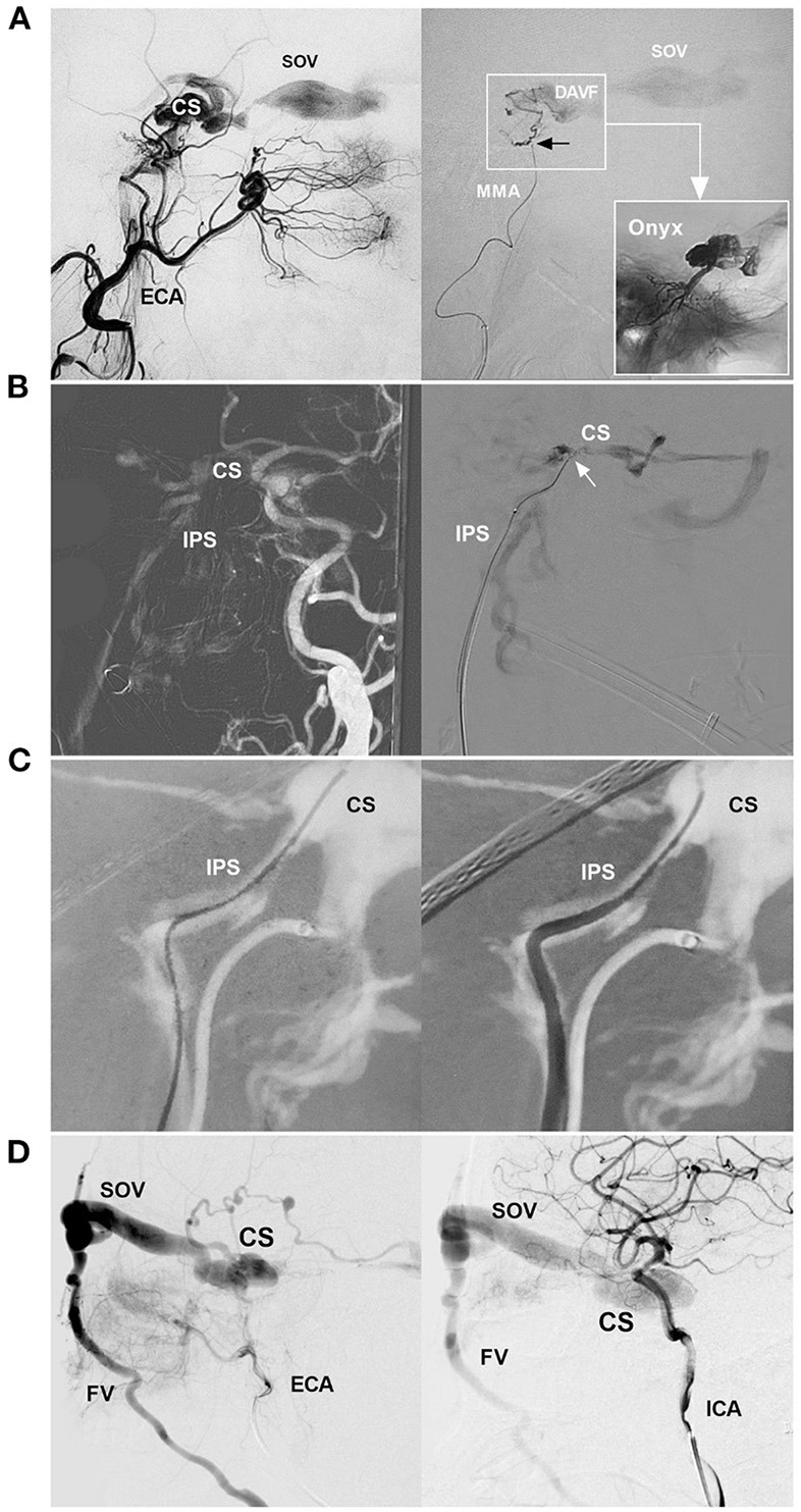
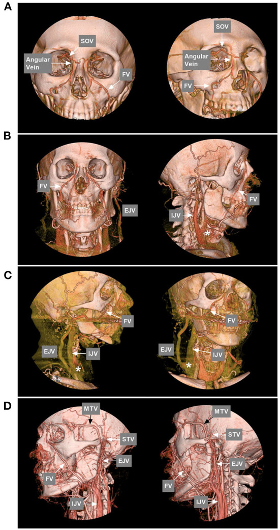
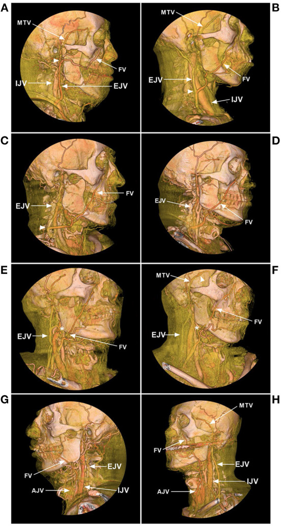
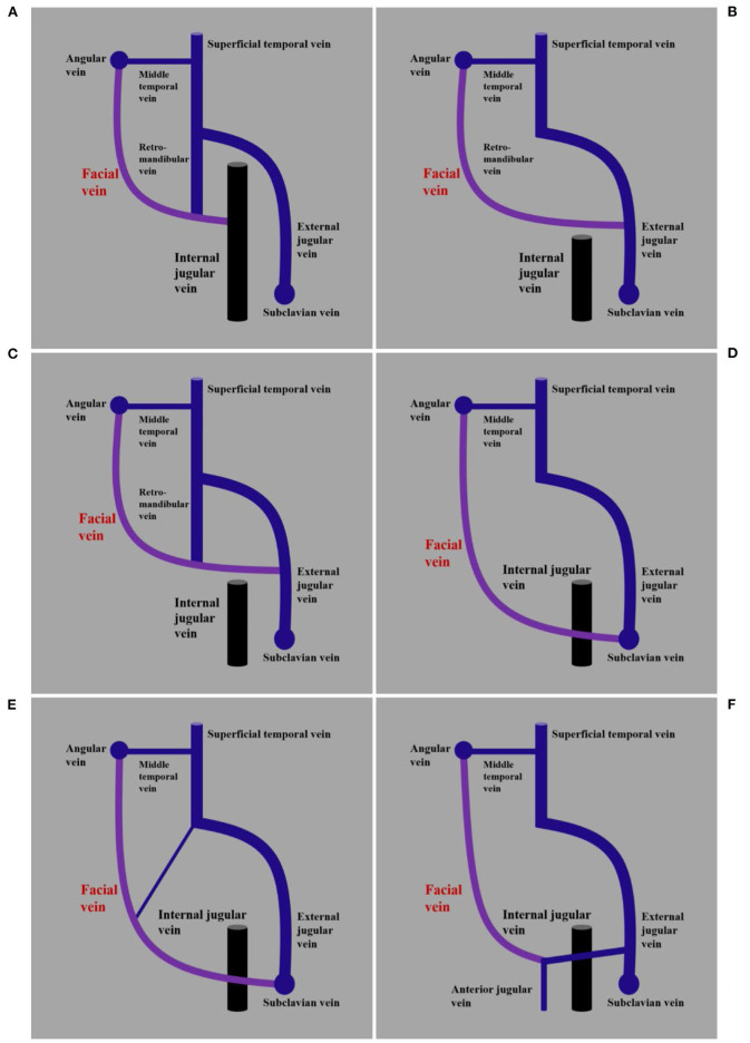
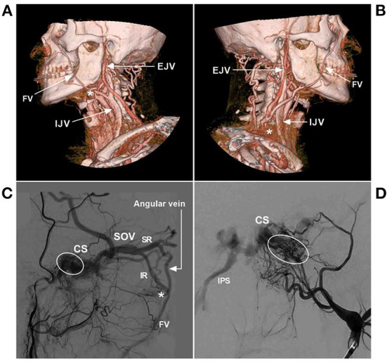
Cavernous sinus dural arteriovenous fistula (CS-DAVF) is an abnormal communication between the CS and dural arteries from the internal carotid artery and external carotid artery. CS-DAVFs are not uncommon. The preferred treatment for most CS-DAVFs is transvenous embolization (TVE), which can achieve a high cure rate with few complications. The trans-inferior petrous sinus (IPS) route from the internal jugular vein to the CS is the favorite and most direct route to perform TVE in the great majority of CS-DAVFs. However, when the trans-IPS route fails and if the facial vein (FV) is patent and dilated, transfemoral trans-FV-superior ophthalmic vein (SOV) embolization of the CS-DAVF can be attempted. However, the transfemoral trans-FV-SOV route to embolize CS-DAVFs is often challenging, and there is insufficient knowledge about it. Therefore, an updated review of the transfemoral trans-FV-SOV route to embolize CS-DAVFs is necessary, and this review includes our experience. The images in this review are from our institute without the dispute of copyright. Issues regarding the transfemoral trans-FV-SOV route to embolize CS-DAV were discussed, including the FV anatomy and variation, various TVE routes to access CS-DAVF, the procedure of the transfemoral trans-FV-SOV route to embolize CS-DAVF, difficulty, and solution of the transfemoral trans-FV-SOV route to embolize CS-DAVF, and complications and prognosis of transfemoral trans-FV-SOV to embolize CS-DAVF. By reviewing the transfemoral trans-FV-SOV route to embolize CS-DAVFs, we found that this route provides a valuable alternative to the other transvenous routes. A good prognosis can be obtained with the transfemoral trans-FV-SOV route to embolize CS-DAVFs in select cases.

Keywords: cavernous sinus; dural arteriovenous fistula; embolization; facial vein; review.


Conflict of interest statement

The authors declare that the research was conducted in the absence of any commercial or financial relationships that could be construed as a potential conflict of interest.

Figures

Figure 1
Figure 1
EVT routes of CS-DAVF. (A) EVT for CS-DAVF via the MMA. Left: angiography of the ECA shows the CS-DAVF draining into the SOV; Right: superselective angiography of the MMA confirmed that the microcatheter obtained a wedged position (black arrow) and accessed the fistula (frame); the picture in a picture (right arrow) shows the casting of Onyx in the CS. (B) EVT for CS-DAVF via IPS. Left: Navigation image of the road map shows that the CS-DAVF drained into the IPS; Right: Superselective angiography confirmed that the microcatheter (arrow) was positioned in the CS. (C) Catheterization via IPS. Navigation image of the road map shows the guidewire into the CS via the IPS (left); then, the catheter went into the IPS following the guidewire (right). (D) CS-DAVF with the SOV as the main draining vein. Angiographies of the ECA (left) and ICA (right) show a CS-DAVF drained via the SOV and then to the FV. The CS-DAVF was appropriated for the trans-FV-SOV route to perform EVT. CS, cavernous sinus; DAVF, dural arteriovenous fistula; ECA, external carotid artery; EVT, endovascular treatment; FV, facial vein; ICA, internal carotid artery; IPS, inferior petrous sinus; MMA, middle meningeal artery; SOV, superior ophthalmic vein.
Figure 2
Figure 2
FV anatomy of CTA. (A) Anterior–posterior view (left) and oblique (right) view of CTA show that the SOV from the CS connected with the angular vein; then, the angular vein continued as the FV. (B) Anterior–posterior view (left) and oblique (right) view of CTA show that the typical FV descends obliquely in a straight line to continue the common FV and flow with the IJV. The asterisk (right) indicates the junction of the FV with the IJV. (C) Lateral view (left) and oblique (right) view of CTA show the FV together with the EJV into the IJV. The asterisks (left and right) indicate the junction of the FV into the IJV. (D) Lateral view (left) and oblique (right) view of CTA show that the MTV joins the STV to form the EJV and then connects with the FV into the IJV; in the face, many tidy veins join into the FV. CS, cavernous sinus; CTA, computed tomography angiography; EJV, external jugular vein; FV, facial vein; IJV, internal jugular vein; MTV, middle temporal vein; SOV, superior ophthalmic vein; STV, superficial temporal vein.
Figure 3
Figure 3
FV variance of CTA. (A) Lateral view CTA shows that the FV courses posteriorly and combines with the MTV at the mandibular joint (arrowhead) from the EJV. (B) Lateral view CTA shows that the FV descends obliquely and curves around the inferior edge of the mandible and flows into the EJV at the middle cervical region (arrowhead). (C) Lateral view CTA shows that the FV flows into the EJV in the low cervical region (arrowhead). (D) Lateral view CTA shows that the FV descends obliquely and curves around the inferior edge of the mandible and flows into the subclavian vein without connection with the IJV or EJV. (E, F) Oblique view CTAs shows that the FV flows into the subclavian vein with the connection with the EJV (asterisks). (G, H) Oblique view CTAs shows that the FVs mainly flow into the AJVs, with the connection with the EJVs. AJV, anterior jugular vein; CTA, computed tomography angiography; EJV, external jugular vein; FV, facial vein; IJV, internal jugular vein; MTV, middle temporal vein.
Figure 4
Figure 4
Illustrations of normal anatomy and variation of the FV. (A) Imaging shows the FV together with the retromandibular vein into the IJV. (B) Imaging shows the FV without the connection of the retromandibular vein into the EJV. (C) Imaging shows the FV without the connection of the retromandibular vein into the EJV. (D) Imaging shows the FV without the connection of the retromandibular vein into the subclavian vein. (E) Imaging shows the FV with the connection of the retromandibular vein into the subclavian vein. (F) Imaging shows the FV with the connection of other veins into the anterior jugular vein. AJV, anterior jugular vein; EJV, external jugular vein; FV, facial vein; IJV, internal jugular vein; MTV, middle temporal vein.
Figure 5
Figure 5
Forward and reverse venous roadmaps. (A) Forward roadmap: after the contrast medium was slowly injected into the carotid artery, the IJV was shown clearly to help the guiding catheter (asterisk) navigate in the IJV, and the arrows indicate the direction of blood flow. (B) Reverse roadmap: immediately after the guiding catheter (asterisk) injected the contrast medium into the AJV, the AJV and nearby veins were shown clearly, and the arrows indicate the direction of blood flow. AJV, anterior jugular vein; IJV, internal jugular vein.
Figure 6
Figure 6
Preoperative angiography of CS-DAVF embolized via the trans-IJV-FV-SOV route. (A, B) Right ICA (A) and ECA (B) angiographies show the dural branches of the ICA and ECA supplied to the DAVF in the right CS. The left FV served as the draining vein into the IJV. (C, D) Left ICA (C) and ECA (D) angiographies show the dural branches of the ICA and ECA supplied to the DAVF. The bilateral FVs served as the draining veins. CS, cavernous sinus; DAVF, dural arteriovenous fistula; ECA, external carotid artery; FV, facial vein; ICA, internal carotid artery; IJV, internal jugular vein; L, left; R, right; SOV, superior ophthalmic vein.
Figure 7
Figure 7
Operative angiography of CS-DAVF embolized via the trans-IJV-FV-SOV route. (A) Angiography of the left ECA via the transarterial diagnostic catheter showing that the transvenous therapeutic guiding catheter (asterisk) was placed into the IJV. (B) Angiography of the left ECA shows that the microcatheter was advanced into the FV (asterisks) via the transfemoral trans-IJV-FV route. (C) Venous navigation roadmap shows that the microcatheter passed through the angular vein and SOV into the CS (arrow). (D) Unsubtracted angiography showing coiling (frame) was performed; the picture in the picture (right angle arrow) shows the combined use of Onyx. (E, F) Post-TVE angiographies of the left carotid artery (E) and right carotid artery (F) show complete occlusion of the DAVF. CS, cavernous sinus; DAVF, dural arteriovenous fistula; ECA, external carotid artery; EJV, external jugular vein; FV, facial vein; IJV, internal jugular vein; L, left; R, right; SOV, superior ophthalmic vein; TVE, transvenous embolization.
Figure 8
Figure 8
Preoperative angiography of CS-DAVF embolized via the trans-EJV-FV-SOV route. (A, B) Angiographies of right (A) and left (B) ICAs show the dural branches of the ICA supplied to the DAVF. (C) Angiography of the left ECA shows the dural branches of the ECA supplied to the DAVF. (D) Venous phase angiography of the right ICA shows that the right FV served as the draining vein of the DAVF into the EJV. CS, cavernous sinus; DAVF, dural arteriovenous fistula; ECA, external carotid artery; EJV, external jugular vein; FV, facial vein; ICA, internal carotid artery; L, left; R, right; SOV, superior ophthalmic vein.
Figure 9
Figure 9
Operative angiography of CS-DAVF embolized via the trans-EJV-FV-SOV route. (A) Venous phase angiography of the right ICA shows the right FV into the EJV. (B) Venous navigation roadmap shows that the microcatheter passed through the right angular vein into the SOV. (C) X-ray image shows the microcatheter (asterisks) passing through the intercavernous sinus from the right into the left CS to perform coiling (arrow). (D) Unsubtracted angiography shows the combined use of Onyx to occlude the CS-DAVF. (E, F) Post-TVE angiographies of the left carotid artery (E) and right carotid artery (F) show complete occlusion of the DAVF. CS, cavernous sinus; DAVF, dural arteriovenous fistula; EJV, external jugular vein; FV, facial vein; ICA, internal carotid artery; L, left; R, right; SOV, superior ophthalmic vein; TVE, transvenous embolization.
Figure 10
Figure 10
Difficulty of transfemoral trans-FV-SOV to embolize CS-DAVF. (A) Lateral view CTA shows the FV jointed into the IJV. The asterisk indicates the looping of the common FV, and catheterization was difficult. (B) Lateral view CTA shows the FV jointed into the EJV. The asterisk indicates the junction of the EJV and subclavian vein; here, venous valves exist. (C) Lateral view angiography of the ECA shows the veins of the orbit region. The asterisk indicates that venous valves exist at the inferior root of the SOV, and the circle indicates the fistula point of the CS-DAVF. (D) Anterior–posterior view angiography of the ECA shows the fistula point (circle) of the CS-DAVF. CS, cavernous sinus; DAVF, dural arteriovenous fistula; ECA, external carotid artery; EJV, external jugular vein; FV, facial vein; IJV, internal jugular vein; IPS, inferior petrous sinus; IR, inferior root; SOV, superior ophthalmic vein; SR, superior root.

Similar articles

Cited by

References

    1. Alexandre AM, Sturiale CL, Bartolo A, Romi A, Scerrati A, Flacco ME, et al. . Endovascular treatment of cavernous sinus dural arteriovenous fistulas. Institutional series, systematic review and meta-analysis. Clin Neuroradiol. (2022) 32:761–71. 10.1007/s00062-021-01107-0 - DOI - PMC - PubMed
    1. Ghobrial GM, Marchan E, Nair AK, Dumont AS, Tjoumakaris SI, Gonzalez LF, et al. . Dural arteriovenous fistulas: a review of the literature and a presentation of a single institution's experience. World Neurosurg. (2013) 80:94–102. 10.1016/j.wneu.2012.01.053 - DOI - PubMed
    1. Kim DJ, Kim DI, Suh SH, Kim J, Lee SK, Kim EY, et al. . Results of transvenous embolization of cavernous dural arteriovenous fistula: a single-center experience with emphasis on complications and management. AJNR Am J Neuroradiol. (2006) 27:2078–82. - PMC - PubMed
    1. Matsumoto A, Okauchi M, Shindo A, Kawanishi M, Tamiya T. Cavernous sinus dural arteriovenous fistula treated by facial vein direct puncture: case report and review of the literature. Interv Neuroradiol. (2017) 23:301–6. 10.1177/1591019917693413 - DOI - PMC - PubMed
    1. Hou K, Li G, Luan T, Xu K, Yu J. Endovascular treatment of the cavernous sinus dural arteriovenous fistula: current status and considerations. Int J Med Sci. (2020) 17:1121–30. 10.7150/ijms.45210 - DOI - PMC - PubMed

LinkOut - more resources