

Accuracy of Classifying Lung Carcinoma Using Immunohistochemical Markers on Limited Biopsy Material: A Two-Center Study
- PMID: 36712764
- PMCID: PMC9875635
- DOI: 10.7759/cureus.32956
Accuracy of Classifying Lung Carcinoma Using Immunohistochemical Markers on Limited Biopsy Material: A Two-Center Study
Abstract
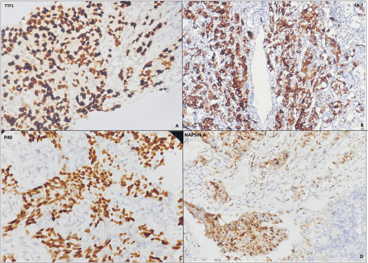
Introduction Accurate classification of lung cancer into primary and metastatic carcinomas is critical for treatment approaches. Immunohistochemistry (IHC) has always been pivotal in unveiling the diverse cell differentiation lineages present in lung cancer by using specific biomarkers such as TTF1 and p63/p40, which closely reflect the relationship between genotype and phenotype.. Methods A retrospective cross-sectional study was conducted to evaluate 57 Tru-Cut biopsies over two years, from 2020-2022. Tumour morphology was evaluated, and IHC for TTF-1, Napsin A, CK-7, P-63, P-40, and CD-56 was performed in two steps. Results Of the lung cancer cases, 58.5% were adenocarcinoma (ADC), 24.5% were squamous cell carcinoma (SCC), 9.4% were small cell carcinoma, and 7.5% were poorly differentiated carcinoma. TTF1 stain had sensitivity and specificity of 78.9% and 50% in 33 cases of ADC, respectively, while CK7 and Napsin A had 100% sensitivity. P63 stain had 77% sensitivity and 50% specificity in 15 cases of SCC, while P-40 had 100% sensitivity. The CD56 stain was 100% sensitive in five cases of small cell carcinoma. Conclusion IHC staining on small lung biopsies allows accurate sub-classification of poorly differentiated lung cancers; however, there is still significant variability. Surgical resection specimens can be further classified due to architectural features that biopsies lack. Morphological findings would be beneficial in the development of an algorithm for sub-classifying lung carcinoma using a variety of markers.
Keywords: adenocarcinoma; immunohistochemistry; lc; sensitivity; small cell carcinoma; specificity; squamous cell carcinoma; trucut biopsy.
Copyright © 2022, Hassan et al.
Conflict of interest statement
The authors have declared that no competing interests exist.
Figures
References
-
- Classification and pathology of lung cancer. Zheng M. Surg Oncol Clin N Am. 2016;25:447–468. - PubMed
-
- Lung cancer detection using CT scan images. Makaju S, Prasad P, Alsadoon A, et al. Procedia Comput Sci. 2018;125:107–114.
-
- The 2015 World Health Organization classification of lung tumors: impact of genetic, clinical and radiologic advances since the 2004 classification. Travis WD, Brambilla E, Nicholson AG, et al. J Thorac Oncol. 2015;10:1243–1260. - PubMed
LinkOut - more resources
Full Text Sources
Research Materials