. 2023 Jan 18:2023:8408574.
doi: 10.1155/2023/8408574. eCollection 2023.

TRIM72 Alleviates Muscle Inflammation in mdx Mice via Promoting Mitophagy-Mediated NLRP3 Inflammasome Inactivation

Affiliations

TRIM72 Alleviates Muscle Inflammation in mdx Mice via Promoting Mitophagy-Mediated NLRP3 Inflammasome Inactivation

Mengli Wu et al. Oxid Med Cell Longev. .

Abstract

Chronic muscle inflammation exacerbates the pathogenesis of Duchenne muscular dystrophy (DMD), which is characterized by progressive muscle degeneration and weakness. NLRP3 (nucleotide-binding domain and leucine-rich repeat pyrin domain containing 3) inflammasome plays a key role in the inflammatory process, and its abnormal activation leads to a variety of inflammatory or immune diseases. TRIM72 (MG53) is a protective myokine for tissue repair and regeneration. However, little is known about the potential impact of TRIM72 in the crosstalk between mitophagy and inflammatory process of DMD. Here, 10-week-old male mdx mice were injected intramuscularly with adeno-associated virus (AAV-TRIM72) to overexpress TRIM72 protein for 6 weeks. Then, skeletal muscle samples were collected, and relevant parameters were measured by histopathological analysis and molecular biology techniques. C2C12 cell line was transfected with lentivirus (LV-TRIM72) to overexpress or siRNA (si-TRIM72) to suppress the TRIM72 expression for the following experiment. Our data firstly showed that the TRIM72 expression was decreased in skeletal muscles of mdx mice. Then, we observed the increased NLRP3 inflammasome and impaired mitophagy in mdx mice compared with wild type mice. In mdx mice, administration of AAV-TRIM72 alleviated the accumulation of NLRP3 inflammasome and the consequent IL-18 and IL1β maturation by inducing autophagy, while this protective effect was reversed by chloroquine. Mitochondrial reactive oxygen species (mtROS), as a recognized activator for NLRP3 inflammasome, was attenuated by TRIM72 through the induction of mitophagy in C2C12 cells. Additionally, we proposed that the TRIM72 overexpression might promote mitophagy through both the early stage by PI3K-AKT pathway and the late stage by autolysosome fusion. In conclusion, the current study suggests that TRIM72 prevents DMD inflammation via decreasing NLRP3 inflammasomes and enhancing mitophagy. Collectively, our study provides insight into TRIM72 as a promising target for therapeutic intervention for DMD.


Conflict of interest statement

All authors declare that there are no competing financial interests exist.

Figures
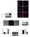
Figure 1
Figure 1
TRIM72 overexpression and knockdown in vivo and vitro. (a) Western blot analysis of tibial anterior muscle extracts using antibodies against TRIM72 after AAV-EV or AAV-TRIM72 transfection in mdx mice. AAV-TRIM72 was designed with 3xflag tag. (b) Representative immunofluorescence images of TRIM72 in tibial anterior muscle of mdx mice transfected with or without AAV. Scale bar, 100 μm. (c) Fluorescence indicated the successful transfection of LV-TRIM72 in C2C12 cells. Scale bar, 50 μm. (d) Western blot analysis of C2C12 cell extracts using antibodies against Flag after lentivirus transfection. LV-TRIM72 was designed with 3xflag tag. (e) The expression of TRIM72 mRNA in C2C12 cells with or without lentivirus transfection for 72 h. (f) C2C12 cells were transfected with si-TRIM72 for 72 h, and western blot was performed with antibodies against TRIM72. (g) The expression of TRIM72 mRNA in C2C12 cells with or without siRNA transfection for 72 h. Data were expressed as mean ± SEM. p < 0.05, ∗∗p < 0.01, and ∗∗∗p < 0.001. AAV-EV: adeno-associated virus with empty vector; LV-EV: lentivirus with empty vector.
Figure 2
Figure 2
Decreased NLRP3 inflammasomes and increased mitophagy in AAV-TRIM72 mdx mice. (a) Representative HE staining images of tibial anterior muscles in mdx mice with or without AAV transfection. Scale bar, 100 μm. (b) Immunofluorescence was performed with specific antibody targeting NLRP3. As shown by arrows in the magnification inset, NLRP3 dots were reduced in AAV-TRIM72 mdx mice. Scale bar, 50 μm. (c) Serum IL-1β level detected by ELISA assay, n = 4. (d, e) Western blot analysis and quantification of NLRP3, ASC, IL18, caspase-1, and IL-1β in three mdx groups, n = 4. (f, g) Immunoblot analysis and quantification of P62, LC3, BNIP3, and PARKIN in lysates of three mdx groups, n = 4. (h) Representative immunofluorescence images of LC3, MitoTracker, and the merged images in three mdx groups. Scale bar, 50 μm. Data were expressed as mean ± SEM. p < 0.05, ∗∗p < 0.01.
Figure 3
Figure 3
Inhibition of autophagy abrogated the inhibitory effects of TRIM72 on NLRP3 activation. (a) Representative histology images and inflammatory response reflected by HE staining in tibial anterior muscles of AAV-TRIM72 mdx mice with or without CQ treatment. Scale bar, 100 μm. (b, c) Western blot and quantification of inflammasome-related proteins NLRP3, ASC, and IL1β expression in tibial anterior muscles of AAV-TRIM72 mdx mice with or without CQ treatment, n = 4. (d, e) Western blot and quantification of autophagy-related proteins P62 and LC3 expression in AAV-TRIM72 mdx mice with or without CQ treatment, n = 4. Data were expressed as mean ± SEM. p < 0.05, ∗∗p < 0.01, and ∗∗∗p < 0.001. CQ: chloroquine.
Figure 4
Figure 4
TRIM72 could scavenge mtROS through mitophagy induction in C2C12 cells. (a, b) Representative images and statistical data of MitoSOX Red fluorescence in C2C12 cells, LV-TRIM72 cells, and LV-TRIM72 + si-TRIM72 cells. Cells were cocultured with or without antimycin A (AA, 10 μM) and/or chloroquine (CQ, 50 μM) for 24 h. Scale bar, 50 μm. Data were obtained from 15 randomly selected cells from 3 independent experiments. (c) Representative immunofluorescence images of LC3, MitoTracker, and the merged images in C2C12 cells. Cells were cultured with or without CCCP (50 μM), LV-TRM72, or si-TRIM72 for 12 h. Scale bar, 20 μm. (d, e) Statistical data of colocalized LC3 and MitoTracker was analyzed by Pearson's coefficient or dots per cell. Data were obtained from 24 randomly selected cells from 3 independent experiments. Data were expressed as mean ± SEM. p < 0.05, ∗∗p < 0.01, and ∗∗∗p < 0.001. AA: antimycin A; CCCP: carbonyl cyanide m-chlorophenylhydrazone.
Figure 5
Figure 5
TRIM72 facilitated the late stage of mitophagy flux. (a–c) Western blot and statistical analysis of P62 and LC3 in C2C12 cells. Cells were cultured with or without CQ (10 μM), LV-TRM72, or si-TRIM72 for 72 h, n = 4. (d) Representative immunofluorescence images of LAMP1, LC3, and the merged images in C2C12 cells. Cells were cultured with or without CQ (50 μM), LV-TRM72, or si-TRIM72 for 24 h. Scale bar, 20 μm. (e, f) Statistical data of colocalized LAMP1 and LC3 was analyzed by Pearson's coefficient or dots per cell. (g) Representative immunofluorescence images of LAMP1, MitoTracker, and the merged images in C2C12 cells. Cells were cultured with or without CQ (50 μM), LV-TRM72, or si-TRIM72 for 24 h. Scale bar, 20 μm. (h, i) Statistical data of colocalized LAMP1 and MitoTracker was analyzed by Pearson's coefficient or dots per cell. (j, k) Western blot and quantification of LAMP1 in tibial anterior muscle of WT and mdx mice, n = 6. (l, m) Western blot and quantification of LAMP1 in tibial anterior muscle of blank, AAV-EV and AAV-TRIM72 treated mdx mice, n = 4. Scale bar, 50 μm. In (e, f) and (h, i), data were obtained from 24 randomly selected cells from 3 independent experiments. Data were expressed as mean ± SEM. p < 0.05, ∗∗p < 0.01, and ∗∗∗p < 0.001.
Figure 6
Figure 6
TRIM72 might regulate the early stage of mitophagy through PI3K-AKT pathway. (a–c) Protein levels of PI3K, AKT, and their phosphorylated forms in tibial anterior muscles of blank, AAV-EV, and AAV-TRIM72-treated mdx mice, n = 4. (d, e) Western blot and quantification of ATG5 in tibial anterior muscles of WT and mdx mice, n = 6. (f, g) Western blot and quantification of ATG5 in tibial anterior muscles of three mdx groups, n = 4. Data were expressed as mean ± SEM. p < 0.05.
Figure 7
Figure 7
Schematic representation of TRIM72 regulating mitophagy, mtROS, and NLRP3 inflammasome in mdx mice. Muscle damage and membrane instability of mdx mice result in damaged mitochondria, which produce endogenous activator of NLRP3 inflammasome such as mtROS. On the one hand, TRIM72 could scavenge mtROS through mitophagy induction. On the other hand, TRIM72 might facilitate autophagy both in the early and late stages, an indispensable process in removing inflammasome activators (mtROS), inflammasome components, and cytokines. Red lines represent the role of TRIM72. Sharp arrow and round head represent the positive and negative effects, respectively.

Similar articles

Cited by

References

    1. Dalkilic I., Kunkel L. M. Muscular dystrophies: genes to pathogenesis. Current Opinion in Genetics & Development . 2003;13(3):231–238. doi: 10.1016/S0959-437X(03)00048-0. - DOI - PubMed
    1. Sebori R., Kuno A., Hosoda R., Hayashi T., Horio Y. Resveratrol decreases oxidative stress by restoring mitophagy and improves the pathophysiology of dystrophin-deficient mdx mice. Oxidative Medicine and Cellular Longevity . 2018;2018:13. doi: 10.1155/2018/9179270.9179270 - DOI - PMC - PubMed
    1. De Paepe B., De Bleecker J. L. Cytokines and chemokines as regulators of skeletal muscle inflammation: presenting the case of Duchenne muscular dystrophy. Mediators of Inflammation . 2013;2013:10. doi: 10.1155/2013/540370.540370 - DOI - PMC - PubMed
    1. Verhaart I. E. C., Aartsma-Rus A. Therapeutic developments for Duchenne muscular dystrophy. Nature Reviews Neurology . 2019;15(7):373–386. doi: 10.1038/s41582-019-0203-3. - DOI - PubMed
    1. Guo H., Callaway J. B., Ting J. P. Inflammasomes: mechanism of action, role in disease, and therapeutics. Nature Medicine . 2015;21(7):677–687. doi: 10.1038/nm.3893. - DOI - PMC - PubMed

LinkOut - more resources