. 2023 Jan 20:2023:2766250.
doi: 10.1155/2023/2766250. eCollection 2023.

Microbiota from Exercise Mice Counteracts High-Fat High-Cholesterol Diet-Induced Cognitive Impairment in C57BL/6 Mice

Affiliations

Microbiota from Exercise Mice Counteracts High-Fat High-Cholesterol Diet-Induced Cognitive Impairment in C57BL/6 Mice

Rui Li et al. Oxid Med Cell Longev. .

Abstract

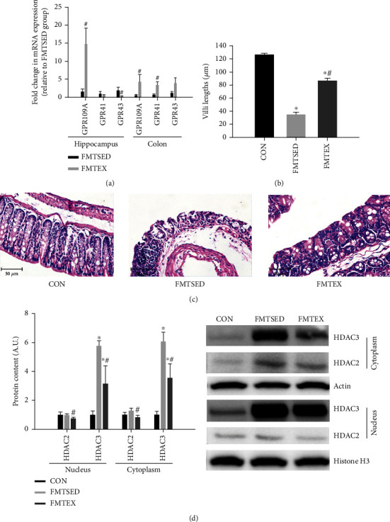
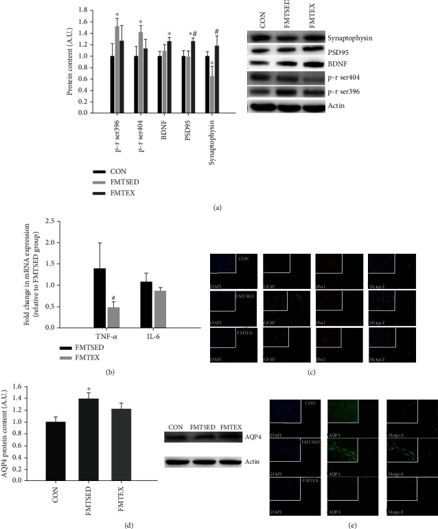
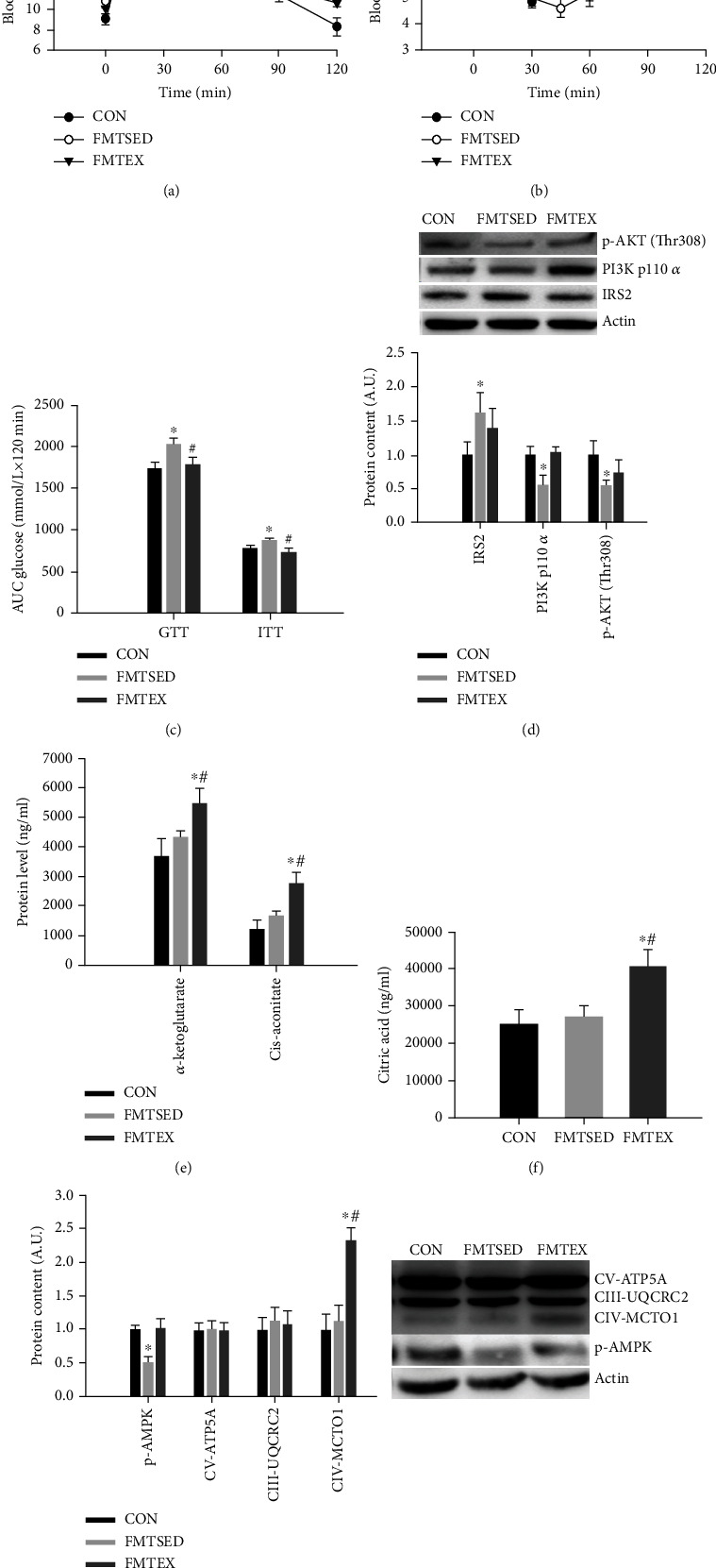
Gut microbes may be the critical mediators for the cognitive enhancing effects of exercise. Via fecal microbiota transplantation (FMT), this study is aimed at determining the mechanism of how voluntary exercise improved learning and memory ability impairment post a high-fat, high-cholesterol (HFHC) diet. The learning and memory abilities assessed via the Morris water maze in the FMT recipient group of voluntary exercising mice were improved compared to sedentary group. 16S rRNA gene sequencing results indicated that exercise-induced changes in gut microbiota distribution were transmissible, mainly in terms of elevated Lactobacillus, Lactobacillus, and Eubacterium nodatum, as well as decreased Clostrida_UCG-014 and Akkermansia after FMT. The neuroprotective effects of FMT were mainly related to the improved insulin signaling pathway (IRS2/PI3K/AKT) and mitochondrial function; inhibition of AQP4; decreased p-Tau at serine 396 and 404; increased BDNF, PSD95, and synaptophysin in the hippocampus; and also decreased HDAC2 and HDAC3 protein expressions in the nuclear and cytoplasmic fractions of the hippocampus. The findings of qRT-PCR suggested that exercise-induced gut microbes, on the one hand, elevated GPR109A and decreased GPR43 and TNF-α in the hippocampus. On the other hand, it increased GPR109A and GPR41 expressions in the proximal colon tissue. In addition, total short-chain fatty acid (SCFA), acetic acid, propionic acid, isobutyric acid, valeric acid, and isovaleric acid contents were also elevated in the cecum. In conclusion, exercise-induced alterations in gut microbiota play a decisive role in ameliorating HFHC diet-induced cognitive deficits. FMT treatment may be a new considerable direction in ameliorating cognitive impairment induced by exposure to HFHC diet.


Conflict of interest statement

The authors declare no conflicts of interest.

Figures

Figure 1
Figure 1
Cognitive function measured via MWM for the C57BL/6J mice. (a) Mean escape latency to hidden platform on the first 5 training days of navigation test. (b) Number of crossing the previous platform location, (c) time spent, (d) swimming distance in the target quadrant, and (e) proximity during probe test. (f) Representative swimming paths of mice from each group. Data were presented as means ± SEM (N = 8). p < 0.05 versus CON and #p < 0.05 versus FMTSED.
Figure 2
Figure 2
Gut microbial comparisons between donor and recipient mice and microbiota characterization of recipient mice. Relative abundance of the top 10 microbiota determined using 16S rRNA sequences in DSED, DEX, FMTSED, and FMTEX at the phylum (a) and genera (b) levels. (c) Relative abundance (% of total bacteria) of bacterial genera that were differentially represented by the exercise group in donor mice and were remained differentially abundant in recipient mice after transplant. p < 0.05 versus DSED and FMTSED, respectively. The α-diversity indices including ACE, Chao1 (d), Shannon, and Simpson (e) indices, β-diversity indices (f), a cladogram of linear discriminant analysis (g) were shown. Data were presented as means ± SEM (N = 8). p < 0.05 versus CON in (d) and (e).
Figure 3
Figure 3
SCFA receptor mRNA expression from both hippocampus and colon, HE staining of colon tissues, and HDAC protein expression from hippocampus. (a) The mRNA expression of GPR41, GPR43, and GPR109A in the hippocampus and proximal colon tissues were measured by qPCR. Colon villi lengths (b) and cross-sections of the mouse colon (c) were shown (H&E stained cross sections; ×20 objective). (d) Protein expression of HDAC2 and HDAC3 in the nuclei and cytosol of hippocampus was measured by western blot. p < 0.05 versus CON and #p < 0.05 versus FMTSED.
Figure 4
Figure 4
Tau phosphorylation, synaptic function, and neuroinflammation-related markers from hippocampus. (a) Protein expression including p-tau ser396 and 404, BDNF, PSD95, and synaptophysin of hippocampus was measured by western blot. (b) TNF-α and IL-6 mRNA expressions from hippocampus were measured by qPCR. (c) Representative double immunofluorescence images of GFAP (in green) and Ibα1 (in red) from mouse cerebral cortex (×50 objective). AQP4 protein expression was measured by western blot (d) and immunofluorescence (e) (×50 objective), respectively. Data were presented as means ± SEM (N = 8). p < 0.05 versus CON and #p < 0.05 versus FMTSED.
Figure 5
Figure 5
Glucose and insulin tolerance test, hippocampal insulin signaling, and mitochondrial function-related markers, and Krebs cycle metabolites from cerebral cortex. The blood glucose levels at indicated timepoints for GTT (a) and ITT (b), respectively, and (c) AUC of GTT and ITT. (d) IRS2, PI3K p110α, and p-AKT (Thr308) protein expressions from hippocampus were measured by western blot. The Krebs cycle intermediate metabolites were measured by GC-MS with α-ketoglutarate, cis-aconitate (e), and citric acid (f) as significantly altered metabolites. (g) Protein expression including CV-ATP5A, CIII-UQCRC2, CIV-MTCO1, and p-AMPK from hippocampus was measured by western blot. Data were presented as means ± SEM (N = 8). p < 0.05 versus CON and #p < 0.05 versus FMTSED.

Similar articles

Cited by

References

    1. Sujkowski A., Hong L., Wessells R. J., Todi S. V. The protective role of exercise against age-related neurodegeneration. Ageing Research Reviews . 2022;74, article 101543 doi: 10.1016/j.arr.2021.101543. - DOI - PMC - PubMed
    1. Gubert C., Kong G., Renoir T., Hannan A. J. Exercise, diet and stress as modulators of gut microbiota: implications for neurodegenerative diseases. Neurobiology of Disease . 2020;134, article 104621 doi: 10.1016/j.nbd.2019.104621. - DOI - PubMed
    1. Kang S. S., Jeraldo P. R., Kurti A., et al. Diet and exercise orthogonally alter the gut microbiome and reveal independent associations with anxiety and cognition. Molecular Neurodegeneration . 2014;9(1):p. 36. doi: 10.1186/1750-1326-9-36. - DOI - PMC - PubMed
    1. Abraham D., Feher J., Scuderi G. L., et al. Exercise and probiotics attenuate the development of Alzheimer’s disease in transgenic mice: role of microbiome. Experimental Gerontology . 2019;115:122–131. doi: 10.1016/j.exger.2018.12.005. - DOI - PubMed
    1. Vendrik K. E. W., Ooijevaar R. E., de Jong P. R. C., et al. Fecal microbiota transplantation in neurological disorders. Frontiers in Cellular and Infection Microbiology . 2020;10:p. 98. doi: 10.3389/fcimb.2020.00098. - DOI - PMC - PubMed

LinkOut - more resources