. 2023 Jan 11:13:1113237.
doi: 10.3389/fmicb.2022.1113237. eCollection 2022.

Marine vampires: Persistent, internal associations between bacteria and blood-feeding marine annelids and crustaceans

Affiliations

Marine vampires: Persistent, internal associations between bacteria and blood-feeding marine annelids and crustaceans

Shana K Goffredi et al. Front Microbiol. .

Abstract

Persistent bacterial presence is believed to play an important role in host adaptation to specific niches that would otherwise be unavailable, including the exclusive consumption of blood by invertebrate parasites. Nearly all blood-feeding animals examined so far host internal bacterial symbionts that aid in some essential aspect of their nutrition. Obligate blood-feeding (OBF) invertebrates exist in the oceans, yet symbiotic associations between them and beneficial bacteria have not yet been explored. This study describes the microbiome of 6 phylogenetically-diverse species of marine obligate blood-feeders, including leeches (both fish and elasmobranch specialists; e.g., Pterobdella, Ostreobdella, and Branchellion), isopods (e.g., Elthusa and Nerocila), and a copepod (e.g., Lernanthropus). Amplicon sequencing analysis revealed the blood-feeding invertebrate microbiomes to be low in diversity, compared to host fish skin surfaces, seawater, and non-blood-feeding relatives, and dominated by only a few bacterial genera, including Vibrio (100% prevalence and comprising 39%-81% of the average total recovered 16S rRNA gene sequences per OBF taxa). Vibrio cells were localized to the digestive lumen in and among the blood meal for all taxa examined via fluorescence microscopy. For Elthusa and Branchellion, Vibrio cells also appeared intracellularly within possible hemocytes, suggesting an interaction with the immune system. Additionally, Vibrio cultivated from four of the obligate blood-feeding marine taxa matched the dominant amplicons recovered, and all but one was able to effectively lyse vertebrate blood cells. Bacteria from 2 additional phyla and 3 families were also regularly recovered, albeit in much lower abundances, including members of the Oceanospirillaceae, Flavobacteriacea, Porticoccaceae, and unidentified members of the gamma-and betaproteobacteria, depending on the invertebrate host. For the leech Pterobdella, the Oceanospirillaceae were also detected in the esophageal diverticula. For two crustacean taxa, Elthusa and Lernanthropus, the microbial communities associated with brooded eggs were very similar to the adults, indicating possible direct transmission. Virtually nothing is known about the influence of internal bacteria on the success of marine blood-feeders, but this evidence suggests their regular presence in marine parasites from several prominent groups.

Keywords: Vibrio; hematophagous; marine leech; obligate blood-feeding; parasitic copepod; parasitic isopod; symbiosis.


Conflict of interest statement

The authors declare that the research was conducted in the absence of any commercial or financial relationships that could be construed as a potential conflict of interest.

Figures
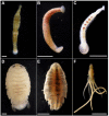
Figure 1
Figure 1
Marine blood-feeding invertebrates examined in this study. The bony fish leeches (A) Pterobdella occidentalis and (B) Ostreobdella californiana. Scale bars 1 mm. (C) The elasmobranch leech Branchellion lobata. Scale bar 5 mm. (D) The isopods Elthusa vulgaris and (E) Nerocila californica. Scale bars 2 mm. (F) The copepod Lernanthropus latis. Scale bar 4 mm. Photo credits: S. Goffredi.
Figure 2
Figure 2
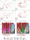
Microbiome diversity analysis of obligate blood feeding (OBF) crustaceans and leeches, based on 16S rRNA gene sequence similarity, using Non-metric multidimensional scaling (NMDS) ordination plots based on Bray–Curtis similarity resemblance, at the (A,B) broad category level, including blood-feeding versus comparison samples of non-blood feeding (NonBF) taxa, biological surfaces, and seawater (SW) and at the (C,D) specific blood-feeding taxa level. (E,F) Relative abundance of bacterial community structure at the genus level, from marine blood-feeders collected primarily from southern California coastal waters, including isopods Elthusa and Nerocila, the copepod Lernanthropus (Lerna), and leeches Branchellion, Ostreobdella, and Pterobdella (specific specimens are listed in Supplementary Table S1 in the same order as shown in the bar charts). Assigned bacterial taxa are color-coded as shown below. Taxa in dark gray were only found in the non-blood feeder (NBF) or seawater (SW) samples. Taxa in light gray were minor taxa in all specimens. See Supplementary Table S4 for a full key.
Figure 3
Figure 3
Relative abundances of the 7 most prevalent Vibrio ribotypes (based on 99% 16S rRNA gene sequence similarity) from obligate marine blood-feeders including the isopods Elthusa and Nerocila (Nero), copepod Lernanthropus (Lerna), and leeches Branchellion, Ostreobdella, and Pterobdella, compared to non-blood-feeding crustaceans (Non-BF), swabs of fish skin, and seawater (SW). Bars are scaled according to percent abundance of each Vibrio ribotype as a function of the entire microbial community in that specimen (for reference, occasional numbers indicate the % abundance of that bar). A total of 135 Vibrionaceae ribotypes were recovered from all samples in total, but the 7 portrayed here accounted for 80% of the total Vibrionaceae diversity. See Supplementary Figure S1 for the phylogenetic position of these ribotypes. Note the pooling of Vibrio ribotypes 19,850 with 213,579 (+) common only in the Nerocila specimens via amplicon sequencing. Occasional symbols note distinction within OBF taxa, including Pterobdella occidentalis recovered from the goby (#), and P. abditovesiculata from Hawaii (*), as well as Branchellion collected from rays other than the pacific ray (°), and Elthusa collected from killifish (+).
Figure 4
Figure 4
Fluorescent visualization and localization of Vibrio in (A) the parasitic copepod Lernanthropus latis, with clasping hooks and egg strings noted. Image taken with a Pentax WG-III handheld camera. Scale bar 2 mm. (B) Cross section of the specimen after being embedded in Steedman’s wax and sectioned. Scale bar 2 mm. (C) A Vibrio-specific fluorescent probe revealed comma-shaped bacterial cells, shown in orange via Cy3, within the digestive lumen space among bloodmeal. Scale bar 20 μm. (inset) A magnified view of the Vibrio cells, near copepod nuclei, shown in blue via DAPI. Scale bar 5 μm. exo, autofluorescent exoskeleton.
Figure 5
Figure 5
Fluorescent visualization and localization of Vibrio in (A) the parasitic isopod Elthusa vulgaris. (B) Ventral dissection showing the enlarged digestive ceca. Scale bar 5 mm. (C) Excised digestive ceca and intestine. Scale bar, 4 mm. A-C were taken with a Pentax WG-III handheld camera. (D) intestine sectioned and Wright stained. Scale bar, 1 mm. Square indicates region in F. (E) ceca sectioned and Wright stained. Scale bar, 1 mm. (D,E) Imaged via light microscopy. (F) A Vibrio-specific fluorescent probe, shown in orange with Cy3, revealed a strong signal within a darkly stained area of the intestine, near the ceca junction. Scale bar 100 μm. (G) Vibrio cells, shown in orange, observed inside of cells, at a specific junction between the midgut and the anterior digestive ceca. Scale bar 20 μm. Isopod nuclei are shown in blue, via DAPI. int., intestine.
Figure 6
Figure 6
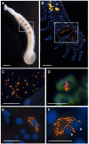
Fluorescent visualization and localization of Vibrio in (A) the elasmobranch leech Branchellion lobata. Square indicates regions of the crop filled with blood, as in B. Scale bar 1 mm. Taken with a Pentax WG-III handheld camera. (B) A partial longitudinal section through an individual, showing fluorescent hybridization using a Vibrio-specific probe to illuminate bacteria cells (shown in orange, via Cy3), with leech cell nuclei shown in blue (via DAPI). Square indicates region in C–F. Scale bar 100 μm. (C,D) A Vibrio-specific fluorescent probe revealed rod-shaped bacterial cells, shown in orange via Cy3, within the lumen space of the crop among bloodmeal (fish host blood cell shown in green, autofluorescence). (D) Vibrio cells appear to be on top of host cell and nucleus. (E,F) Within leech cells, sometimes in proximity to multiple nuclei (arrowheads), shown in blue via DAPI. C-F scale bars are 10 μm. rt., autofluorescent reproductive tissues.
Figure 7
Figure 7
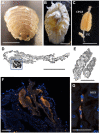
The bony fish leech Pterobdella occidentalis (A) on the fins and body of the longjaw mudsucker (Gillichthys mirabilis), indicated by the arrow. Scale bar 1 cm. (B) Whole specimen, taken with a Pentax WG-III handheld camera. (C) Specimen embedded in Steedman’s wax and sectioned, imaged via light microscopy. Square indicates region in D. Scale bar, 1 mm. (D) Longitudinal section through the anterior end, showing general bacteria cells in a pair of mycetomes (shown in orange, via Eub338-Cy3), counterstained with DAPI, shown in blue. Square indicates region in E. Scale bar, 200 μm. (E) Close-up of bacterial cells, shown in orange, attached to the putative mycetome epithelia. Scale bar is 10 μm. (F) Transmission electron microscopy of a putative mycetome, revealing bacteria-like cells, some which appear to be dividing (arrows). Scale bar, 1 μm. (G) Longitudinal section through a near complete leech specimen, stained with DAPI. Scale bar, 1 mm. (H) Vibrio-specific cells within the crop (shown in orange, via Cy3), counterstained with DAPI, shown in blue. Scale bar, 200 μm. (I). Close-up of bacterial cells, shown in orange, within the crop. Scale bar, 10 μm. myc, mycetomes.

References

    1. Aft R. L., Mueller G. C. (1984). Hemin-mediated oxidative-degradation of proteins. J. Biol. Chem. 259, 301–305. doi: 10.1016/S0021-9258(17)43657-X - DOI - PubMed
    1. Akman L., Yamashita A., Watanabe H., Oshima K., Shiba T., Hattori M., et al. (2002). Genome sequence of the endocellular obligate symbiont of tsetse, Wigglesworthia glossinidia. Nat. Genet. 32, 402–407. doi: 10.1038/ng986, PMID: - DOI - PubMed
    1. Amann R. I., Krumholz L., Stahl D. A. (1990). Fluorescent-oligonucleotide probing of whole cells for determinative, phylogenetic, and environmental studies in microbiology. J. Bacteriol. 172, 762–770. doi: 10.1128/jb.172.2.762-770.1990, PMID: - DOI - PMC - PubMed
    1. Boxshall G. A. (1986). A new genus and two new species of Pennellidae (Copepoda: Siphonostomatoida) and an analysis of evolution within the family. Syst. Parasitol. 8, 215–225. doi: 10.1007/BF00009890 - DOI
    1. Brusca R. C. (1978). Studies on the Cymothoid fish symbionts of the eastern Pacific (isopoda, Cymothoidae) I Biology of Nerocila californica. Crustaceana 34, 141–154.

LinkOut - more resources