. 2023 Jan 9;8(3):2907-2914.
doi: 10.1021/acsomega.2c05040. eCollection 2023 Jan 24.

Rapid In Vitro Quantification of a Sensitized Gadolinium Chelate via Photoinduced Triplet Harvesting

Affiliations

Rapid In Vitro Quantification of a Sensitized Gadolinium Chelate via Photoinduced Triplet Harvesting

James A Tranos et al. ACS Omega. .

Abstract

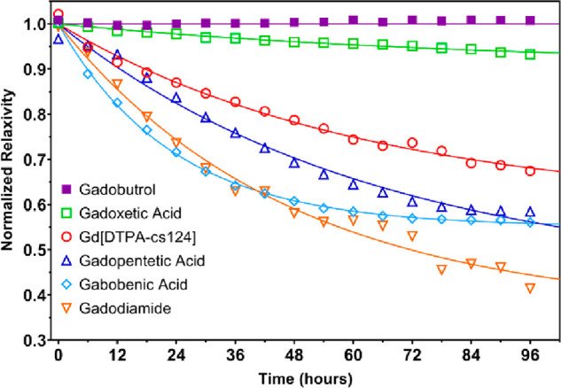
Gadolinium (Gd) based contrast agents (GBCAs) are widely used in magnetic resonance imaging (MRI) and are paramount to cancer diagnostics and tumor pharmacokinetic analysis. Accurate quantification of gadolinium concentration is essential to monitoring the biodistribution, clearance, and pharmacodynamics of GBCAs. However, current methods of quantifying gadolinium in blood or plasma (biological media) are both low throughput and clinically unavailable. Here, we have demonstrated the use of a sensitized gadolinium chelate, Gd[DTPA-cs124], as an MRI contrast agent that can be used to measure the concentration of gadolinium via luminescence quantification in biological media following transmetalation with a terbium salt. Gd[DTPA-cs124] was synthesized by conjugating carbostyril-124 (cs124) to diethylenetriaminepentaacetic acid (DTPA) and chelating to gadolinium. We report increases in both stability and relaxivity compared to the clinically approved analog Gd[DTPA] (gadopentetic acid or Magnevist). In vivo MRI experiments were conducted using C57BL6 mice in order to further illustrate the performance of Gd[DTPA-cs124] as an MRI contrast agent in comparison to Magnevist. Our results indicate that similar chemical modification to existing clinically approved GBCA may likewise provide favorable property changes, with the ability to be used in a gadolinium quantification assay. Furthermore, our assay provides a straightforward and high-throughput method of measuring gadolinium in biological media using a standard laboratory plate reader.


Conflict of interest statement

The authors declare no competing financial interest.

Figures

Figure 1
Figure 1
Mechanism of the transmetalation assay response to free aqueous Tb3+. Following excitation at 330 nm, the gadolinium complex is shown emitting blue light (365 nm) from the carbostyril-124 moiety, and the terbium complex is shown emitting green light (545 nm) from the terbium ion due to intramolecular energy transfer between carbostyril-124 and Tb3+ following the transmetalation reaction.
Figure 2
Figure 2
Fluorescence lifetime plots of (A) Gd[DTPA-cs124] and (B) Tb[DTPA-cs124], including the fluorescence lifetime (τ). Note that the Tb chelate exhibits a 106× increase in emission lifetime properties.
Figure 3
Figure 3
Standard curves for quantifying Gd[DTPA-cs124] via a transmetalation assay. Measurements were performed in an aqueous medium, 5% human plasma, and 5% human blood.
Figure 4
Figure 4
Competitive cation stability assay between various GBCA chelates and equimolar Zn2+. Measurements acquired for gadoteric acid and gadoteridol are not featured in the plot, and exhibit highly similar trends to gadobutrol.
Figure 5
Figure 5
In vivo serial images illustrating the time course of GBCA clearance from full-body MRI scans of C57BL/6J mice (n = 3 for Gd[DTPA-cs124] and n = 1 for gadopentetic acid). (A) Horizontal 2D section covering the whole mouse from head to hindlimbs prior to and following administration of a bolus injection with Gd[DTPA-cs124] (0.1 mmol/kg) via the tail vein. (B) The corresponding time plot illustrating the contrast clearance of Gd[DTPA-cs124] monitored serially. (B, D) The absolute concentration of GBCA was obtained via a region of interest drawn within the descending aortic artery using MRI, as shown in the zoomed-in region in red. In comparison, the same time-imaging protocol and analysis was performed on a C57BL/6J mouse given an IV bolus injection of gadopentetic acid (dose = 0.1 mmol/kg). The clinically approved GBCA shows both similar (C) contrast enhancement patterns and (D) GBCA clearance, as measured using MRI via the descending aortic artery.
Figure 6
Figure 6
Scatter plot for the assessment of plasma clearance performed in C57Bl6 mice (n = 5) within 2 h of the bolus injection of the sensitized GBCA, i.e., Gd[DTPA-cs124].

References

    1. Choyke P. L.; Dwyer A. J.; Knopp M. V. Functional tumor imaging with dynamic contrast-enhanced magnetic resonance imaging. J. Magn. Reson. Imaging 2003, 17 (5), 509–520. 10.1002/jmri.10304. - DOI - PubMed
    1. Ramalho J.; Semelka R. C.; Ramalho M.; Nunes R. H.; AlObaidy M.; Castillo M. Gadolinium-Based Contrast Agent Accumulation and Toxicity: An Update. Am. J. Neuroradiol. 2016, 37 (7), 1192–1198. 10.3174/ajnr.A4615. - DOI - PMC - PubMed
    1. Bharadwaj Das A.; Tranos J. A.; Zhang J.; Zaim Wadghiri Y.; Kim S. G. Estimation of Contrast Agent Concentration in DCE-MRI Using 2 Flip Angles. Invest. Radiol. 2022, 57 (5), 343–351. 10.1097/RLI.0000000000000845. - DOI - PMC - PubMed
    1. Knowles B. R.; Batchelor P. G.; Parish V.; Ginks M.; Plein S.; Razavi R.; Schaeffter T. Pharmacokinetic modeling of delayed gadolinium enhancement in the myocardium. Magn. Reson. Med. 2008, 60 (6), 1524–1530. 10.1002/mrm.21767. - DOI - PubMed
    1. Harrigan C. J.; Peters D. C.; Gibson C. M.; Maron B. J.; Manning W. J.; Maron M. S.; Appelbaum E. Hypertrophic cardiomyopathy: Quantification of late gadolinium enhancement with contrast-enhanced cardiovascular MR imaging. Radiology 2011, 258 (1), 128–133. 10.1148/radiol.10090526. - DOI - PubMed