

Diagnostic validation of a portable whole slide imaging scanner for lymphoma diagnosis in resource-constrained setting: A cross-sectional study
- PMID: 36714453
- PMCID: PMC9874079
- DOI: 10.1016/j.jpi.2023.100188
Diagnostic validation of a portable whole slide imaging scanner for lymphoma diagnosis in resource-constrained setting: A cross-sectional study
Abstract
Background: Telepathology utilizing high-throughput static whole slide image scanners is proposed to address the challenge of limited pathology services in resource-restricted settings. However, the prohibitive equipment costs and sophisticated technologies coupled with large amounts of space to set up the devices make it impractical for use in resource-limited settings. Herein, we aimed to address this challenge by validating a portable whole slide imaging (WSI) device against glass slide microscopy (GSM) using lymph node biopsies from suspected lymphoma cases from Sub-Saharan Africa.
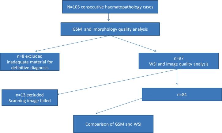
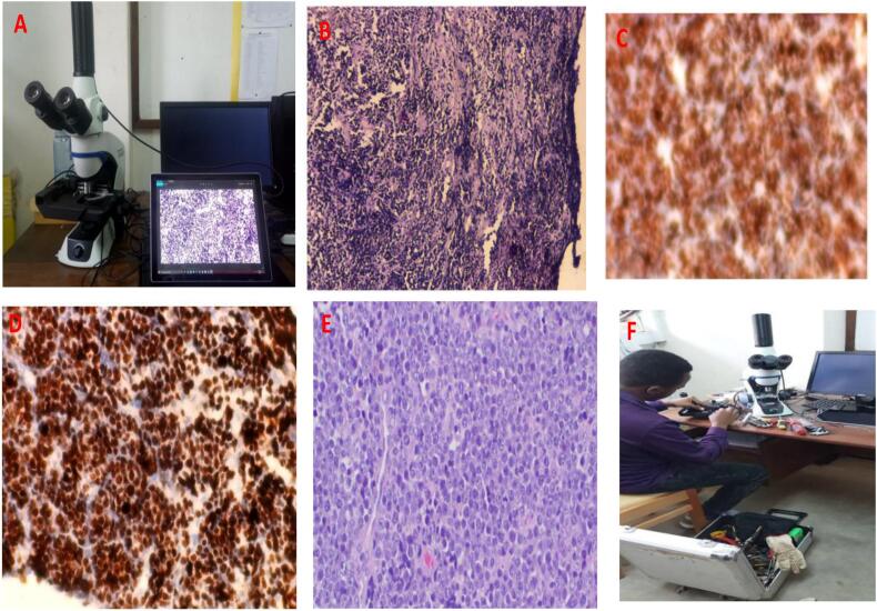
Material and methods: This was part of a multicenter prospective case-control head-to-head comparison study of liquid biopsy against conventional pathology. For the portable WSI scanner validation, the study pathologists evaluated 105 surgical lymph node specimens initially confirmed by gold-standard pathology between February and December 2021. The tissues were processed according to standard protocols for Hematoxylin and Eosin (H&E) and Immunohistochemistry (IHC) staining by well-trained histotechnicians, then digitalized the H& E and IHC slides at each center. The digital images were anonymized and uploaded to a HIPAA-compliant server by the histotechnicians. Three study pathologists independently accessed and reviewed the images after a 6-week washout. The agreement between diagnoses established on GSM and WSI across the pathologists was described and measured using Cohens' kappa coefficient (κ).
Results: On GSM, 65.5% (n=84) of specimens were lymphoma; 25% were classified as benign, while 9.5% were metastatic. Morphological quality assessment on GSM and WSI established that 79.8% and 53.6% of cases were of high quality, respectively. When diagnoses by GSM were compared to WSI, the overall concordance for various diagnostic categories was 93%, 100%, and 86% for lymphoma, metastases, and benign conditions respectively. The sensitivity and specificity of WSI for the detection of lymphoma were 95.2% and 85.7%, respectively, with an overall inter-observer agreement (κ) of 0.86; 95% CI (0.70-0.95).
Conclusions: We demonstrate that mobile whole slide imaging (WSI) is non-inferior to conventional glass slide microscopy (GSM) for the primary diagnosis of malignant infiltration of lymph node specimens. Our results further provide proof of concept that mobile WSI can be adapted to resource-restricted settings for primary surgical pathology and would significantly improve patient outcomes.
Keywords: BL, Burkitt Lymphoma; CAP, College of American Pathologists; DLBCL, Diffuse Large B-cell Lymphoma; GSM, Glass slide microscopy; H&E, Hematoxylin and Eosin staining; HL, Hodgkin’s Lymphoma; IHC, Immunohistochemistry; LMICs, Low-and-middle income countries; Lymphoma diagnosis; NPV, Negative predictive value; PPV, Positive predictive value; Portable whole slide imaging scanner; Resource-limited setting; Validation; WSI, Whole slide imaging.
© 2023 The Authors.
Conflict of interest statement
None declared.
Figures
References
-
- Sung H., Ferlay J., Siegel R.L., Laversanne M., Soerjomataram I., Jemal A., et al. Global Cancer Statistics 2020: GLOBOCAN Estimates of Incidence and Mortality Worldwide for 36 Cancers in 185 Countries. CA Cancer J Clin. 2021;71(3):209–249. doi: 10.3322/caac.21660. Epub 2021 Feb 4. PMID: 33538338. - DOI - PubMed
-
- Hitchcock C.L. The future of telepathology for the developing world. Arch Pathol Lab Med. 2011;135:211–214. - PubMed
LinkOut - more resources
Full Text Sources
Miscellaneous
