. 2022 Aug 24;1(4):pgac166.
doi: 10.1093/pnasnexus/pgac166. eCollection 2022 Sep.

Vascular endothelial growth factor from retinal pigment epithelium is essential in choriocapillaris and axial length maintenance

Affiliations

Vascular endothelial growth factor from retinal pigment epithelium is essential in choriocapillaris and axial length maintenance

Yan Zhang et al. PNAS Nexus. .

Erratum in

Abstract

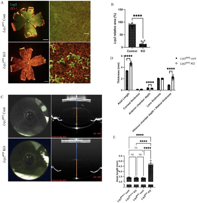
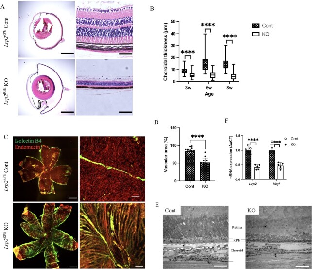
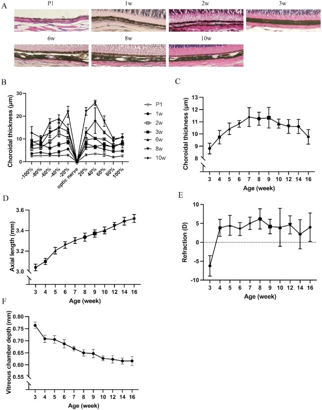
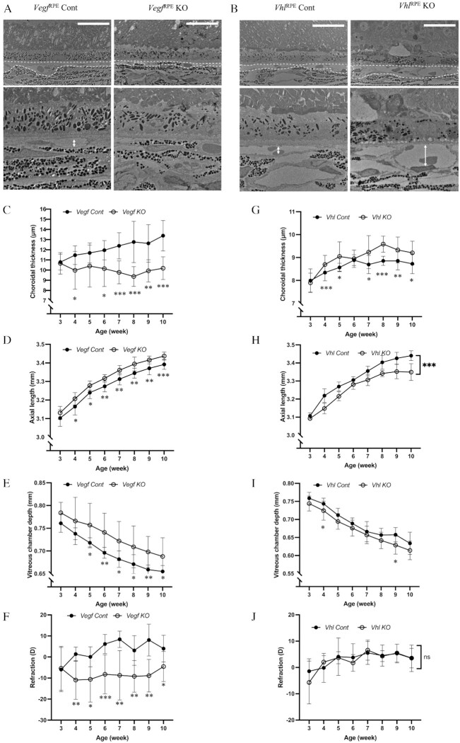
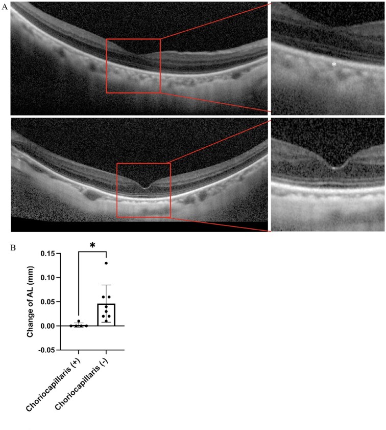
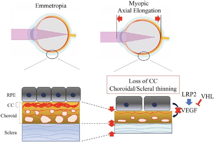
Myopia, which prevalence is rapidly increasing, causes visual impairment; however, the onset mechanism of pathological axial length (AL) elongation remains unclear. A highly vascularized choroid between the retinal pigment epithelium (RPE) and sclera not only maintains physiological activities, but also contributes to ocular development and growth regulation. Vascular endothelial growth factor (VEGF) secreted from the RPE to the choroid is essential for retinal function and maintenance of the choriocapillaris. Herein, we demonstrated that the loss of VEGF secreted from the RPE caused abnormal choriocapillaris development and AL elongation, with features similar to those of the lens-induced myopia (LIM) mouse model, whereas VEGF overexpression by knocking-out von Hippel-Lindau (VHL) specific to the RPE expands the choriocapillaris and shortens the AL. Additionally, LDL Receptor Related Protein 2 (LRP2) deletion in the RPE downregulated VEGF expression and leads to pathological AL elongation. Furthermore, high-myopia patients without choriocapillaris demonstrated longer ALs than did those with preserved choriocapillaris. These results suggest that physiological secretion of VEGF from the RPE is required for proper AL development by maintaining the choriocapillaris. The pinpoint application of VEGF to the choriocapillaris may become a potential intervention for the prevention and treatment of axial myopia progression.

Keywords: VEGF; axial length; choriocapillaris; myopia.


Figures

Fig. 1.
Fig. 1.
Lrp2 gene in RPE cells but not in retinal neurons are required for the proper ocular biometric development. (A) Representative immunohistochemistry of choroid flat-mount of 8-week-old Lrp2RPE Cont and Lrp2RPE KO mouse (green: LRP2, red: ZO and 1). Enlargement and deformation of RPEs and less LRP2 expression are observed in Lrp2RPE KO mouse (bottom) compared to Cont mouse (upper). Scale bar: 1 mm (left panels), 100 µm (right panels). (B) Quantification of LRP2 relative area in RPEs of the choroid of 8-week-old Lrp2RPE KO and Lrp2RPE Cont mouse. n = 3. ****P < 0.0001, two-tailed Student's t-tests. (C) Representative OCT image of the whole eye in 8-week-old Lrp2RPE Cont and Lrp2RPE KO mouse (vitreous chamber depth + retinal thickness; blue line, lens thickness; orange line, anterior chamber depth; gray line; corneal thickness; and yellow line). Scale bar in red: 1 mm. (D) Quantification of different axial parameters in 8-week-old Lrp2RPE Cont and Lrp2RPE KO mouse. Significant differences are shown in axial length, vitreous chamber depth + retinal thickness, and anterior chamber depth. n = 4 Cont, n = 8 KO. ****P < 0.0001, two-tailed Student's t-tests. (E) Axial length comparison between different cre-transgenic Lrp2floxed/floxed mice (Lrp2Retina Cont, Lrp2Retina KO; Lrp2RPE Cont, Lrp2RPE KO). No significant difference is seen in Lrp2Retina KO and its control (Lrp2Retina Cont) group. n = 4. ****P < 0.0001, one-way ANOVA tests. Graphs present as mean ± SD.
Fig. 2.
Fig. 2.
Decrease of Vegf expression from RPEs and abnormal development of choriocapillaris are observed in Lrp2RPE KO mice. (A) The chordal region in the representative H&E-stained cross-sections of enucleated eyes from 6-week-old Lrp2RPE Cont and Lrp2RPE KO mouse showing enlarged overall eye size and thinner choroidal thickness in KO mouse (bottom). Scale bar: 1 mm (left panels), 50 µm (right panels). (B) Choroidal thickness measurements in 3-, 6-, and 8-week-old Lrp2RPE Cont and Lrp2RPE KO mouse showing reduction of choroidal thickness in KO mouse. (C) Representative immunohistochemistry of choroid flat-mount (green: isolectin B4, red: endomucin) and (D) quantification of vascular area in choroid of 8-week-old Lrp2RPE Cont and Lrp2RPE KO mouse showing reduction of choriocapillaris in KO mouse. Scale bar: 1 mm (left panels), 20 µm (right panels). (E) Transmission electron microscope observation of choroids of 8-week-old Lrp2RPE Cont and Lrp2RPE KO mouse. Choriocapillaris highlighted by dashed line in Cont (left) is disappeared in KO (right), resulting in choroidal thinning. Scale bar: 100 µm. (F) Lrp2 and Vegf mRNA expression from primary RPEs in 6-week-old Lrp2RPE Cont and Lrp2RPE KO mouse. Graphs represent as mean ± SD. n = 5. ***P < 0.001, ****P < 0.0001, two-tailed Student's t-tests.
Fig. 3.
Fig. 3.
Choroidal thickness and axial length development in C57B6/J mice. (A) Representative H&E-stained cross-sections of enucleated eyes from wild-type C57B6/J mice and (B) measurement of choroidal thickness according to its distance from the optic nerve. Thickness of choroid is relatively thicker in the posterior pole than the anterior pole n = 3. (C) Choroidal thickness measurement by OCT images shows that choroidal thickness gradually increases and reaches a plateau at about 7 to 9 weeks old after then the thickness gradually become thinner along with the age n = 3. (D) Axial length, (E) refraction error, and (F) vitreous chamber depth with growth showing axial length increase with age and emmetropic refractive status. Numbers between each time point in D indicate slope n = 6. Graphs represent as mean ± SD.
Fig. 4.
Fig. 4.
Vegf RPE KO mice reveal myopic features while VhlRPE KO mice reveal hyperopic features. (A and B) Transmission electron microscope observation of choroids in 10-week-old VegfRPE Cont and VegfRPE KO (A), and VhlRPE Cont and VhlRPE KO (B) mouse showing choriocapillaris thinning in VegfRPE KO and choriocapillaris vasodilatation in VhlRPE KO. Choriocapillaris highlighted by dashed line. Scale bar: 20 µm. (C to J) Choroidal thickness (C and G), axial length (D and H), vitreous chamber depth (E and I) and refraction error (F and J) measurement of VegfRPE Cont and VegfRPE KO (C to F), and VhlRPE Cont and VhlRPE KO mouse (G to J) with growth n = 5. *P < 0.05, **P < 0.01, ***P < 0.001, two-tailed Student's t-tests.
Fig. 5.
Fig. 5.
Axial length of high myopic patients without choriocapillaris increased more compared with high myopic patients who have preserved choriocapillaris. (A) OCT of a 70-year-old female high myopic patient with an axial length of 27.22 mm is shown in the upper panel of A (choriocapillaris highlighted by *), while OCT of a 70-year-old female high myopic patient with an axial length of 28.72 and absent of choriocapillaris are shown in the lower panel of A. High myopic patients with choriocapillaris degeneration [choriocapillaris (-) increased more compared with high myopic patients who have preserved choriocapillaris (+)] in a follow-up for more than 6 months. *P < 0.05, two-tailed Student's t-tests (B) (n = 5 in choriocapillaris group, n = 8 in choriocapillaris degeneration group, respectively). Error bars indicate mean ± SD.
Fig. 6.
Fig. 6.
VEGF derived from RPE is necessary for choriocapillaris development and maintenance, which is essential for overall ocular size development and maintenance. The development of emmetropic eyes requires VEGF derived from RPE to promote the development of choriocapillaris and maintain their physiological thickness. The decrease of VEGF derived from RPE can cause choriocapillaris dysplasia, and the thinner choroid thickness will promote axial length elongation and result in myopia. In a word, generous physiological secretion of VEGF is essential for choriocapillaris and axial length maintenance, while in the case of hypoxia in high-myopia, local pathological secretion of VEGF results in the formation of myopic choroidal neovascularization.

References

    1. Ohno-Matsui K, Lai TYY, Lai CC, Cheung CMG. 2016. Updates of pathologic myopia. Prog Retin Eye Res. 52:156–187. - PubMed
    1. Wong Y-L, Saw S-M. 2016. Epidemiology of Pathologic Myopia in Asia and Worldwide. Asia-Pacific J Ophthalmol. 5(6):394–402. - PubMed
    1. Wong CW et al. 2021. Digital screen time during the COVID-19 Pandemic: risk for a further Myopia Boom?. Am J Ophthalmol. 223:333–337. - PMC - PubMed
    1. Mertz JR, Wallman J. 2000. Choroidal retinoic acid synthesis: a possible mediator between refractive error and compensatory eye growth. Exp Eye Res. 70(4):519–527. - PubMed
    1. Wallman J et al. 1995. Moving the retina: choroidal modulation of refractive state. Vision Res. 35(1):37–50. - PubMed