. 2022 Aug 9;1(4):pgac130.
doi: 10.1093/pnasnexus/pgac130. eCollection 2022 Sep.

C25-modified rifamycin derivatives with improved activity against Mycobacterium abscessus

Affiliations

C25-modified rifamycin derivatives with improved activity against Mycobacterium abscessus

Laura Paulowski et al. PNAS Nexus. .

Abstract

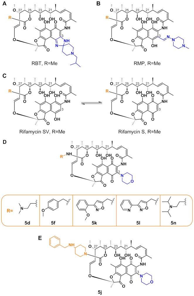
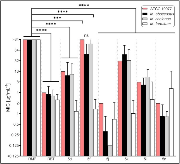
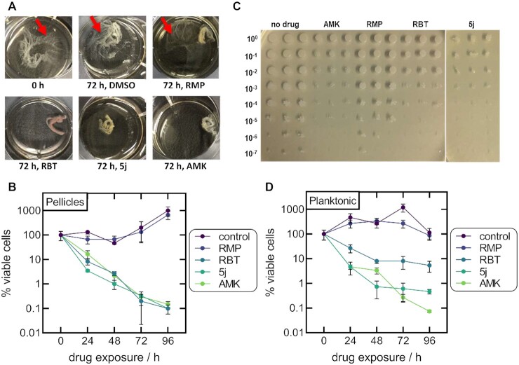
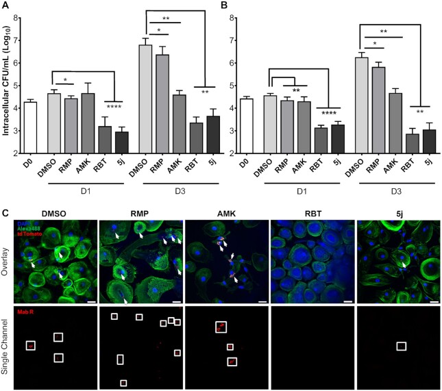
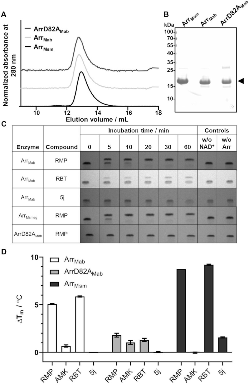
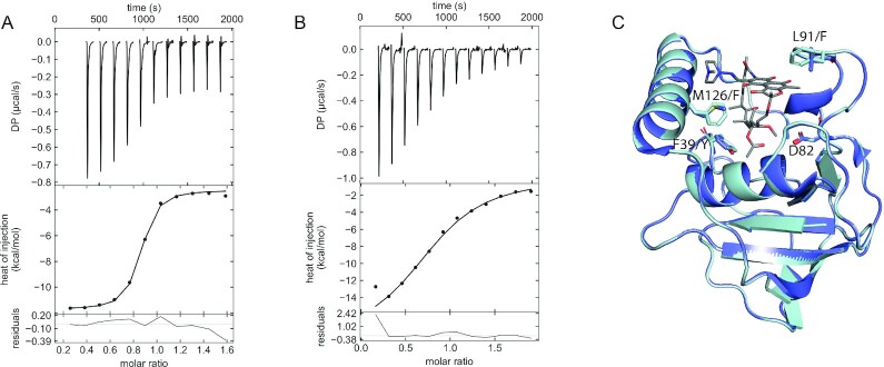
Infections caused by Mycobacterium abscessus are difficult to treat due to its intrinsic resistance to most antibiotics. Formation of biofilms and the capacity of M. abscessus to survive inside host phagocytes further complicate eradication. Herein, we explored whether addition of a carbamate-linked group at the C25 position of rifamycin SV blocks enzymatic inactivation by ArrMab, an ADP-ribosyltransferase conferring resistance to rifampicin (RMP). Unlike RMP, 5j, a benzyl piperidine rifamycin derivative with a morpholino substituted C3 position and a naphthoquinone core, is not modified by purified ArrMab. Additionally, we show that the ArrMab D82 residue is essential for catalytic activity. Thermal profiling of ArrMab in the presence of 5j, RMP, or rifabutin shows that 5j does not bind to ArrMab. We found that the activity of 5j is comparable to amikacin against M. abscessus planktonic cultures and pellicles. Critically, 5j also exerts potent antimicrobial activity against M. abscessus in human macrophages and shows synergistic activity with amikacin and azithromycin.

Keywords: ADP-ribosylation; Mycobacterium abscessus; antimicrobial resistance; rifabutin; rifampicin.


Figures

Fig. 1.
Fig. 1.
Illustration of the chemical structures of rifamycins and rifamycin derivatives. Structures of RBT (A), RMP (B), rifamycin SV and rifamycin S (C), C25-modified rifamycin S derivatives used in this work (D; Table S1, Supplementary Material), and 5j (E) (34, 40, 41).
Fig. 2.
Fig. 2.
Minimal inhibitory concentrations of RMP, RBT, and rifamycin derivatives against M. abscessus ATCC 19977 and clinical NTM isolates. MICs were generated in cation-adjusted Muller–Hinton medium based on the procedure outlined in CLSI document M24 (61). MIC plates were read after 3 days of incubation at 30°C. MIC values were obtained for M. abscessus (MabA, n = 9; MabB, n = 7; and MabM, n = 8), M. chelonae (n = 8), and M. fortuitum (n = 8). Data are plotted as mean ± SD. Data for the MabA ATCC 19977 reference strain are depicted by red bars. ***, P < 0.001; ****P < 0.0001; ns, not significant; and CI 99%.
Fig. 3.
Fig. 3.
Inhibitory effect of rifamycins on M. abscessus pellicles and planktonic cultures. (A) Examples of the visual appearance of Mab pellicles prior to drug exposure and following 72 h of exposure to 10 µg/mL of 5j, RBT, RMP, or AMK. Pellicles treated with RMP retained a reticular formation at the air–liquid interface similar to the DMSO control (red arrows). (B) Viable cell counts of Mab pellicles exposed to 10 µg/mL of 5j, RBT, RMP, or AMK for up to 96 h without shaking. Data are plotted as mean ± SD of three independent experiments. CFU counts were normalized to the CFU count prior to the addition of antibiotics. (C) Example of CFU counts from Mab pellicles obtained following 72 h of drug exposure (three technical replicates per drug). (D) Viable cell counts of planktonic cultures incubated with 10 µg/mL of the respective drug with shaking (three technical replicates per drug).
Fig. 4.
Fig. 4.
Intracellular survival of M. abscessus smooth and rough morphotypes within human macrophages treated with 5j, RMP, RBT, or amikacin. THP-1 macrophages were infected with Mab CIP104536 smooth (A) or rough variant (B) at a MOI of 2:1 for 4 h. Data are plotted as mean ± SD of four independent experiments (n = 4). (C) Confocal microscopy of human monocyte-derived macrophages (HMDM) infected for 6 h with Mab R expressing tdTomato at a MOI of 10:1. Infected cells were treated with a final concentration of 5 µg/mL of RMP, AMK, RBT, or 5j, respectively, for 72 h prior to fixation and staining. Bacterial cells appear in red and are additionally highlighted by white arrows in the overlay images (upper panel) and by white boxes in the single channel images (lower panel). HMDM nuclei were stained with DAPI (blue). Actin staining was done with Alexa488-conjugated Phalloidin (green) (54). Scale bars apply to all images shown and correspond to 25 µm.
Fig. 5.
Fig. 5.
Interaction of purified ArrMab, ArrD82AMab, and ArrMsm with RMP, RBT, and 5j. (A) Representative size exclusion profile of purified ArrMab, the catalytically inactive mutant (ArrD82AMab) and ArrMsm, with the corresponding peak fraction separated by SDS–PAGE. (B) The arrow indicates the predominant band corresponding to the Arr variants. (C) ADP-ribosylation reactions stopped after the indicated incubation times, analyzed by rpTLC. Reactions without NAD+ or without the enzymes were added as negative controls (n = 3). (D) Change in melting temperature (ΔTm) of Arr variants in the presence of selected compounds. The ΔTm value was derived by subtracting mean Tm of the control (1% DMSO) from the mean Tm in the presence of ligand observed in NanoDSF. Melting experiments were performed in triplicate (n = 3), RMP, 300 µM RMP; RBT, 300 µM RBT; AMK, 300 µM amikacin; and 5j, 300 µM compound 5j.
Fig. 6.
Fig. 6.
Arr active site and quantification of RMP binding. (A) and (B) ITC thermogram of RMP titrated into ArrMsm (A) and ArrMab (B), (n = 3). (C) Overlay of ArrMsm crystal structure (PDB 2HW2) with the ArrMab model, shown in dark blue and light blue, respectively. Residue numbering is according to ArrMsm. The catalytic aspartate (D82) has been shown in stick representation along with other residues which differ in the ArrMab RMP binding site. RMP is shown as a stick model.

References

    1. Nigam A, et al. . 2014.; Modification of rifamycin polyketide backbone leads to improved drug activity against rifampicin-resistant Mycobacterium tuberculosis. J Biol Chem. 289:21142–21152. - PMC - PubMed
    1. Daley CL, et al. . 2020. Treatment of nontuberculous mycobacterial pulmonary disease: an official ATS/ERS/ESCMID/IDSA clinical practice guideline: executive summary. Clin Infect Dis. 71:e1–e36. - PMC - PubMed
    1. Johansen MD, Herrmann JL, Kremer L, 2020. Non-tuberculous mycobacteria and the rise of Mycobacterium abscessus. Nat Rev Microbiol. 18:392–407. - PubMed
    1. Maurer F, et al. . 2014.; Postsurgical wound infections due to rapidly growing mycobacteria in Swiss medical tourists following cosmetic surgery in Latin America between 2012 and 2014. Eurosurveillance. 19:20905. - PubMed
    1. Ryan K, Byrd TF. 2018. Mycobacterium abscessus: shapeshifter of the mycobacterial World. Front Microbiol. 9:2642. - PMC - PubMed

LinkOut - more resources