. 2022 Aug 18;1(4):pgac151.
doi: 10.1093/pnasnexus/pgac151. eCollection 2022 Sep.

Correction of large jawbone defect in the mouse using immature osteoblast-like cells and a 3D polylactic acid scaffold

Affiliations

Correction of large jawbone defect in the mouse using immature osteoblast-like cells and a 3D polylactic acid scaffold

Shigeto Suzuki et al. PNAS Nexus. .

Abstract

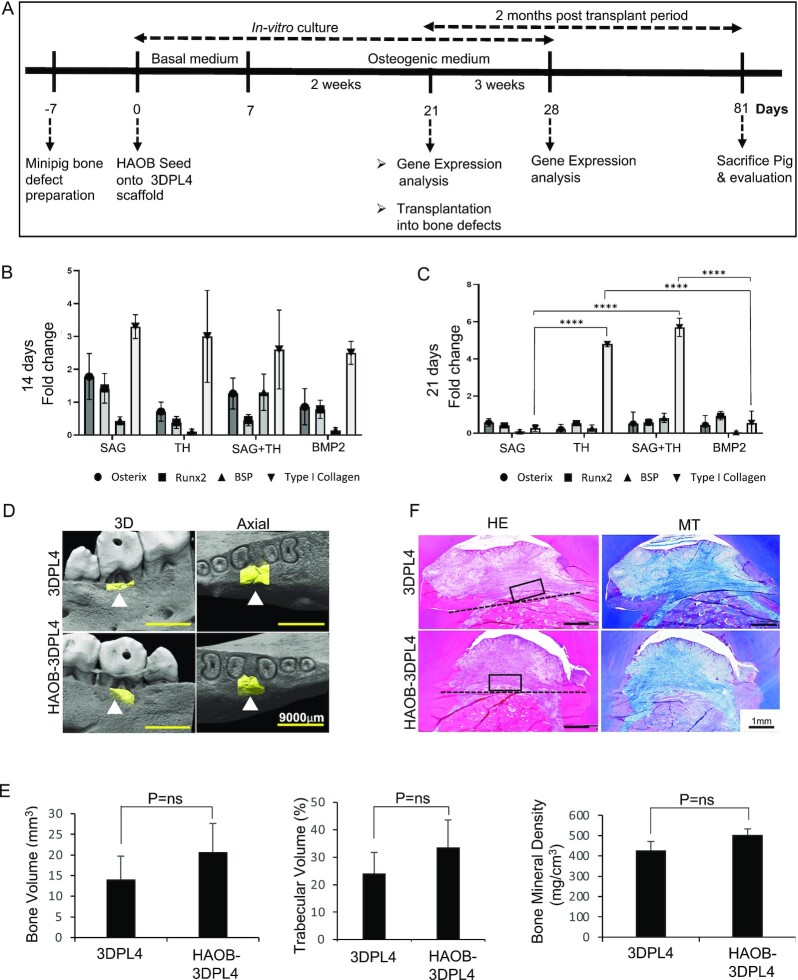
Bone tissue engineering has been developed using a combination of mesenchymal stem cells (MSCs) and calcium phosphate-based scaffolds. However, these complexes cannot regenerate large jawbone defects. To overcome this limitation of MSCs and ceramic scaffolds, a novel bone regeneration technology must be developed using cells possessing high bone forming ability and a scaffold that provides space for vertical bone augmentation. To approach this problem in our study, we developed alveolar bone-derived immature osteoblast-like cells (HAOBs), which have the bone regenerative capacity to correct a large bone defect when used as a grafting material in combination with polylactic acid fibers that organize the 3D structure and increase the strength of the scaffold material (3DPL). HAOB-3DPL constructs could not regenerate bone via xenogeneic transplantation in a micromini pig alveolar bone defect model. However, the autogenic transplantation of mouse calvaria-derived immature osteoblast-like cells (MCOBs) isolated using the identical protocol for HAOBs and mixed with 3DPL scaffolds successfully regenerated the bone in a large jawbone defect mouse model, compared to the 3DPL scaffold alone. Nanoindentation analysis indicated that the regenerated bone had a similar micromechanical strength to native bone. In addition, this MCOB-3DPL regenerated bone possesses osseointegration ability wherein a direct structural connection is established with the titanium implant surface. Hence, a complex formed between a 3DPL scaffold and immature osteoblast-like cells such as MCOBs represents a novel bone tissue engineering approach that enables the formation of vertical bone with the micromechanical properties required to treat large bone defects.

Keywords: bone regeneration; functional bone; human alveolar osteoblasts; mice calvaria osteoblasts; polylactic acid scaffold.


Figures

Fig. 1.
Fig. 1.
Synthesis of PLLA/gelatin floccular scaffolds for bone regeneration therapy. (A) Schematic illustration of the fabrication process used to produce PLLA/gelatin floccular scaffolds, based on PLLA/gelatin floccular fibers, using emulsion electrospinning. (B) Photograph of as-formed floccular PLLA/gelatin fabrics on the target electrode and (C) cut PLLA/gelatin floccular fabrics. (D) Photograph of the PLLA/gelatin floccular scaffolds. (E to I) SEM images of the PLLA/ gelatin floccular scaffolds (E, 3DPL1) (F, 3DPL2), (G, 3DPL4), (H, 3DPL6), and 2D sheet scaffolds (I, 2DPL). Scale bar, 50 μm. (J) Fiber diameter distributions of the PLLA/ gelatin floccular fibers from laser confocal microscopy (LCM) observations. (K) IR spectra of the PLLA/gelatin floccular fibers (i) and their chemically etched residue (ii) with IR spectra of the PLLA (iii) and gelatin (iv) included for comparison. (L) Tensile stress–strain curves of the floccular PLLA/gelatin nonwoven fabrics. (M) Degradation of the PLLA/ gelatin floccular scaffolds under a wet environment. (N) Compression stress–strain curves of floccular PLLA/gelatin nonwoven scaffolds (3DPL4).
Fig. 2.
Fig. 2.
HAOB-3DPL4 constructs fail to regenerate bone following xenotransplantation into a micromini pig furcation defect model. (A) Overview of the timeline for defect preparation and HAOB-3DPL4 scaffold construct preparation for in-vitro osteogenic characterization and transplantation into a bone defect. (B, C) Osteogenic-related gene expression of HAOB-3DPL4 scaffold constructs after 14- and 21-days incubation under osteogenic medium. (D) Three dimensional reconstruction of micro-CT images and axial view of the furcation defect following HAOB-3DPL4 constructs and 3DPL4 scaffold transplantation into the established micromini pig furcation defect model. (E) Quantification of the regenerated alveolar bone in terms of bone volume, trabecular bone, and bone mineral density from reconstructed 3D micro-CT images. Representative sections of 3D4PL4 and HAOB-3D4PL4 scaffold constructs examined by hematoxylin–eosin (HE) and Masson’s trichrome (MT) staining. (ns; not significant). (F) Representative sections of HAOB-3DPL4 constructs and 3DPL4 scaffold transplant samples were examined by Haematoxylin and Eosin (H&E) staining.
Fig. 3.
Fig. 3.
Analysis of the osteoblast differentiation ability of MCOB. (A) Representative images of alizarin red and ALP staining of MCOB at day 10 without (upper panel) and with (lower panel) ODM treatment. Relative positive alizarin and ALP activity detected only in MCOB conditioned with ODM. (B) The relative mRNA expression levels of Osterix, Osteocalcin, and Runx2 of MCOB treated with ODM for 10 days were shown as fold change by real time-PCR analysis. (C) SEM images of MCOB seeded in 3DPL4 scaffold (left). Higher magnification (right) showing external cell–cell and cell–3DPL4 contact indicated by red arrows. (D) SEM images of 3DPL4 scaffold (left). Higher magnification (right) showing 3D arrangement of polylactic acid fibers. (E) The photographs of H&E staining of MCOB-3DPL4 and 3DPL4 (F) implant groups at 4 weeks (upper) and 8 weeks (lower) after the subcutaneous transplantation into mouse. Boxed areas are shown at higher magnification. Higher magnified view of the MCOB-3DPL4 groups showed ectopic bone formation within 3DPL4 scaffold at both 4- and 8-weeks post-implantation, highlighted by yellow arrow heads. Highly magnified view of the 3DPL4 groups showed connective formation within 3DPL4 scaffold and yellow arrow heads indicate the PL fibers.
Fig. 4.
Fig. 4.
Bone regeneration capacity in the mice alveolar bone defect evaluated by microCT (μCT) and histological analysis. (A) Two dimensional μCT images of the defect areas, filled with MCOB-3DPL4, 3DPL4, Cytrans, and empty defect groups at 4- and 8-week post-transplantation. (B) μCT quantification of new bone volumes in the mice alveolar defect areas. (C) 3D-reconstructed images of the defect area. (D) Representative H&E–stained images of the low (Upper row) and high (framed regions of upper row) magnifications of the defect areas at 8-week post-transplantation. The yellow arrow heads indicate 3DPL4 fibers, red arrow heads indicate cytrans granules, and the M3: maxillary third molar, M2: maxillary second molar, and M1 area: maxillary first molar area/defect areas, black arrow indicates the mesial direction of the jaw, yellow-dotted line represents the outer edge of the regenerated bone, blue-dotted line represents the CEJ line, and the area below it indicates bone defect area.
Fig. 5.
Fig. 5.
Biomechanical and functional properties of regenerated bone at 8-week post-transplantation. (A) Hardness and (B) elastic modulus of regenerated bone at a maximum load. (C) Two dimensional and (D) 3D μCT images of a mouse alveolar defect area at 4-week post-implant placement. (E) H&E staining of the mouse alveolar bone defect at 4 week after implant placement. Boxed regions are shown as highly magnified sections in the middle and lower rows. M2: maxillary second molar, M1 area: maxillary first molar area/regenerated bone area, and black arrow indicates mesial direction of the jaw.

References

    1. Hayashi K., Kishida R., Tsuchiya A., Ishikawa K., 2020. Granular honeycombs composed of carbonate apatite, hydroxyapatite, and β-tricalcium phosphate as bone graft substitutes: effects of composition on bone formation and maturation. ACS Applied Bio Materials. 3(3): 1787–1795. - PubMed
    1. Sato N, et al. , 2020.Comparison of the vertical bone defect healing abilities of carbonate apatite, β-tricalcium phosphate, hydroxyapatite and bovine-derived heterogeneous bone. Dent Mater J. 39(2): 309–318. - PubMed
    1. Sotome S, et al. , 2016.Efficacy and safety of porous hydroxyapatite/type 1 collagen composite implantation for bone regeneration: a randomized controlled study. J Orthop Sci. 21(3): 373–380. - PubMed
    1. Funayama T., Noguchi H., Tsukanishi T., Sakane M., 2012.Histological analysis of bone bonding and ingrowth into connected porous hydroxyapatite spacers in spinal surgery. Key Eng Mater. 529-530: 309–312.
    1. Wang T.-Y., Xu S.-L., Wang Z.-P., Guo J.-Y., 2020.Mega-oss and mega-TCP versus Bio-Oss granules fixed by alginate gel for bone regeneration. BDJ Open. 6(1): 14. - PMC - PubMed