. 2023 Jan 1:48:bjad001.
doi: 10.1093/chemse/bjad001.

Neural suppression in odor recognition memory

Affiliations

Neural suppression in odor recognition memory

Tom Eek et al. Chem Senses. .

Abstract

Little is known about the neural basis of lower- and higher-order olfactory functions such as odor memory, compared with other sensory systems. The aim of this study was to explore neural networks and correlates associated with 3 functions: passive smelling (PS), odor encoding (OE), and in particular odor recognition memory (ORM). Twenty-six healthy participants were examined using functional magnetic resonance imaging conducted across 3 sessions, one for each function. Independent component analysis revealed a difference between sessions where a distinct ORM component incorporating hippocampus and posterior cingulate showed delayed triggering dissociated from odor stimulation and recognition. By contrasting Hit for ORM (target odors correctly recognized as old) and a combination of PS and detected odors from OE, we found significantly lower activations in amygdala, piriform cortex, insula, thalamus, and the inferior parietal lobule. Region of interest analysis including anterior insula, posterior cingulate gyrus, dentate gyrus, left middle frontal gyrus, amygdala, and piriform cortex demonstrated that Hit were associated with lower activations compared with other memory responses. In summary, our findings suggest that successful recognition of familiar odors (odor familiarity) is associated with neural suppression in the abovementioned regions of interest. Additionally, network including the hippocampus and posterior cingulate is engaged in a postrecognition process. This process may be related to incidental encoding of less familiar and more novel odors (odor novelty) and should be subject for future research.

Keywords: episodic memory; familiarity; functional magnetic resonance imaging (fMRI); independent component analysis (ICA); olfaction; region of interest analysis (ROI).


Figures

Fig. 1.
Fig. 1.
Schematic representation of fMRI paradigm. Olfactory stimulation was performed in blocks for all 3 sessions: PS, OE, and ORM. Visual cueing was only used during OE and ORM.
Fig. 2.
Fig. 2.
Spatial representation of ROIs included in the post hoc ROI analysis. The following ROIs: anterior insula, medial dorsal thalamus, middle frontal gyrus, posterior cingulate gyrus, precuneus, dentate gyrus of hippocampus, amygdala, and posterior piriform cortex were extracted bilaterally, generating 16 ROIs in total.
Fig. 3.
Fig. 3.
Results from ICA of PS. One independent component was associated with functional connectivity among parts of amygdala and posterior piriform cortex (z = −16), basal ganglia, thalamus, and insula (z = −4), as well as the secondary somatosensory cortex (z = 20). The signal change for this network (temporal mode) coincided with olfactory stimulation. The default threshold of P > 0.5 was used for testing the alternative hypothesis post statistically. Color bars are given in terms of T-statistic. Left and right in radiological convention.
Fig. 4.
Fig. 4.
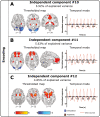
Results from ICA of OE. A) Independent component #10 was associated with functional connectivity among hippocampus (z = −12, y = −34), ventral striatum (z = −4), and posterior cingulate gyrus (y = −34). B) Another component (#11) was associated with functional connectivity among caudate nucleus (z = 0), the medial part of thalamus (z = 12), and the prefrontal cortex (z = 0, z = 12, x = 10). C) Independent component #12 was associated with functional connectivity among amygdala and posterior piriform cortex (z = −16, z = −12), as well as basal ganglia, thalamus, and anterior insula (z = −4). The signal change for all these networks (temporal mode) coincided with olfactory stimulation. The default threshold of P > 0.5 was used for testing the alternative hypothesis post statistically. Color bars are given in terms of T-statistic. Left and right in radiological convention.
Fig. 5.
Fig. 5.
Results from ICA of ORM. A) Independent component #8 was associated with functional connectivity among amygdala and posterior piriform cortex (z = −16), basal ganglia, thalamus, and anterior insula (z = 4), as well the left middle frontal gyrus (z = 34). The signal change for this network (temporal mode) coincided with olfactory stimulation. B) Another component (#11) was associated with functional connectivity among hippocampus (y = −34), as well as posterior cingulate gyrus and precuneus (x = −12, x = 6). Signal change for this network is dissociated with stimulation, with positive signal change occurring during the resting time. The default threshold of P > 0.5 was used for testing the alternative hypothesis post statistically. Color bars are given in terms of T-statistic. Left and right in radiological convention.
Fig. 6.
Fig. 6.
Results from GLM analysis. A) During ORM, anterior insula, pallidum, and thalamus bilaterally (z = 0), parts of hippocampus, particularly on the right hemisphere (y = −36), as well as the middle frontal gyrus and the supplementary motor area bilaterally (y = −2) showed significantly higher activation compared with the combination of PS and OE. B) For Hit during ORM, amygdala and piriform cortex bilaterally (z = −16), parts of insula (mostly posteriorly), and thalamus (z = −38), as well as the inferior parietal lobule (y = −38) showed significantly lower activation compared with the combination of PS and perceived odors during OE. Left and right in radiological convention; P < 0.05 family-wise error (FWE) corrected.
Fig. 7.
Fig. 7.
FIR event-related time courses for anterior insula, middle frontal gyrus, posterior cingulate gyrus, dentate gyrus, amygdale, and posterior piriform cortex (left hemisphere) during the odor recollection memory task. For insula, posterior cingulate gyrus, dentate gyrus, amygdala, and posterior piriform cortex peak signal change of Hit was significantly lower than the one of FA (repeated measures ANOVA, Table 2). Additionally, peak signal change of Hit was significantly lower than the one of CR in middle frontal gyrus, posterior cingulate gyrus, amygdala, and posterior piriform cortex (repeated measures ANOVA, Table 2). Peak signal change of Hit was significantly lower than the one of Miss only in posterior cingulate gyrus (repeated measures ANOVA, Table 2).

References

    1. Arshamian A, Iannilli E, Gerber JC, Willander J, Persson J, Seo HS, Hummel T, Larsson M.. The functional neuroanatomy of odor evoked autobiographical memories cued by odors and words. Neuropsychologia. 2013;51(1):123–131. - PubMed
    1. Beckmann CF, Smith SM.. Probabilistic independent component analysis for functional magnetic resonance imaging. IEEE Trans Med Imaging. 2004;23(2):137–152. - PubMed
    1. Beckmann CF, Smith SM.. Tensorial extensions of independent component analysis for multisubject FMRI analysis. Neuroimage. 2005;25(1):294–311. - PubMed
    1. Bird CM. The role of the hippocampus in recognition memory. Cortex. 2017;93:155–165. - PubMed
    1. Boesveldt S, de Muinck Keizer RJ, Wolters E, Berendse HW.. Odor recognition memory is not independently impaired in Parkinson’s disease. J Neural Transm. 2009;116(5): 575–578. - PubMed

Publication types