Review
. 2023 Jan 30;13(1):4295-4319.
doi: 10.1002/cphy.c220010.

Tensions in Taxonomies: Current Understanding and Future Directions in the Pathobiologic Basis and Treatment of Group 1 and Group 3 Pulmonary Hypertension

Affiliations
Review

Tensions in Taxonomies: Current Understanding and Future Directions in the Pathobiologic Basis and Treatment of Group 1 and Group 3 Pulmonary Hypertension

Sue Gu et al. Compr Physiol. .

Abstract

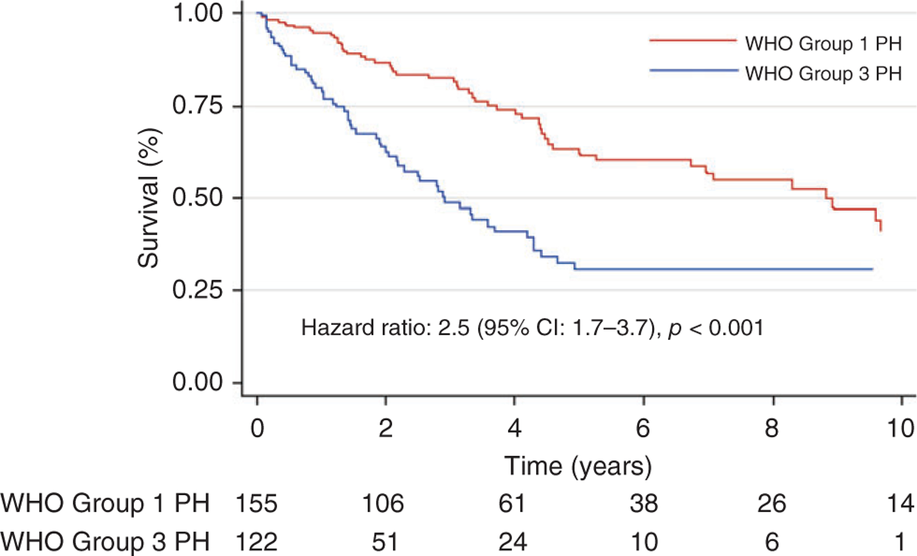
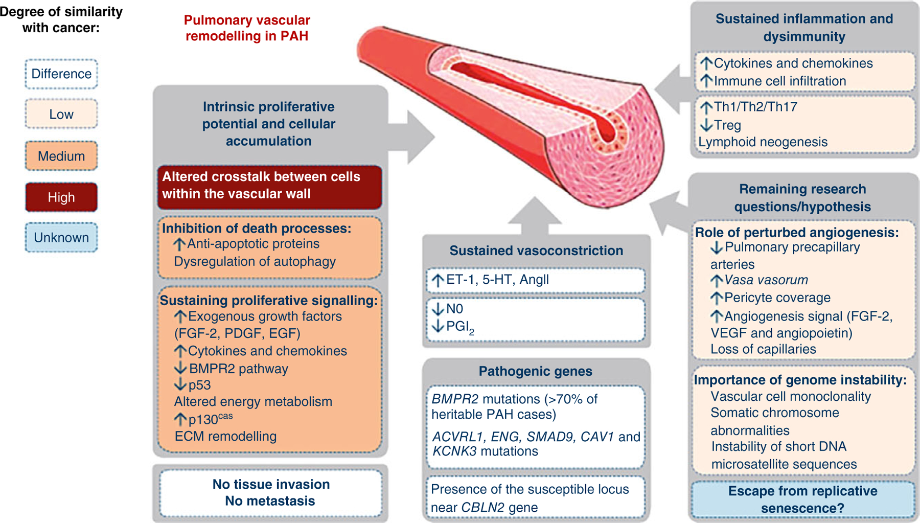
In the over 100 years since the recognition of pulmonary hypertension (PH), immense progress and significant achievements have been made with regard to understanding the pathophysiology of the disease and its treatment. These advances have been mostly in idiopathic pulmonary arterial hypertension (IPAH), which was classified as Group 1 Pulmonary Hypertension (PH) at the Second World Symposia on PH in 1998. However, the pathobiology of PH due to chronic lung disease, classified as Group 3 PH, remains poorly understood and its treatments thus remain limited. We review the history of the classification of the five groups of PH and aim to provide a state-of-the-art review of the understanding of the pathogenesis of Group 1 PH and Group 3 PH including insights gained from novel high-throughput omics technologies that have revealed heterogeneities within these categories as well as similarities between them. Leveraging the substantial gains made in understanding the genomics, epigenomics, proteomics, and metabolomics of PAH to understand the full spectrum of the complex, heterogeneous disease of PH is needed. Multimodal omics data as well as supervised and unbiased machine learning approaches after careful consideration of the powerful advantages as well as of the limitations and pitfalls of these technologies could lead to earlier diagnosis, more precise risk stratification, better predictions of disease response, new sub-phenotype groupings within types of PH, and identification of shared pathways between PAH and other types of PH that could lead to new treatment targets. © 2023 American Physiological Society. Compr Physiol 13:4295-4319, 2023.


Figures

Figure 1
Figure 1
Survival in Group 1 and Group 3 PH. Reused, with permission, from Prins KW, et al., 2018 (138), American Thoracic Society.
Figure 2
Figure 2
Pathogenic concepts of pulmonary arterial hypertension: Differences and similarities with cancer. FGF, fibroblast growth factor; PDGF, platelet-derived growth factor; EGF, epidermal growth factor; BMPR2, bone morphogenetic protein receptor 2; ECM, extracellular matrix; ET-1, endothelin-1; 5-HT, serotonin; NO, nitric oxide; PGI2, prostacyclin; Th, T-helper cell; Treg, regulatory T-cell; VEGF, vascular endothelial growth factor. Reused, with permission, from Guignabert C, et al., 2013 (66), European Respiratory Society.
Figure 3
Figure 3
Inflammatory components in plexiform type lesions of PAH. Inflammatory cells are highlighted by the anti-CD45 immunostaining (arrowheads) (A), B-cell marker (UL-26) (B), T-cell marker (UCHSC) (C), and macrophage marker (CD68) (D and E) (arrowheads). Note infiltration of the outer portion of the plexogenic vessel (D) and vessel obliterated by concentric proliferation (E) by macrophages (immunoperoxidase, A-C, E 500X; D: 200x). I-Vascular lumen. Reused, with permission, from Tuder RM, et al., 1994 (199), from Elsevier.
Figure 4
Figure 4
Inflammatory and fibrotic processes in usual interstitial pneumonia with marked pulmonary vascular remodeling. (A) Low magnification of subpleural region showing marked subpleural fibrosis (arrow), extending irregularly toward central regions of the lung with conglomerates of lymphoid cells (arrowhead) (Hematoxilin and Eosin). (B) Marked perivascular (arrows) and airway (yellow arrow) fibrosis and inflammation, forming a continuous process along the bronchovascular and parencymal structures. The pulmonary arteries show intima thickening, with significant obliteration of the vascular lumina (Pentachrome). (C) Exudative fibroblastic focus (arrowhead) next to small pulmonary artery (arrowhead), accompanied by scattered inflammatory cells (Pentachrome). (D) Active bridging fibrosis along alveolar and bronchovascular (yellow arrow) associated with exudative process (arrow) and inflammation (arrowhead). Note the marked obliteration of an adjacent pulmonary artery (long arrow) (Pentachrome). (E) Active inflammation and interstitial fibrosis in the advancing edge of usual interstitial pneumonia (yellow arrow) (Hematoxilin and eosin). (F) Marked media and intima thickening in muscular artery (arrows) in a region not immediately affected by UIP (arrows) (Hematoxilin and eosin).
Figure 5
Figure 5
Survival analysis of based on a panel of plasma proteins and REVEAL score in an observational cohort of idiopathic and heritable PAH patients. Kaplan-Meier survival estimates in patients with different panel scores, in all patients with idiopathic pulmonary arterial hypertension from (A) cohorts 1 and 2 and (B) cohort 4. ROC analysis of Cox models before and after addition of the prognostic protein panel to the established equation, in all patients with idiopathic pulmonary arterial hypertension from (C) cohorts 1 and 2 and (D) cohort 4. ROC, receiver operating characteristic; AUC, area under the curve. Reused, with permission, from Rhodes CJ, et al., 2017 (153)/Elsevier/CC BY 4.0.
Figure 6
Figure 6
Metabolic pathways and potential therapeutic interventions in PH. CHC, a-cyano-4-hydroxycinnamic acid; DCA, dichloroacetate; PDK, pyruvate dehydrogenase kinase. Reused, with permission, from D’Alessandro A, et al., (38)/with permission of Mary Ann Liebert.

References

    1. Abdul-Salam VB, Wharton J, Cupitt J, Berryman M, Edwards RJ, Wilkins MR. Proteomic analysis of lung tissues from patients with pulmonary arterial hypertension. Circulation 122: 2058–2067, 2010. - PubMed
    1. Abe K, Toba M, Alzoubi A, Ito M, Fagan KA, Cool CD, Voelkel NF, McMurtry IF, Oka M. Formation of plexiform lesions in experimental severe pulmonary arterial hypertension. Circulation 121: 2747–2754, 2010. - PubMed
    1. Abid S, Marcos E, Parpaleix A, Amsellem V, Breau M, Houssaini A, Vienney N, Lefevre M, Derumeaux G, Evans S, Hubeau C, Delcroix M, Quarck R, Adnot S, Lipskaia L. CCR2/CCR5-mediated macrophage-smooth muscle cell crosstalk in pulmonary hypertension. Eur Respir J 54: 1802308, 2019. - PubMed
    1. Acharya AP, Tang Y, Bertero T, Tai YY, Harvey LD, Woodcock CC, Sun W, Pineda R, Mitash N, Konigshoff M, Little SR, Chan SY. Simultaneous pharmacologic inhibition of yes-associated protein 1 and glutaminase 1 via inhaled poly(lactic-co-glycolic) acid-encapsulated microparticles improves pulmonary hypertension. J Am Heart Assoc 10: e019091, 2021. - PMC - PubMed
    1. Aldred MA, Comhair SA, Varella-Garcia M, Asosingh K, Xu W, Noon GP, Thistlethwaite PA, Tuder RM, Erzurum SC, Geraci MW, Coldren CD. Somatic chromosome abnormalities in the lungs of patients with pulmonary arterial hypertension. Am J Respir Crit Care Med 182: 1153–1160, 2010. - PMC - PubMed