. 2023 Feb 22;51(3):1173-1188.
doi: 10.1093/nar/gkad015.

Unexpected moves: a conformational change in MutSα enables high-affinity DNA mismatch binding

Affiliations

Unexpected moves: a conformational change in MutSα enables high-affinity DNA mismatch binding

Susanne R Bruekner et al. Nucleic Acids Res. .

Abstract

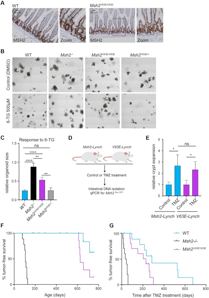
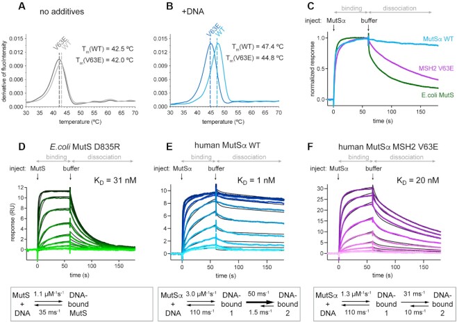
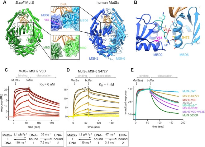
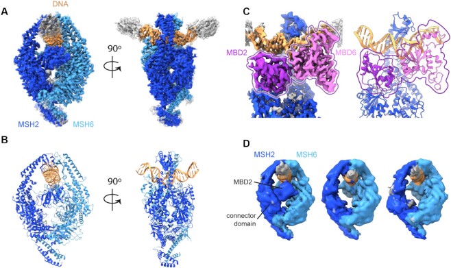
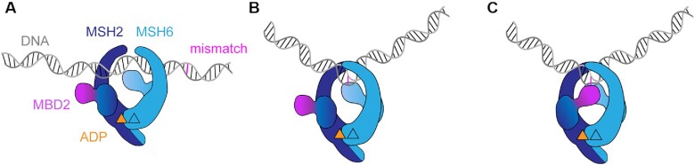
The DNA mismatch repair protein MutSα recognizes wrongly incorporated DNA bases and initiates their correction during DNA replication. Dysfunctions in mismatch repair lead to a predisposition to cancer. Here, we study the homozygous mutation V63E in MSH2 that was found in the germline of a patient with suspected constitutional mismatch repair deficiency syndrome who developed colorectal cancer before the age of 30. Characterization of the mutant in mouse models, as well as slippage and repair assays, shows a mildly pathogenic phenotype. Using cryogenic electron microscopy and surface plasmon resonance, we explored the mechanistic effect of this mutation on MutSα function. We discovered that V63E disrupts a previously unappreciated interface between the mismatch binding domains (MBDs) of MSH2 and MSH6 and leads to reduced DNA binding. Our research identifies this interface as a 'safety lock' that ensures high-affinity DNA binding to increase replication fidelity. Our mechanistic model explains the hypomorphic phenotype of the V63E patient mutation and other variants in the MBD interface.


Figures

Figure 1.
Figure 1.
The MSH2 V63E variant shows a weak pathogenic phenotype in vivo. (A) Immunohistochemical staining for MSH2 on small intestines from WT or Msh2V63E/V63E mice (n = 3). (B) Representative images of WT, Msh2/, Msh2V63E/V63E and Msh2V63E/+ organoids treated with control or 500 nM 6-TG. (C) Quantification of panel (B). Bars represent the average and standard deviation (SD) of four independent experiments. Asterisks indicate a significance level of P < 0.01 and P < 0.0001, respectively (one-way ANOVA with Tukey’s correction for multiple comparisons). (D) Experimental setup. Loss of WT MSH2 activity in Msh2-Lynch mice and V63E-Lynch mice was induced with tamoxifen. Then, mice were treated with control solution (n = 3) or TMZ (n = 12) for 10 days. Mice were sacrificed 2 weeks after the treatment and a qPCR for the Msh2floxOFF allele was performed on small intestinal DNA to measure intestinal crypt expansion. (E) Relative crypt expansion after TMZ treatment in Msh2-Lynch and V63E-Lynch mice. Bars represent average and SD. Asterisks indicate a significance level of P < 0.05 (one-way ANOVA with Tukey’s correction for multiple comparisons). (F) Tumor-free survival of WT, Msh2/− and Msh2V63E/V63E mice. (G) Tumor-free survival of WT, Msh2/− and Msh2V63E/V63E mice after 5-day TMZ treatment.
Figure 2.
Figure 2.
MutSα DNA binding involves a conformational change (A, B) Thermostability of MutSα WT versus V63E in nanoDSF assay: (A) without additives and (B) in the presence of GT mismatched DNA. (C) Comparison of DNA binding and release for MutSα WT, MSH2 V63E and E. coli MutS WT visualized from normalized SPR profiles at 128 nM protein concentration [full titration in panels (E) and (F), and Supplementary Figure S2G]. (D–F) Curve profiles of protein titrations ranging from 1 to 256 nM in 2-fold dilutions (from light to dark color). Binding and dissociation were fitted using CLAMP (29) (black curves) and resulting values shown in scheme below. (D) Escherichia coli MutS D835R curves fitted with 1:1 model. MutSα curves required fitting with a two-state reaction model (see Supplementary Figure S2K–M for controls of the model): (E) MutSα WT and (F) MSH2 V63E.
Figure 3.
Figure 3.
Interface between the MBDs is responsible for high affinity DNA binding (A) In MutSα, the MBD of MSH2 (dark blue) and MBD of MSH6 (light blue) form an interface. V63 is located in the MBD of MSH2 and highlighted in magenta. It is not involved in DNA binding but part of a hydrophobic network that forms the interface with MBD6. DNA is colored in gold. Escherichia coli MutS, with monomers in light and dark green, has no interface between the MBDs. (B) MBD interface residues and selected mutations. Residues involved in the interface according to PISA are shown as sticks. V3 (red) on MSH2 side points toward MSH6. S472 (yellow) is in the middle of the MSH6 interface helix. MSH2 residues 2–7 are highlighted in cyan. (C, D) SPR mismatch DNA binding profiles of protein titrations ranging from 1 to 256 nM (C) or from 1 to 128 nM (D) in 2-fold dilutions (from light to dark color). Binding and dissociation periods were fitted using CLAMP and values given in scheme below. Curves were fitted with a two-state reaction model (black curves): (C) MutSα MSH2 V3D and (D) MutSα MSH6 S472Y. (E) Comparison of SPR normalized dissociation curves of all tested mutants at 128 nM protein concentration.
Figure 4.
Figure 4.
CryoEM structures reveal mobility of MBD2 in V63E. CryoEM density maps and model for MutSα WT and MSH2 V63E. (A) CryoEM map of MutSα WT colored as in Figure 3A. (B) Atomic model of MutSα, colored as in Figure 3A. (C) Map (left) and model (right) of MBD interface with MBD2 in dark purple and MBD6 in light purple for better visibility. (D) The subclasses of the MSH2 V63E dataset show flexibility of the MBD2 domain. Left panel: closed MBD2 reminiscent of MutSα WT; middle panel: disordered MBD2; right panel: disordered MBD2 and dragged-out connector domain.
Figure 5.
Figure 5.
Model of MutSα DNA mismatch binding involving a conformational change (A) MutSα scans DNA and (B) initial mismatch binding through MBD6. The insertion of phenylalanine generates the low-affinity DNA-bound state. (C) In a second step, a conformational change occurs. In this step, closing of the MBD2 forms the interface with MBD6 stabilizing the high-affinity DNA-bound state. In all states, MSH2 has ADP bound, while MSH6 is nucleotide-free.

References

    1. Kunkel T.A., Erie D.A.. DNA mismatch repair. Annu. Rev. Biochem. 2005; 74:681–710. - PubMed
    1. Jiricny J. Postreplicative mismatch repair. Cold Spring Harb. Perspect. Biol. 2013; 5:a012633. - PMC - PubMed
    1. Lynch H.T., Snyder C.L., Shaw T.G., Heinen C.D., Hitchins M.P.. Milestones of Lynch syndrome: 1895–2015. Nat. Rev. Cancer. 2015; 15:181–194. - PubMed
    1. Abedalthagafi M. Constitutional mismatch repair-deficiency: current problems and emerging therapeutic strategies. Oncotarget. 2018; 9:35458–35469. - PMC - PubMed
    1. Nik-Zainal S., Morganella S.. Mutational signatures in breast cancer: the problem at the DNA level. Clin. Cancer Res. 2017; 23:2617–2629. - PMC - PubMed

Publication types