

Medial joint space narrowing progresses after pullout repair of medial meniscus posterior root tear
- PMID: 36715713
- PMCID: PMC10522731
- DOI: 10.1007/s00264-023-05701-4
Medial joint space narrowing progresses after pullout repair of medial meniscus posterior root tear
Abstract
Purpose: The extent to which arthropathic changes progress after medial meniscus posterior root tear (MMPRT) repair remains controversial. This retrospective study assessed medial joint space (MJS) narrowing progression after pullout repair for MMPRT and identified the correlating factors.
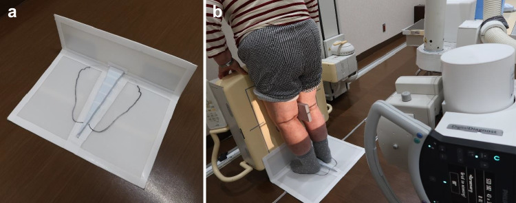
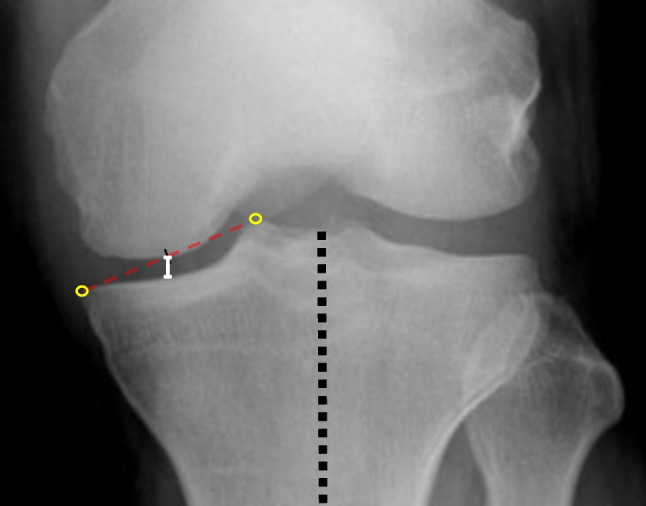
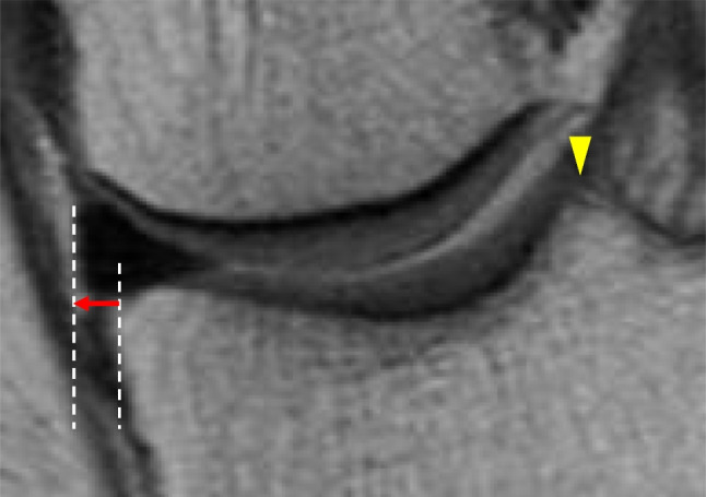
Methods: We included 56 patients who underwent pullout repair for MMPRT. The MJS of the bilateral knees was assessed with radiography using the fixed-flexion view. A second-look arthroscopy was performed one year post-operatively for all patients. The baseline characteristics, clinical scores, Kellgren-Lawrence (KL) grade, and medial meniscus extrusion (MME) were identified. Statistical comparisons and correlation analyses were conducted.
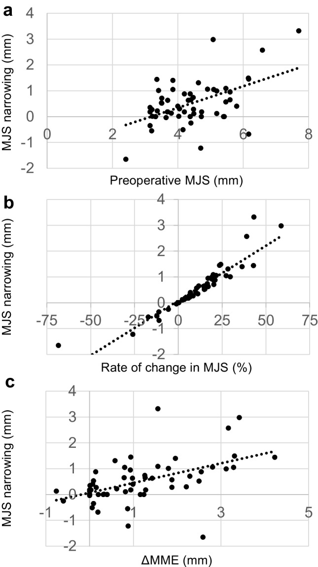
Results: The MJS narrowing width was significantly larger in MMPRT knees than in contralateral knees (0.51 ± 0.85 mm vs. 0.09 ± 0.49 mm, p < 0.001). KL grade progression was observed in 23.2% (13/56) of patients. There was a significant difference between pre- and post-operative MME values, indicating MME progression (p < 0.001). Each clinical score showed significant improvement one year post-operatively (p < 0.001). Positive correlations were found between MJS narrowing and pre-operative MJS (coefficient = 0.510, p < 0.001), rate of change in MJS (coefficient = 0.929, p < 0.001), and increase in MME (ΔMME) (coefficient = 0.506, p < 0.001).
Conclusion: Knees that underwent pullout repair for MMPRT showed progression of MJS narrowing by 0.51 mm at one year post-operatively, although clinical scores markedly improved. Correlating factors for MJS narrowing were pre-operative MJS, rate of change in MJS, and ΔMME. Preventing MME progression is essential for preventing arthropathic changes.
Keywords: Fixed-flexion view; Medial joint space; Medial meniscus extrusion; Meniscus; Posterior root tear; Pullout repair.
© 2023. The Author(s).
Conflict of interest statement
The authors declare no competing interests.
Figures

References
-
- Krych AJ, Reardon PJ, Johnson NR, Mohan R, Peter L, Levy BA, Stuart MJ. Non-operative management of medial meniscus posterior horn root tears is associated with worsening arthritis and poor clinical outcome at 5-year follow-up. Knee Surg Sports Traumatol Arthrosc. 2017;25:383–389. doi: 10.1007/s00167-016-4359-8. - DOI - PubMed
-
- Feucht MJ, Kühle J, Bode G, Mehl J, Schmal H, Südkamp NP, Niemeyer P. Arthroscopic transtibial pullout repair for posterior medial meniscus root tears: a systematic review of clinical, radiographic, and second-look arthroscopic results. Arthroscopy. 2015;31:1808–1816. doi: 10.1016/j.arthro.2015.03.022. - DOI - PubMed
LinkOut - more resources
Full Text Sources
Miscellaneous
