Review
. 2023 Nov;165(11):3299-3323.
doi: 10.1007/s00701-022-05473-7. Epub 2023 Jan 30.

Should individual timeline and serial CT/MRI panels of all patients be presented in acute brain insult cohorts? A pilot study of 45 patients with decompressive craniectomy after aneurysmal subarachnoid hemorrhage

Affiliations
Review

Should individual timeline and serial CT/MRI panels of all patients be presented in acute brain insult cohorts? A pilot study of 45 patients with decompressive craniectomy after aneurysmal subarachnoid hemorrhage

Anniina H Autio et al. Acta Neurochir (Wien). 2023 Nov.

Abstract

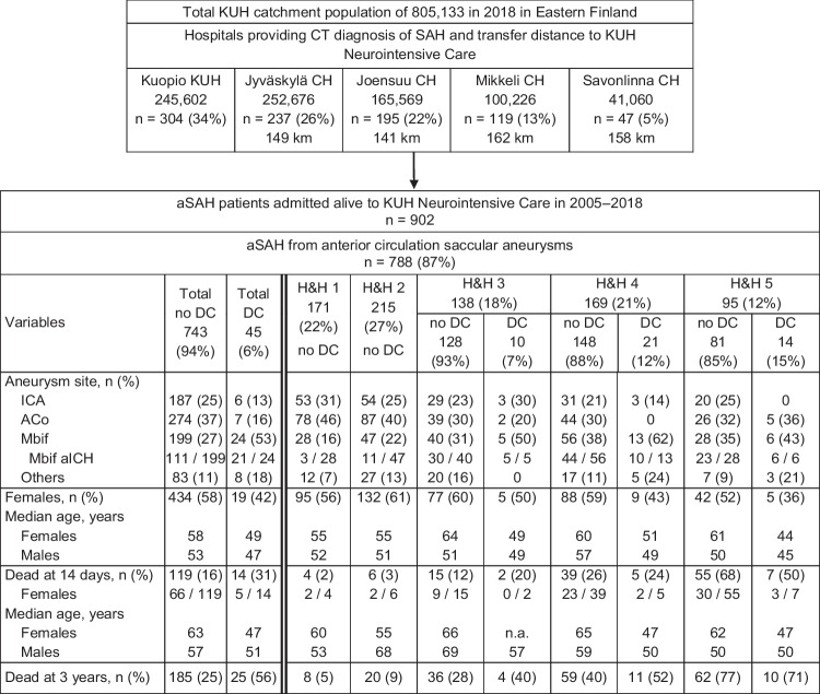
Purpose: Our review of acute brain insult articles indicated that the patients' individual (i) timeline panels with the defined time points since the emergency call and (ii) serial brain CT/MRI slice panels through the neurointensive care until death or final brain tissue outcome at 12 months or later are not presented.

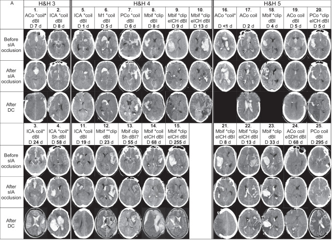
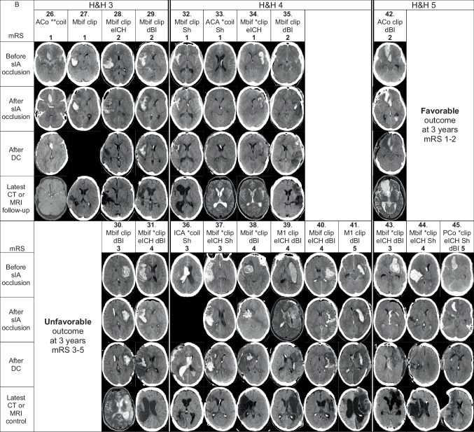
Methods: We retrospectively constructed such panels for the 45 aneurysmal subarachnoid hemorrhage (aSAH) patients with a secondary decompressive craniectomy (DC) after the acute admission to neurointensive care at Kuopio University Hospital (KUH) from a defined population from 2005 to 2018. The patients were indicated by numbers (1.-45.) in the pseudonymized panels, tables, results, and discussion. The timelines contained up to ten defined time points on a logarithmic time axis until death ([Formula: see text]; 56%) or 3 years ([Formula: see text]; 44%). The brain CT/MRI panels contained a representative slice from the following time points: SAH diagnosis, after aneurysm closure, after DC, at about 12 months (20 survivors).

Results: The timelines indicated re-bleeds and allowed to compare the times elapsed between any two time points, in terms of workflow swiftness. The serial CT/MRI slices illustrated the presence and course of intracerebral hemorrhage (ICH), perihematomal edema, intraventricular hemorrhage (IVH), hydrocephalus, delayed brain injury, and, in the 20 (44%) survivors, the brain tissue outcome.

Conclusions: The pseudonymized timeline panels and serial brain imaging panels, indicating the patients by numbers, allowed the presentation and comparison of individual clinical courses. An obvious application would be the quality control in acute or elective medicine for timely and equal access to clinical care.

Keywords: Aneurysmal subarachnoid hemorrhage; Brain tissue outcome; EMS (emergency medical services); Individual serial brain imaging panels; Individual timeline panels; Neurointensive care.


Conflict of interest statement

The authors declare no competing interests.

Figures

Fig. 1
Fig. 1
Map of Eastern Finland. The defined Eastern Finnish catchment population (805,133 in 2018) of the tertiary Kuopio University Hospital (KUH; black dot) is shown in white. The four referring Central Hospitals (red dots) in Jyväskylä (Jy CH), Joensuu (Jo CH), Mikkeli (Mi CH), and Savonlinna (Sa CH) serve their own districts (borderlines in black) with 24/7 neuroacutology, CT, and intensive care services. There are also three regional hospitals (green dots) in Iisalmi, Pieksämäki, and Varkaus, serving their own subdistricts. The transfer distances in road kilometers (km) by ambulance to KUH are shown in the flowchart (Fig. 2). Abbreviations: KUH, Kuopio University Hospital; Jy CH, Jyväskylä Central Hospital; Jo CH, Joensuu Central Hospital; Mi CH, Mikkeli Central Hospital; Sa CH, Savonlinna Central Hospital; CT, computed tomography; SAH, subarachnoid hemorrhage; GPS, Global Positioning System; EMS, emergency medical services; HEMS, helicopter emergency medical services; km, kilometer
Fig. 2
Fig. 2
Flowchart. A total of 902 consecutive patients were acutely admitted—within 24 h from the CT diagnosis of the first subarachnoid hemorrhage (SAH)—to the neurosurgical and neurointensive care at the tertiary Kuopio University Hospital (KUH) between 2005 and 2018 from a defined Eastern Finnish catchment population (Fig. 1). The overall KUH catchment area contains four Central Hospitals. The transfer distances in road kilometers (km) by ambulance to KUH are denoted. The 788 (87%) aSAH patients with a ruptured anterior circulation saccular aneurysm, including all 114 posterior communicating artery (PCo) aneurysms, were selected for the present analysis, with regard to the 45 cases of decompressive craniectomy (DC). The 114 (13%) posterior circulation aneurysm cases were excluded, including three DC cases (aneurysm sites: vertebral artery, basilar trunk, P3 of posterior cerebral artery). Abbreviations: CT, computed tomography; SAH, subarachnoid hemorrhage; KUH, Kuopio University Hospital; CH, Central Hospital; km, kilometer; aSAH, aneurysmal SAH; PCo, posterior communicating artery; DC, decompressive craniectomy; H&H, Hunt and Hess scale; ICA, internal carotid artery trunk and bifurcation; ACo, anterior communicating artery; Mbif, middle cerebral artery bifurcation; aICH, intracerebral hemorrhage from ruptured saccular aneurysm; n.a., not applicable
Fig. 3
Fig. 3
The cumulative survival rates. The cumulative survival rates at 14 days, 12 months, and three years of the 788 patients acutely admitted for the first verified subarachnoid hemorrhage from an anterior circulation saccular aneurysm (aSAH) to the neurosurgical and neurointensive care at the Kuopio University Hospital (KUH) between 2005 and 2018 from its defined Eastern Finnish catchment population. A total of 45 (6%) patients underwent a secondary decompressive craniectomy (DC). The follow-up time is logarithmic to emphasize the early high mortality. A H&H 1–5 patients without DC (n=743). B H&H 3 patients with DC (n=10) vs. no DC (n=128). C H&H 4 patients with DC (n=21) vs. no DC (n=148). D H&H 5 patients with DC (n=14) vs. no DC (n=81). Abbreviations: aSAH, subarachnoid hemorrhage from anterior circulation saccular aneurysm; KUH, Kuopio University Hospital; DC, decompressive craniectomy
Fig. 4
Fig. 4
Individual timeline panels of the 45 DC patients. Individual timelines through the emergency care and the neurointensive care of the 45 aSAH patients whose condition required a secondary decompressive craniectomy (DC) during their neurointensive care course at the Kuopio University Hospital (KUH) between 2005 and 2018 from a defined population. A The 25 deceased DC patients numbered 1.–25. as in their CT panel in Fig. 5A. B The 20 DC survivors numbered 26.–45. as in their CT panels in Fig. 5B. The timelines start from the emergency phone call (112), if not indicated otherwise. The time scale in minutes is logarithmic to emphasize the EMS, transfer, and early KUH phases. The time points of one minute, 10 min, one hour, one day, one week, one month, and one year are indicated by vertical thin lines. The two timeline panels are zoomable to study details. The red star indicates the time points of either suspected (seizure, unconsciousness, dilated pupil suggestive of tentorial herniation) or CT-verified re-bleeds. The time points on the timelines are as follows: 112 = 112 call; GCS = ambulance arrival (Glasgow Coma Scale points 3–15 denoted); CT = diagnostic computed tomography at the first hospital; TI = tracheal intubation; KUH = KUH arrival; E = extraventricular drainage installation and start of ICP monitoring; OC = start of ruptured sIA occlusion; ICH = intracerebral hemorrhage removal; DC = decompressive craniectomy; D = death; CP = cranioplasty. Five patients (15. 27. 31. 38. 43.) came to the first medical assessment on their own. Importantly, all abbreviations above can be identified in the two panels using the find command: for example, the time points of all CTs, EVD installations, sIA occlusions, or decompressive craniectomies. In B, however, the find command (CP) shows that two of the 20 survivors (41. 45.) did not receive cranioplasty
Fig. 4
Fig. 4
Individual timeline panels of the 45 DC patients. Individual timelines through the emergency care and the neurointensive care of the 45 aSAH patients whose condition required a secondary decompressive craniectomy (DC) during their neurointensive care course at the Kuopio University Hospital (KUH) between 2005 and 2018 from a defined population. A The 25 deceased DC patients numbered 1.–25. as in their CT panel in Fig. 5A. B The 20 DC survivors numbered 26.–45. as in their CT panels in Fig. 5B. The timelines start from the emergency phone call (112), if not indicated otherwise. The time scale in minutes is logarithmic to emphasize the EMS, transfer, and early KUH phases. The time points of one minute, 10 min, one hour, one day, one week, one month, and one year are indicated by vertical thin lines. The two timeline panels are zoomable to study details. The red star indicates the time points of either suspected (seizure, unconsciousness, dilated pupil suggestive of tentorial herniation) or CT-verified re-bleeds. The time points on the timelines are as follows: 112 = 112 call; GCS = ambulance arrival (Glasgow Coma Scale points 3–15 denoted); CT = diagnostic computed tomography at the first hospital; TI = tracheal intubation; KUH = KUH arrival; E = extraventricular drainage installation and start of ICP monitoring; OC = start of ruptured sIA occlusion; ICH = intracerebral hemorrhage removal; DC = decompressive craniectomy; D = death; CP = cranioplasty. Five patients (15. 27. 31. 38. 43.) came to the first medical assessment on their own. Importantly, all abbreviations above can be identified in the two panels using the find command: for example, the time points of all CTs, EVD installations, sIA occlusions, or decompressive craniectomies. In B, however, the find command (CP) shows that two of the 20 survivors (41. 45.) did not receive cranioplasty
Fig. 5
Fig. 5
Individual CT/MRI panels of the 45 DC patients. Serial CT scan panels of the 45 aneurysmal subarachnoid hemorrhage (aSAH) patients who underwent decompressive craniectomy (DC) after admission within 24 h from the CT diagnosis of SAH to the tertiary Kuopio University Hospital (KUH). The patients (white data box) and the representative CT scan slices are arranged into vertical columns according to the Hunt & Hess scale (H&H 3–5) on admission. The black areas indicate the lack of CT or MRI scan. The white data box contains the patient number; site of the ruptured saccular intracranial aneurysm (sIA); microsurgical (clip; 28/45) or endovascular (coil; 17/45) occlusion; evacuation of aICH (eICH; 16/45) or aSDH (eSDH; 1/45) during the microsurgical clipping (15/28) or after the endovascular occlusion (2/17); delayed brain injury (dBI) seen here in the third CT or MRI of the patient (34/45). dBI? denotes uncertainty between dBI vs. peri-ICH edema. Asterisk (*) indicates the sIA re-bleeding between the ictus and the sIA occlusion (30/45), either clinically suspected (seizure or worsened condition; 24/45) or verified by two CT scans (6/45). Furthermore, there were four re-bleeds during or after the sIA coiling. A Serial CT scan panel of the 25 aSAH patients who died within three years after DC, arranged from left to right according to increasing times (days) from DC to death. For each patient, three CT slices were selected: (1) CT: before clipping or coiling; (2) CT: after clipping or coiling; (3) CT: after DC. Of the deceased DC patients, there were 20 sIA re-bleeds (16 clinical; 4 CT verified) between the ictus and the sIA occlusion. *clip = sIA re-bleed before clipping (9/28); **clip = sIA re-bleed two times before clipping (1/28); *coil = sIA re-bleed before coiling (6/17); coil* = sIA re-bleed during or after coiling (1/17); *coil* = sIA re-bleed before coiling and re-bleed during or after coiling (3/17). Of the 25 patients, two had a ventriculoperitoneal shunt (Sh). B Serial CT scan panel of the 20 aSAH patients who survived after DC, arranged from left to right according to the modified Rankin Scale (mRS) within the H&H (3–5) columns. For each patients, four CT slices were selected: (1) CT: before clipping or coiling; (2) CT: after clipping or coiling; (3) CT: after DC; (4) CT or MRI during follow-up at about 12 months. Of the survived DC patients, there were 11 sIA re-bleeds (8 clinical; 3 CT verified) between the ictus and the sIA occlusion. *clip = sIA re-bleed before clipping (7/28); *coil = sIA re-bleed before coiling (2/17); **coil = sIA re-bleed two times before coiling (1/17). Of the 20 patients, six had a ventriculoperitoneal shunt (Sh). Abbreviations: CT, computed tomography; aSAH, subarachnoid hemorrhage from anterior circulation saccular aneurysm; DC, decompressive craniectomy; SAH, subarachnoid hemorrhage; KUH, Kuopio University Hospital; H&H, Hunt & Hess scale; MRI, magnetic resonance imaging; sIA, saccular intracranial aneurysm; clip, microsurgical occlusion; coil, endovascular occlusion; aICH, intracerebral hemorrhage from ruptured anterior circulation saccular aneurysm; eICH, evacuation of aICH; aSDH, acute subdural hemorrhage from aSAH; eSDH, evacuation of aSDH; dBI, delayed brain injury; ICH, intracerebral hemorrhage; Sh, shunt; mRS, modified Rankin Scale; ACo, anterior communicating artery; ICA, internal carotid artery trunk and bifurcation; M1, M1 segment of the middle cerebral artery; PCo, posterior communicating artery; Mbif, middle cerebral artery bifurcation; ACA, anterior cerebral artery
Fig. 5
Fig. 5
Individual CT/MRI panels of the 45 DC patients. Serial CT scan panels of the 45 aneurysmal subarachnoid hemorrhage (aSAH) patients who underwent decompressive craniectomy (DC) after admission within 24 h from the CT diagnosis of SAH to the tertiary Kuopio University Hospital (KUH). The patients (white data box) and the representative CT scan slices are arranged into vertical columns according to the Hunt & Hess scale (H&H 3–5) on admission. The black areas indicate the lack of CT or MRI scan. The white data box contains the patient number; site of the ruptured saccular intracranial aneurysm (sIA); microsurgical (clip; 28/45) or endovascular (coil; 17/45) occlusion; evacuation of aICH (eICH; 16/45) or aSDH (eSDH; 1/45) during the microsurgical clipping (15/28) or after the endovascular occlusion (2/17); delayed brain injury (dBI) seen here in the third CT or MRI of the patient (34/45). dBI? denotes uncertainty between dBI vs. peri-ICH edema. Asterisk (*) indicates the sIA re-bleeding between the ictus and the sIA occlusion (30/45), either clinically suspected (seizure or worsened condition; 24/45) or verified by two CT scans (6/45). Furthermore, there were four re-bleeds during or after the sIA coiling. A Serial CT scan panel of the 25 aSAH patients who died within three years after DC, arranged from left to right according to increasing times (days) from DC to death. For each patient, three CT slices were selected: (1) CT: before clipping or coiling; (2) CT: after clipping or coiling; (3) CT: after DC. Of the deceased DC patients, there were 20 sIA re-bleeds (16 clinical; 4 CT verified) between the ictus and the sIA occlusion. *clip = sIA re-bleed before clipping (9/28); **clip = sIA re-bleed two times before clipping (1/28); *coil = sIA re-bleed before coiling (6/17); coil* = sIA re-bleed during or after coiling (1/17); *coil* = sIA re-bleed before coiling and re-bleed during or after coiling (3/17). Of the 25 patients, two had a ventriculoperitoneal shunt (Sh). B Serial CT scan panel of the 20 aSAH patients who survived after DC, arranged from left to right according to the modified Rankin Scale (mRS) within the H&H (3–5) columns. For each patients, four CT slices were selected: (1) CT: before clipping or coiling; (2) CT: after clipping or coiling; (3) CT: after DC; (4) CT or MRI during follow-up at about 12 months. Of the survived DC patients, there were 11 sIA re-bleeds (8 clinical; 3 CT verified) between the ictus and the sIA occlusion. *clip = sIA re-bleed before clipping (7/28); *coil = sIA re-bleed before coiling (2/17); **coil = sIA re-bleed two times before coiling (1/17). Of the 20 patients, six had a ventriculoperitoneal shunt (Sh). Abbreviations: CT, computed tomography; aSAH, subarachnoid hemorrhage from anterior circulation saccular aneurysm; DC, decompressive craniectomy; SAH, subarachnoid hemorrhage; KUH, Kuopio University Hospital; H&H, Hunt & Hess scale; MRI, magnetic resonance imaging; sIA, saccular intracranial aneurysm; clip, microsurgical occlusion; coil, endovascular occlusion; aICH, intracerebral hemorrhage from ruptured anterior circulation saccular aneurysm; eICH, evacuation of aICH; aSDH, acute subdural hemorrhage from aSAH; eSDH, evacuation of aSDH; dBI, delayed brain injury; ICH, intracerebral hemorrhage; Sh, shunt; mRS, modified Rankin Scale; ACo, anterior communicating artery; ICA, internal carotid artery trunk and bifurcation; M1, M1 segment of the middle cerebral artery; PCo, posterior communicating artery; Mbif, middle cerebral artery bifurcation; ACA, anterior cerebral artery

References

    1. Adams H, Ban VS, Leinonen V, Aoun SG, Huttunen J, Saavalainen T, Lindgren A, Frosen J, von Und Zu, Fraunberg M, Koivisto T, Hernesniemi J, Welch BG, Jääskeläinen JE, Huttunen TJ. Risk of shunting after aneurysmal subarachnoid hemorrhage: a collaborative study and initiation of a consortium. Stroke. 2016;47:2488–2496. doi: 10.1161/STROKEAHA.116.013739. - DOI - PubMed
    1. Akins PT, Guppy KH. Does impaired glymphatic drainage cause glymphedema? A review tailored to neurocritical care and neurosurgery. Neurocrit Care. 2021;35:545–558. doi: 10.1007/s12028-021-01224-1. - DOI - PMC - PubMed
    1. Allen JW, Prater A, Kallas O, Abidi SA, Howard BM, Tong F, Agarwal S, Yaghi S, Dehkharghani S. Diagnostic performance of computed tomography angiography and computed tomography perfusion tissue time-to-maximum in vasospasm following aneurysmal subarachnoid hemorrhage. J Am Heart Assoc. 2022 doi: 10.1161/JAHA.121.023828. - DOI - PMC - PubMed
    1. Alotaibi NM, Elkarim GA, Samuel N, Ayling OGS, Guha D, Fallah A, Aldakkan A, Jaja BNR, de Oliveira Manoel AL, Ibrahim GM, Macdonald RL. Effects of decompressive craniectomy on functional outcomes and death in poor-grade aneurysmal subarachnoid hemorrhage: a systematic review and meta-analysis. J Neurosurg. 2017;127:1315–1325. doi: 10.3171/2016.9.JNS161383. - DOI - PubMed
    1. Autio AH, Paavola J, Tervonen J, Lång M, Huuskonen TJ, Huttunen J, Kärkkäinen V, von Und Zu, Fraunberg M, Lindgren AE, Koivisto T, Jääskeläinen JE, Kämäräinen OP. Clinical condition of 120 patients alive at 3 years after poor-grade aneurysmal subarachnoid hemorrhage. Acta Neurochir. 2021;163:1153–1166. doi: 10.1007/s00701-021-04725-2. - DOI - PMC - PubMed

Publication types

LinkOut - more resources