. 2023 Mar;46(1):325-337.
doi: 10.1007/s13246-023-01222-x. Epub 2023 Jan 30.

Intraoperative thermal infrared imaging in neurosurgery: machine learning approaches for advanced segmentation of tumors

Affiliations

Intraoperative thermal infrared imaging in neurosurgery: machine learning approaches for advanced segmentation of tumors

Daniela Cardone et al. Phys Eng Sci Med. 2023 Mar.

Abstract

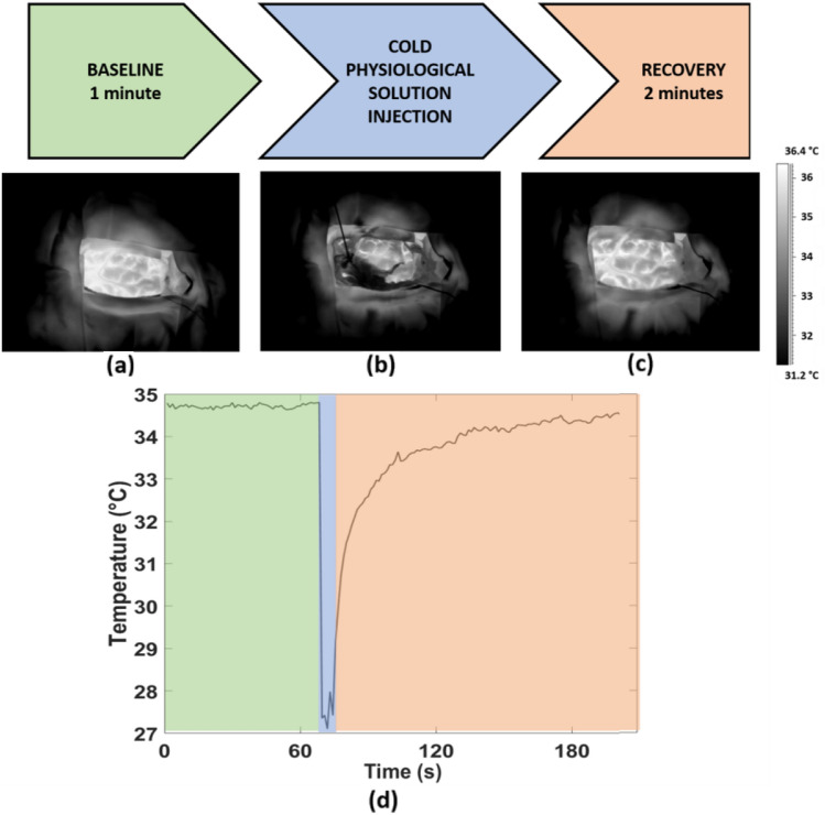
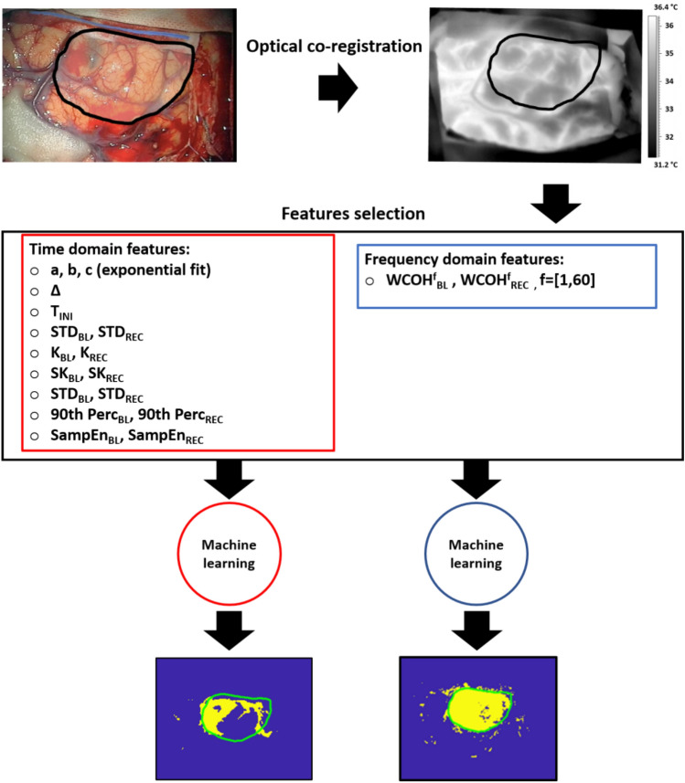
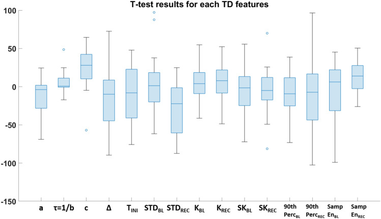
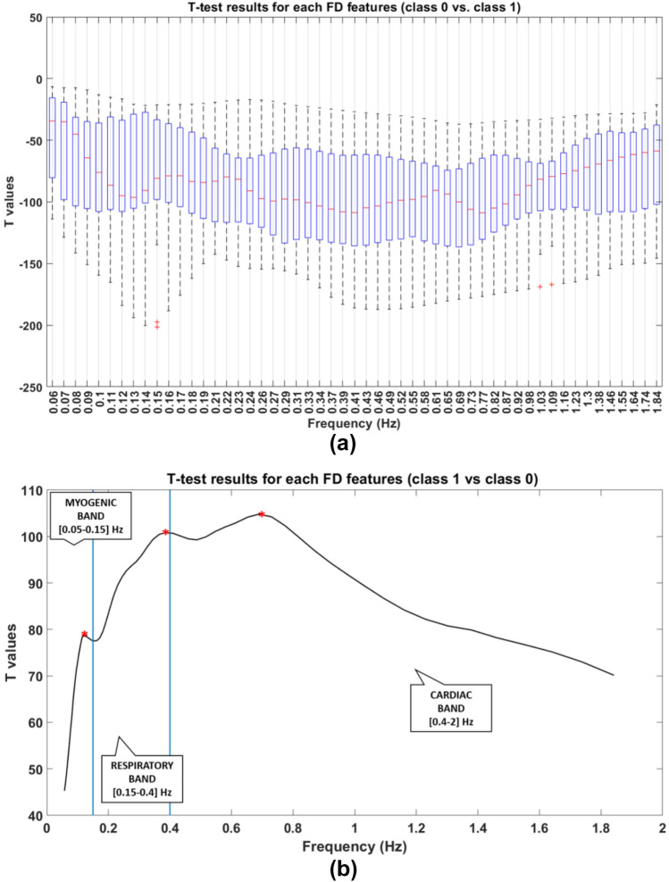
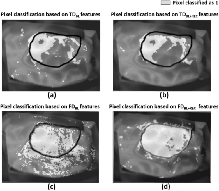
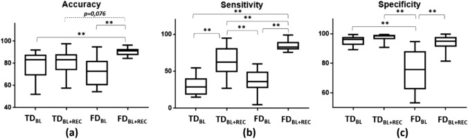
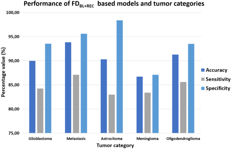
Surgical resection is one of the most relevant practices in neurosurgery. Finding the correct surgical extent of the tumor is a key question and so far several techniques have been employed to assist the neurosurgeon in preserving the maximum amount of healthy tissue. Some of these methods are invasive for patients, not always allowing high precision in the detection of the tumor area. The aim of this study is to overcome these limitations, developing machine learning based models, relying on features obtained from a contactless and non-invasive technique, the thermal infrared (IR) imaging. The thermal IR videos of thirteen patients with heterogeneous tumors were recorded in the intraoperative context. Time (TD)- and frequency (FD)-domain features were extracted and fed different machine learning models. Models relying on FD features have proven to be the best solutions for the optimal detection of the tumor area (Average Accuracy = 90.45%; Average Sensitivity = 84.64%; Average Specificity = 93,74%). The obtained results highlight the possibility to accurately detect the tumor lesion boundary with a completely non-invasive, contactless, and portable technology, revealing thermal IR imaging as a very promising tool for the neurosurgeon.

Keywords: Brain tumor segmentation; Classification; Machine learning; Neurosurgery; Thermal infrared imaging.


Conflict of interest statement

The authors have no relevant financial or non-financial interests to disclose.

Figures

Fig. 1
Fig. 1
Experimental procedure consisting in a baseline (BL) phase (highlighed in green), cold physiological solution injection (highligheted in light blue) and recovery (REC) phase (highligheted in orange); (a),(b),(c) Thermal IR images of the exposed brain tissue relative to BL, injection and REC phases respectively; d) thermal signal of one representive pixel over time
Fig. 2
Fig. 2
Pipeline of the processing approach developed in the present study. The optical co-registration between visible and thermal imaging is necessary to have an indication of the boundary of the cancer area on IRI. Then, TD and FD fetaures are extracted for the only BL and BL + REC phases. Last, supervised machine learning approaches are developed to classify healthy tissue from cancer areas, for each patient
Fig. 3
Fig. 3
Whisker plot of t-values resulted from statistical t-tests for each TD feature. The comparison is between the features values relatively to class 0 vs. class 1 pixels
Fig. 4
Fig. 4
Representation of t-values resulted from statistical t-tests for each FD feature. a Whisker plot of t-values resulted from statistical t-tests for each FD feature. The comparison is between the features values relatively to class 0 vs. class 1 pixels. b Average of t-values among subjects. The comparison is between the features values relatively to class 1 vs. class 0 pixels, in order to have positive values. Maximum values are represented with red asterisks
Fig. 5
Fig. 5
Outcome of classification models for an exemplificative subject relaying on: a TD features of the only BL; b TD features of the whole experiment (BL + REC); c FD features of the only BL; d FD features of the whole experiment (BL + REC). Black boundary is indicative of the tumor area whereas light grey pixels are the ones that the models classify as class 1 (i.e. tumor)
Fig. 6
Fig. 6
Average performances of the developed classifers: a average accuracy, b average sensitivity, c average specificity for the four categories of classifiers. Significant comparison are reported on the graphics (** = p <  < 0.01)
Fig. 7
Fig. 7
Bar plot of the performances indices of the FDBL+REC models relatively to the tumor category

Similar articles

Cited by

References

    1. Hervey-Jumper SL, Berger MS. Maximizing safe resection of low- and high-grade glioma. J Neurooncol. 2016;130:269–282. doi: 10.1007/s11060-016-2110-4. - DOI - PubMed
    1. Willems PW, Taphoorn MJ, Burger H, van Berkelbach der Sprenkel JW, Tulleken CAF. Effectiveness of neuronavigation in resecting solitary intracerebral contrast-enhancing tumors: a randomized controlled trial. J Neurosurg. 2006;104:360–368. doi: 10.3171/jns.2006.104.3.360. - DOI - PubMed
    1. Trevisi G, Barbone P, Treglia G, Mattoli MV, Mangiola A. Reliability of intraoperative ultrasound in detecting tumor residual after brain diffuse glioma surgery: a systematic review and meta-analysis. Neurosurg Rev. 2020;43:1221–1233. doi: 10.1007/s10143-019-01160-x. - DOI - PubMed
    1. Eatz TA, Eichberg DG, Lu VM, Di L, Komotar RJ, Ivan ME. Intraoperative 5-ALA fluorescence-guided resection of high-grade glioma leads to greater extent of resection with better outcomes: a systematic review. J Neurooncol. 2022;156:233–256. doi: 10.1007/s11060-021-03901-9. - DOI - PubMed
    1. Boschi A, Della Puppa A. 5-ALA fluorescence on tumors different from malignant gliomas. review of the literature and our experience. J Neurosurg Sci. 2019;63:661–669. doi: 10.23736/s0390-5616.19.04766-0. - DOI - PubMed

LinkOut - more resources