

Visualizing Fusome Morphology via Tubulin Immunofluorescence in Drosophila Ovarian Germ Cells
- PMID: 36715903
- PMCID: PMC10088872
- DOI: 10.1007/978-1-0716-2970-3_7
Visualizing Fusome Morphology via Tubulin Immunofluorescence in Drosophila Ovarian Germ Cells
Abstract
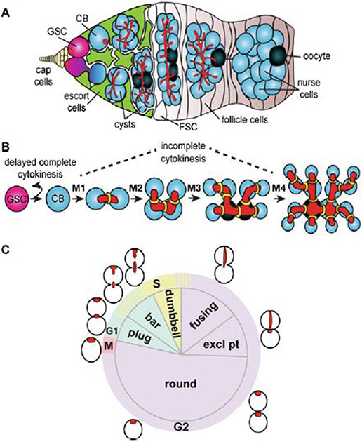
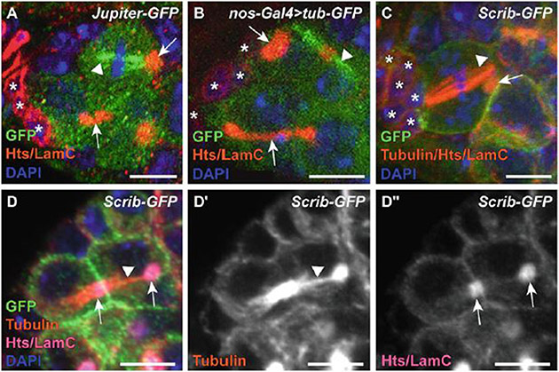
In many species, oocytes are initially formed by the mitotic divisions of germline stem cells and their differentiating daughters. These progenitor cells are frequently interconnected in structures called cysts, which may function to safeguard oocyte quality. In Drosophila, an essential germline-specific organelle called the fusome helps maintain and coordinate the mitotic divisions of both germline stem cells and cyst cells. The fusome also serves as a useful experimental marker to identify germ cells during their mitotic divisions. Fusomes are cytoplasmic organelles composed of microtubules, endoplasmic reticulum-derived vesicles, and a meshwork of membrane skeleton proteins. The fusome branches as mitotic divisions progress, traversing the intercellular bridges of germline stem cell/cystoblast pairs and cysts. Here, we provide a protocol to visualize fusome morphology in fixed tissue by stabilizing microtubules and immunostaining for α-Tubulin and other protein constituents of the fusome. We identify a variety of fluorophore-tagged proteins that are useful for visualizing the fusome and describe how these might be combined experimentally. Taken together, these tools provide a valuable resource to interrogate the genetic control of germline stem cell function, oocyte selection, and asymmetric division.
Keywords: Cyst; Germarium; Germline; Germline stem cell; Microtubules; Oocyte.
© 2023. The Author(s), under exclusive license to Springer Science+Business Media, LLC, part of Springer Nature.
Figures
Similar articles
-
The fusome organizes the microtubule network during oocyte differentiation in Drosophila.Development. 2000 Oct;127(19):4253-64. doi: 10.1242/dev.127.19.4253. Development. 2000. PMID: 10976056
-
Orbit/Mast, the CLASP orthologue of Drosophila, is required for asymmetric stem cell and cystocyte divisions and development of the polarised microtubule network that interconnects oocyte and nurse cells during oogenesis.Development. 2003 Mar;130(5):901-15. doi: 10.1242/dev.00315. Development. 2003. PMID: 12538517
-
Asymmetric germ cell division and oocyte determination during Drosophila oogenesis.Int Rev Cytol. 2001;203:93-138. doi: 10.1016/s0074-7696(01)03005-4. Int Rev Cytol. 2001. PMID: 11131529 Review.
-
The abnormal spindle protein is required for germ cell mitosis and oocyte differentiation during Drosophila oogenesis.Exp Cell Res. 2004 Aug 1;298(1):96-106. doi: 10.1016/j.yexcr.2004.03.054. Exp Cell Res. 2004. PMID: 15242765
-
Asymmetric divisions of germline cells.Prog Mol Subcell Biol. 2007;45:97-120. doi: 10.1007/978-3-540-69161-7_5. Prog Mol Subcell Biol. 2007. PMID: 17585498 Review.
Cited by
-
Fusome morphogenesis is sufficient to promote female germline stem cell self-renewal in Drosophila.bioRxiv [Preprint]. 2025 Mar 13:2025.03.10.642432. doi: 10.1101/2025.03.10.642432. bioRxiv. 2025. PMID: 40161740 Free PMC article. Preprint.
References
-
- Pepling M, Lei L (2018) Germ cell nests and germline cysts. In: Skinner MK (ed) Encyclopedia of reproduction, 2nd edn. Academic Press, Oxford, pp 159–166. 10.1016/B978-0-12-801238-3.64710-4 - DOI
Publication types
MeSH terms
Substances
Grants and funding
LinkOut - more resources
Full Text Sources
Molecular Biology Databases
