. 2023:2626:179-191.
doi: 10.1007/978-1-0716-2970-3_9.

Optimized Fixation and Phalloidin Staining of Basally Localized F-Actin Networks in Collectively Migrating Follicle Cells

Affiliations

Optimized Fixation and Phalloidin Staining of Basally Localized F-Actin Networks in Collectively Migrating Follicle Cells

Mitchell T Anderson et al. Methods Mol Biol. 2023.

Abstract

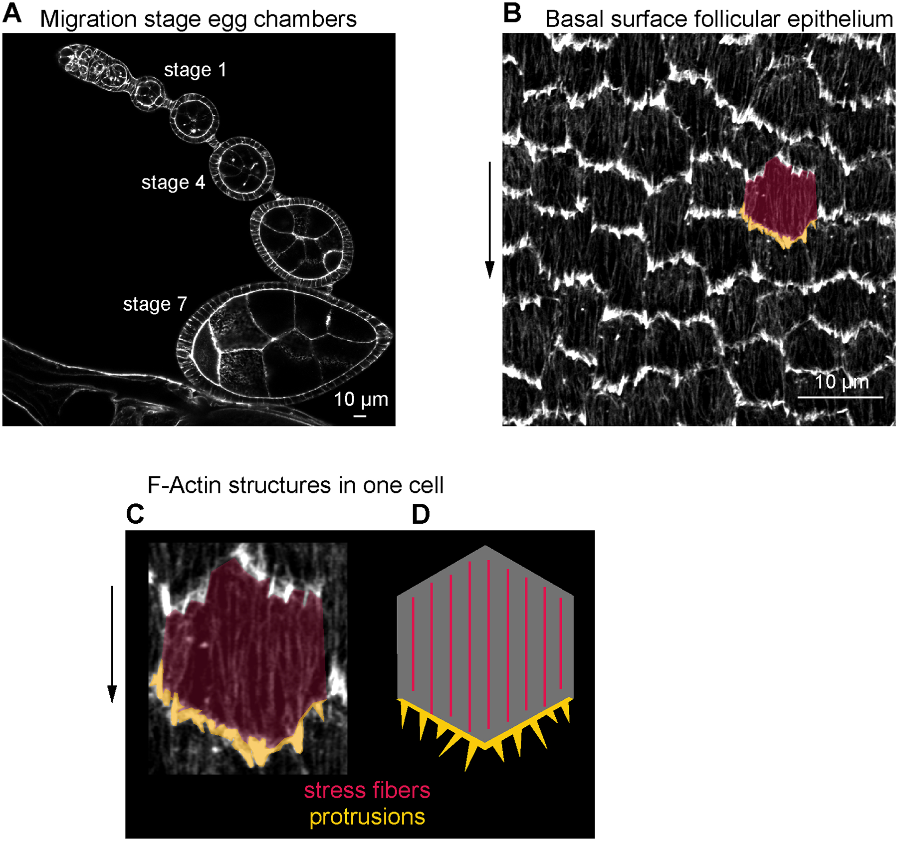
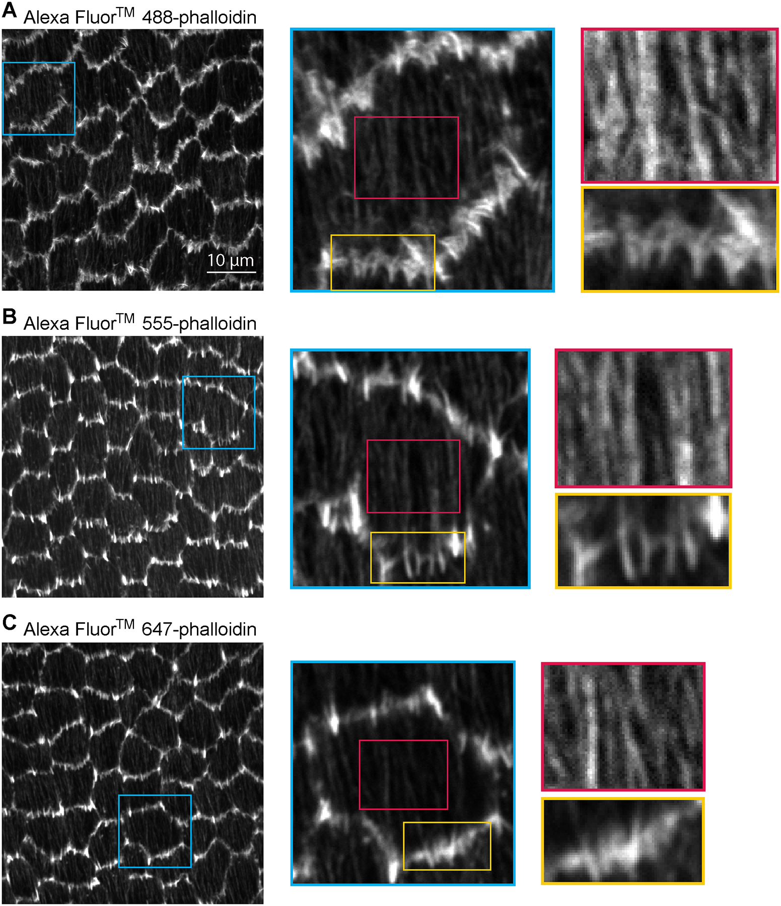
The follicular epithelial cells of the Drosophila egg chamber have become a premier model to study how cells globally orient their actin-based machinery for collective migration. The basal surface of each follicle cell has lamellipodial and filopodial protrusions that extend from its leading edge and an array of stress fibers that mediate its adhesion to the extracellular matrix; these migratory structures are all globally aligned in the direction of tissue movement. To understand how this global alignment is achieved, one must be able to reliably visualize the underlying F-actin; however, dynamic F-actin networks can be difficult to preserve in fixed tissues. Here, we describe an optimized protocol for the fixation and phalloidin staining of the follicular epithelium. We also provide a brief primer on relevant aspects of the image acquisition process to ensure high quality data are collected.

Keywords: Actin; Collective cell migration; Drosophila; Egg chamber; Fixed imaging; Follicle; Morphogenesis; Phalloidin; Protrusions; Staining; Stress fibers.


Figures

Figure 1.
Figure 1.. Phalloidin-stained F-actin structures at the basal surface of the follicular epithelium.
(a) Image of the basal surface of the follicular epithelium at stage 4. (b) Image of the basal surface of the follicular epithelium at stage 7. (c) Image of one follicle cell from (b), highlighting leading-edge protrusions (gold) and stress fibers (red). (d) Image of a transverse section through a developmental array of egg chambers at stages when the follicular epithelium is migrating.
Figure 2.
Figure 2.. Comparison of different fluorophore conjugates of phalloidin.
Images of (a-c) Alexa Fluor 488-phalloidin, (d-f) Alexa Fluor 555-phalloidin, and (g-i) Alexa Fluor 647-phalloidin staining at the basal surface of the follicular epithelium of a stage 8 egg chamber. (a) Image of Alexa Fluor 488-phalloidin staining of a field of follicle cells. (b) Zoom-in of an individual cell in the blue boxed region of (a). (c) Gold and red rectangles show a zoom-in of leading edge protrusions and stress fibers, respectively, in the boxed regions of (b). (d) Image of Alexa Fluor 555-phalloidin staining of a field of follicle cells. (e) Zoom-in of an individual cell in the blue boxed region of (d). (f) Gold and red rectangles show a zoom-in of leading edge protrusions and stress fibers, respectively, in the boxed regions of (e). (g) Image of Alexa Fluor 647-phalloidin staining of a field of follicle cells. (h) Zoom-in of an individual cell int eh blue boxed region of (g). (i) Gold and red rectangles show a zoom-in of leading edge protrusions and stress fibers, respectively, in the boxed regions of (h). Brightness is increased specific to each image in red boxes to better show stress fibers (c, f, i). All images were taken with a 63× 1.4 NA Plan Apochromat oil lens, a digital zoom of 1x, and the pinhole set to 1 Airy Unit. Detailed images in colored boxes are shown scaled by pixel number to illustrate how fluorophore wavelength affects the maximum resolution that can be achieved. Resolution of images in left-most column: (a) 13.4493 pixels/μm. (d) 12.1642 pix/μm. (g) 11.4424 pix/μm.

Similar articles

Cited by

References

    1. Friedl P, Gilmour D (2009) Collective cell migration in morphogenesis, regeneration and cancer. Nat Rev Mol Cell Biol 10(7):445–457. doi:10.1038/nrm2720 - DOI - PubMed
    1. Mayor R, Etienne-Manneville S (2016) The front and rear of collective cell migration. Nat Rev Mol Cell Biol 17(2):97–109. doi:10.1038/nrm.2015.14 - DOI - PubMed
    1. Cetera M, Ramirez-San Juan GR, Oakes PW, Lewellyn L, Fairchild MJ, Tanentzapf G, Gardel MG, Horne-Badovinac S (2014) Epithelial rotation promotes the global alignment of contractile actin bundles during Drosophila egg chamber elongation. Nat Comm 5:5511. doi:10.1038/ncomms6511 - DOI - PMC - PubMed
    1. Gutzeit HO (1991) Organization and in vitro activity of microfilament bundles associated with the basement membrane of Drosophila follicles. Acta Histochem Suppl 41:201–210 - PubMed
    1. Gutzeit HO, Eberhardt W (1991) Laminin and basement membrane-associated microfilaments in wild-type and mutant Drosophila ovarian follicles. J Cell Sci 100(4):781–788. doi:10.1242/jcs.100.4.781 - DOI - PubMed

Publication types

LinkOut - more resources