Optimized Fixation and Phalloidin Staining of Basally Localized F-Actin Networks in Collectively Migrating Follicle Cells
- PMID: 36715905
- PMCID: PMC11229081
- DOI: 10.1007/978-1-0716-2970-3_9
Optimized Fixation and Phalloidin Staining of Basally Localized F-Actin Networks in Collectively Migrating Follicle Cells
Abstract
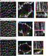
The follicular epithelial cells of the Drosophila egg chamber have become a premier model to study how cells globally orient their actin-based machinery for collective migration. The basal surface of each follicle cell has lamellipodial and filopodial protrusions that extend from its leading edge and an array of stress fibers that mediate its adhesion to the extracellular matrix; these migratory structures are all globally aligned in the direction of tissue movement. To understand how this global alignment is achieved, one must be able to reliably visualize the underlying F-actin; however, dynamic F-actin networks can be difficult to preserve in fixed tissues. Here, we describe an optimized protocol for the fixation and phalloidin staining of the follicular epithelium. We also provide a brief primer on relevant aspects of the image acquisition process to ensure high quality data are collected.
Keywords: Actin; Collective cell migration; Drosophila; Egg chamber; Fixed imaging; Follicle; Morphogenesis; Phalloidin; Protrusions; Staining; Stress fibers.
© 2023. The Author(s), under exclusive license to Springer Science+Business Media, LLC, part of Springer Nature.
Figures


References
-
- Gutzeit HO (1991) Organization and in vitro activity of microfilament bundles associated with the basement membrane of Drosophila follicles. Acta Histochem Suppl 41:201–210 - PubMed
Publication types
MeSH terms
Substances
Grants and funding
LinkOut - more resources
Full Text Sources
Molecular Biology Databases

