. 2023 Mar;22(3):100505.
doi: 10.1016/j.mcpro.2023.100505. Epub 2023 Jan 28.

Increasing Complexity of the N-Glycome During Caenorhabditis Development

Affiliations

Increasing Complexity of the N-Glycome During Caenorhabditis Development

Iain B H Wilson et al. Mol Cell Proteomics. 2023 Mar.

Abstract

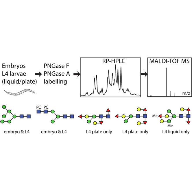
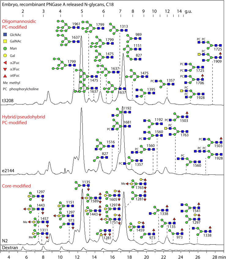
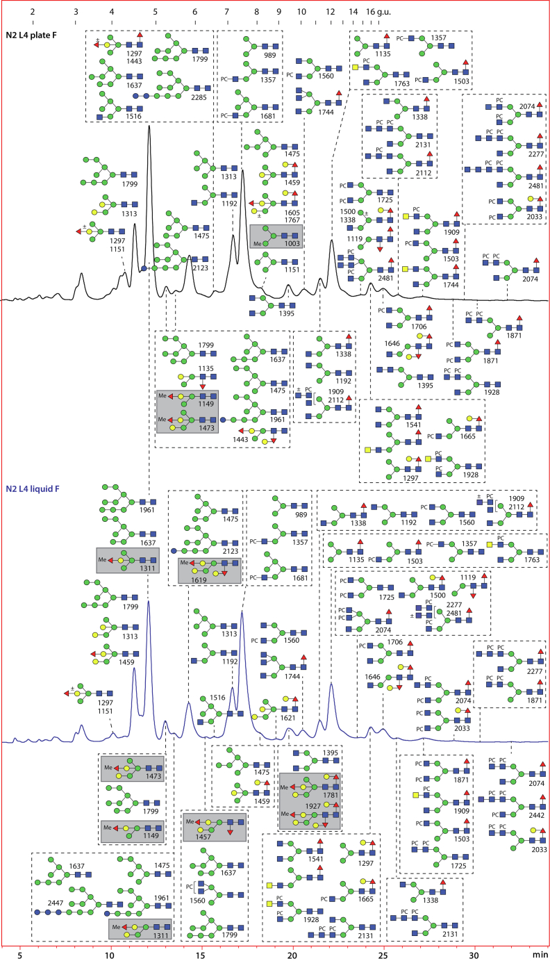
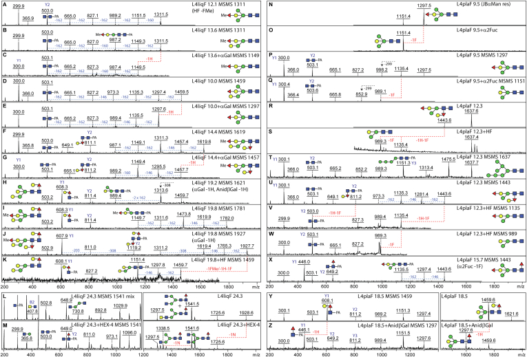
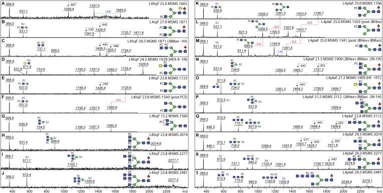
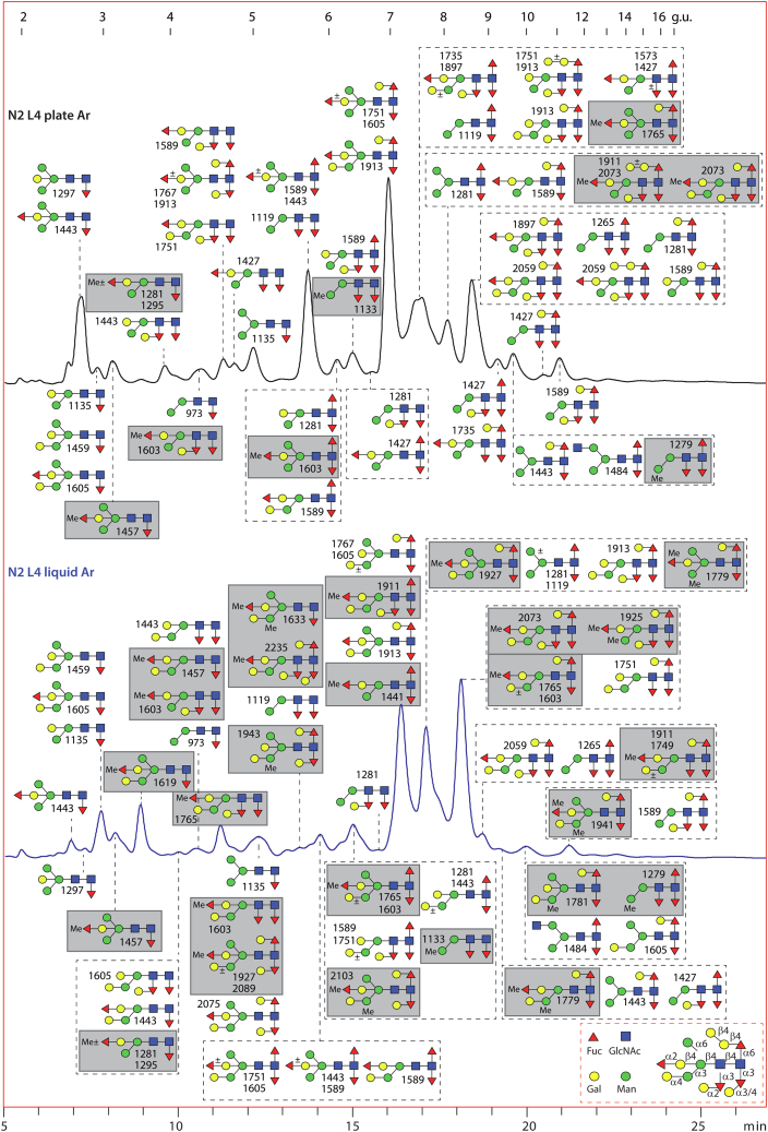
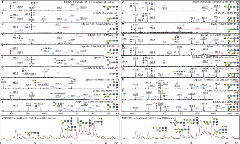
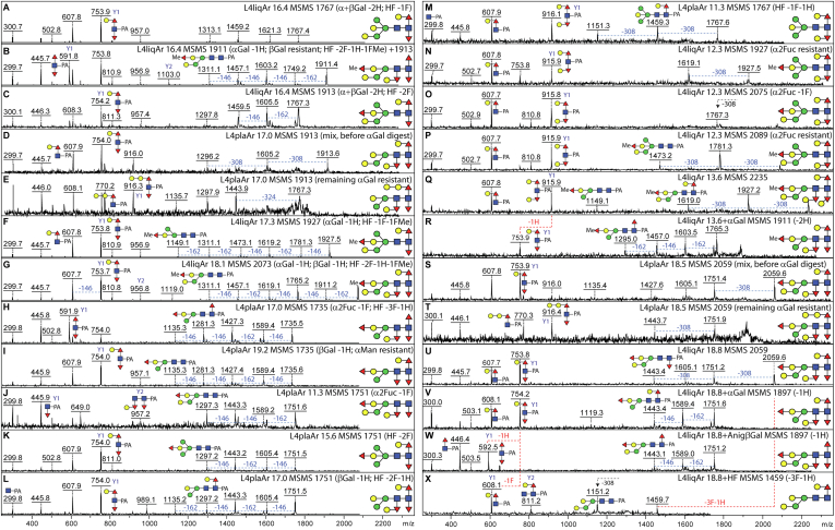
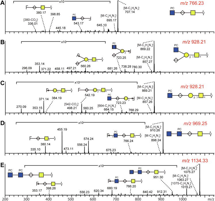
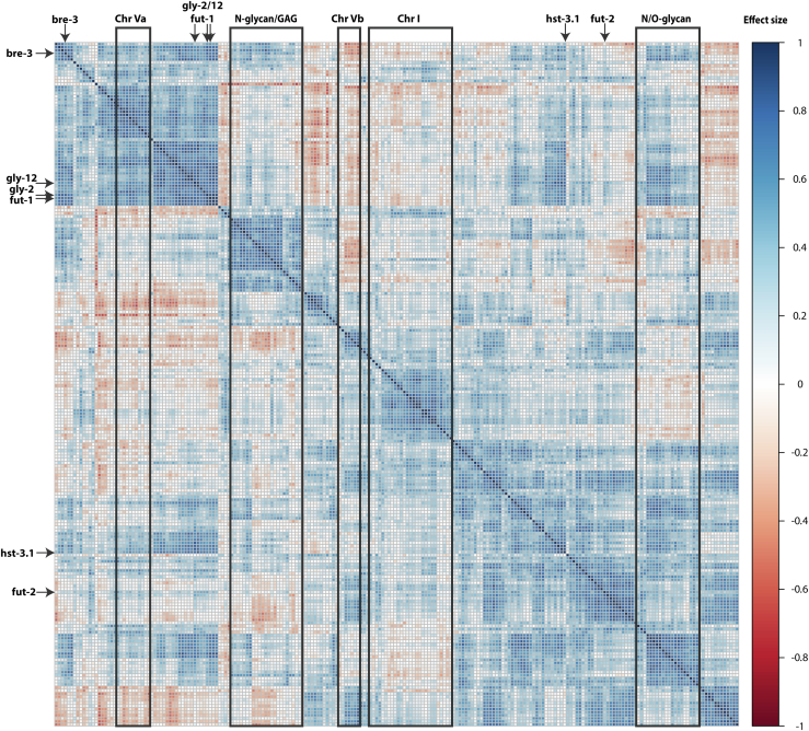
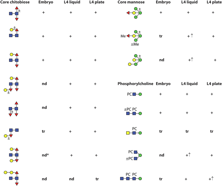
Caenorhabditis elegans is a frequently employed genetic model organism and has been the object of a wide range of developmental, genetic, proteomic, and glycomic studies. Here, using an off-line MALDI-TOF-MS approach, we have analyzed the N-glycans of mixed embryos and liquid- or plate-grown L4 larvae. Of the over 200 different annotatable N-glycan structures, variations between the stages as well as the mode of cultivation were observed. While the embryonal N-glycome appears less complicated overall, the liquid- and plate-grown larvae differ especially in terms of methylation of bisecting fucose, α-galactosylation of mannose, and di-β-galactosylation of core α1,6-fucose. Furthermore, we analyzed the O-glycans by LC-electrospray ionization-MS following β-elimination; especially the embryonal O-glycomes included a set of phosphorylcholine-modified structures, previously not shown to exist in nematodes. However, the set of glycan structures cannot be clearly correlated with levels of glycosyltransferase transcripts in developmental RNA-Seq datasets, but there is an indication for coordinated expression of clusters of potential glycosylation-relevant genes. Thus, there are still questions to be answered in terms of how and why a simple nematode synthesizes such a diverse glycome.

Keywords: fucose; galactose; glycomics; mass spectrometry; phosphorylcholine.


Conflict of interest statement

Conflict of interest The authors declare no competing interests.

Figures

None
Graphical abstract
Fig. 1
Fig. 1
RP-HPLC of PNGase A-released pyridylaminated N-glycans from Caenorhabditis elegans embryos. Chromatograms for the wildtype N2, mutant e2144, and t3208 strains are shown annotated with dextran hydrolysate as external calibrant (in glucose units, g.u.) and with the structures found on the basis of MS, MS/MS, and digestion data; glycans are depicted according to the Standard Nomenclature for Glycans as shown. There are only minor differences in the N-glycans detected in the three strains, for example, some core α1,3-fucosylated Hex3–5HexNAc2Fuc1–3 structures were not detected in the mutants. For clarity, the oligomannosidic structures are shown in the upper panel only, the phosphorylcholine-modified ones in the upper and middle panels, and the various core-modified paucimannosidic ones in the lower panel. See supplemental Fig. S2 for MS of individual fractions of N-glycans from N2 embryos and supplemental Table S1 for a full list. Previous studies have shown that different pyridylaminated Man6–8GlcNAc2 and Man2–3GlcNAc2Fuc1 isomers have distinct RP-HPLC retention times and fragmentation patterns (33, 35, 87). PNGase, peptide:N-glycosidase; RP, reversed-phase.
Fig. 2
Fig. 2
RP-HPLC of PNGase F-released pyridylaminated N-glycans from Caenorhabditis elegans L4 larvae cultivated in liquid or on plates. Chromatograms for the wildtype L4 liquid- and plate-grown larvae are shown annotated with dextran hydrolysate external calibrant (in glucose units, g.u.) and with the structures found on the basis of MS, MS/MS, and digestion data. Native methylated glycans (highlighted with gray boxes) are more frequent in the liquid-grown larvae. See Figures 3 and 4 for MS/MS and digestion data for example glycans, supplemental Fig. S3 for chromatograms of independent preparations, and supplemental Fig. S5 for 2D-HPLC of the plate-grown larvae glycome. PNGase, peptide:N-glycosidase; RP, reversed-phase.
Fig. 3
Fig. 3
Example of MS/MS and digestion data for PNGase F-released N-glycans from Caenorhabditis elegans L4 larvae. RP-HPLC fractionated glycans were subject to MALDI-TOF MS and MS/MS in positive mode before and after chemical or enzymatic treatment; each glycan is annotated with the fraction name (L4liqF or L4plaF and the retention time in minutes), the m/z, the proposed structure, key Y1 or Y2 fragment ions, and sensitivity to α- or β-galactosidase, α1,2-fucosidase, HEX-4 β1,4-N-acetylgalactosaminidase, or hydrofluoric acid (HF), resulting in the indicated losses of fucose (F), hexose (H), or N-acetylhexosamine (N). AK, MS/MS spectra for glycans from larvae cultivated in liquid before or after coffee bean α-galactosidase (αGal) or HF treatment; shown are data for two isomers of m/z 1311 and single isomers of m/z 1459, 1619, 1621, 1781, and 1927, which display differences in the occurrence of α-galactosylation, α1,2-fucosylation, methylation, and core fucosylation; as appropriate, losses of 146 (Fuc), 160 (MeFuc), 162 (Hex), 299 (HexNAc1-PA), or 308 Da (Hex1Fuc1) are indicated. L and M, MS and MS/MS for two coeluting forms of Hex3HexNAc4Fuc1 (m/z 1541); treatment with the C. elegans HEX-4 β-N-acetylgalactosaminidase resulted in partial conversion to m/z 1338 and a loss of the trace m/z 407 (LacdiNAc) B2 fragment ion; the HEX-4-sensitive m/z 1928 glycan in this fraction is PC modified (Fig. 4D). NX, MS and MS/MS spectra for glycans from larvae cultivated on plate before or after microbial α1,2-fucosidase (α2Fuc) or HF treatment; shown are data for a simple bisected glycan of m/z 1297 and a bisected glycan of m/z 1443 with a modified distal GlcNAc, the latter coeluting with an HF-resistant Man7GlcNAc2 structure of m/z 1637, as well as a core fucosylated isomer of m/z 1443. Y and Z, MS and MS/MS for an isoform of Hex5HexNAc2Fuc1 (m/z 1459) with a “GalFuc” epitope on the proximal core GlcNAc; treatment with Aspergillus nidulans β-galactosidase (βGal) resulted in conversion to m/z 1297 and replacement of the m/z 608 Gal1Fuc1GlcNAc1-PA Y1 fragment ion by one at m/z 446. A lack of such m/z 446 Y1-fragments (Fuc1GlcNAc1-PA) for monofucosylated and difucosylated glycans is indicative of an unmodified proximal GlcNAc. HF-sensitive distal GalFuc modifications (F, G, and U) are defined by the presence of m/z 811 Y2 and the absence of m/z 608 Y1 MS/MS fragments; this modification has been characterized by GC–MS and ESI–MS2 (11, 26). Bisecting β-galactose (with or without fucose or methylated fucose) is a motif previously defined by serial chemical/enzymatic digestion, ESI–MS2 and NMR in C. elegans double fut-1;fut-6 and triple fut-1;fut-6;fut-8 knockout strains lacking two or three chitobiose core-modifying α-fucosyltransferases (12, 13). ESI, electrospray ionization; PNGase F, peptide:N-glycosidase; RP, reverse-phase.
Fig. 4
Fig. 4
Example of MS/MS and digestion data for PNGase F-released zwitterionic N-glycans from Caenorhabditis elegans L4 larvae. RP-HPLC fractionated phosphorylcholine (PC)-modified glycans isolated from liquid- or plate-cultivated larvae were subject to MALDI-TOF MS and MS/MS in positive mode before and after enzymatic treatment; the MS/MS spectra are annotated with the fraction name (L4liqF or L4plaF and the retention time), the m/z value, key B fragments, and sensitivity to jack bean α-mannosidase, jack bean β-hexosaminidase, HEX-4 β1,4-N-acetylgalactosaminidase, or phosphorylcholine esterase (PCE), resulting in indicated losses of hexose (H, 162 Da), N-acetylhexosamine (N, 203 Da), or PC (165 Da); a loss of 607 Da (A) correlates with the presence of a reducing-terminal GalFuc modification. Variations in the B fragment ions are indicative of differences in the location and number of the PC moieties (see, e.g., isomers of m/z 1871, 1909, 2074, 2277, 2481; B, C, HJ, N, O, and RT), whereas efficient removal of a mannose residue after combined β-N-acetylhexosaminidase/α-mannosidase (JBHex/JBMan) treatment is indicative that the nonmodified HexNAcHex is α1,3-linked to the core β-mannose (KM). Terminal HexNAc in the context of a HexNAc2–4PC1–2 motif is not sensitive to jack bean β-N-acetylhexosaminidase (N and P), but in one case, it was removed by HEX-4 β-N-acetylgalactosaminidase (D, m/z 1928; Fig. 3, L and M). Whereas HF quantitatively removes PC residues, phosphorylcholine esterase (PCE) is only partially efficient (E and F). Based on previous NMR, GC–MS, and Q-TOF CAD-MS/MS data on nematode glycans, the phosphorylcholine residues are assumed to substitute the C6 of GlcNAc residues (41, 74), which are either nonreducing terminal GlcNAc or within 4-linked HexNAc-based chito-oligomer and LacdiNAc motifs (39). PNGase F, peptide:N-glycosidase; RP, reverse-phase.
Fig. 5
Fig. 5
RP-HPLC of PNGase Ar-released pyridylaminated N-glycans from Caenorhabditis elegans L4 larvae cultivated in liquid or on plates. Chromatograms for the wildtype L4 liquid- and plate-grown larvae are shown annotated with dextran hydrolysate as external calibrant (in glucose units, g.u.) and with the structures found on the basis of MS, MS/MS, and digestion data. The key (bottom right) indicates linkages proposed from previous LC- or GC–MS data (11, 25, 26). Residual oligomannosidic and paucimannosidic glycans found in the PNGase F digests are not annotated on these chromatograms. There are approximately three times more structures with α-galactosylation of mannose and/or methylation in the liquid-grown glycome as compared with the plate-grown (34 and 30 compared with 9 and 10), suggestive of a stress-related glycomic shift. See Figures 6 and 7 and S9–S11 for example of MS/MS and digestion data, supplemental Fig. S6 for chromatograms of independent preparations, and supplemental Fig. S8 for 2D-HPLC of the plate-grown larvae glycome. It is estimated that approximately 10% of the total N-glycomes were released with PNGase Ar. PNGase F, peptide:N-glycosidase; RP, reverse-phase.
Fig. 6
Fig. 6
Example of MS/MS and digestion data for PNGase Ar-released N-glycans from Caenorhabditis elegans L4 larvae. RP-HPLC fractionated glycans were subject to MALDI-TOF MS and MS/MS in positive mode before and after chemical or enzymatic treatment. The MS/MS spectra are annotated with the fraction name (L4liqAr or L4plaAr and the retention time), the m/z value, key fragments and summarized or shown sensitivity to α- or β-galactosidase, α-mannosidase, α1,2-fucosidase, or hydrofluoric acid, resulting in the indicated losses of fucose (F), methylated fucose (FMe), or hexose (H). AJ, comparison of five isomers of m/z 1281 (Hex3HexNAc2Fuc2) and two isomers of m/z 1443 (Hex4HexNAc2Fuc2) with indications of differences in core Y1 fragments and effects of enzymatic or chemical treatments. KM, comparison of two isomers of m/z 1589 (Hex4HexNAc2Fuc3) found in the L4 liquid glycome and the effect of HF treatment of one of them resulting in replacement of the proximal Y1 difucosylated fragment at m/z 592 with a monofucosylated one at m/z 446 in addition to loss of the distal GalFuc motif. NT, comparison of five isomers of m/z 1589 in the L4 plate glycome and the effect of α1,2-fucosidase on two of them; the summed evidence shows variations in the positions of the fucose and galactose residues. The occurrence of difucosylated and trifucosylated chitobiose cores is in accordance with the defined activities of C. elegans FUT-1, FUT-6, and FUT-8 (14), the absence of such cores from the fut-1;fut-6;fut-8 triple knockout strain (12), and others’ ESI–MSn data on permethylated C. elegans N-glycans (21, 25). U and V, depiction of the separation of Hex3HexNAc2Fuc2 and Hex4HexNAc2Fuc3 isomers by RP-HPLC; the overlaid liquid and plate chromatograms in Figure 5 are shown in red and black, respectively. ESI, electrospray ionization; RP, reverse phase.
Fig. 7
Fig. 7
Example of MS/MS and digestion data for PNGase Ar-released N-glycans from Caenorhabditis elegans elegans L4 larvae. RP-HPLC fractionated glycans, indicated with m/z value, and the fraction name (L4liqAr or L4plaAr and the retention time), were subject to MALDI-TOF MS and MS/MS in positive mode before and after chemical or enzymatic treatment. The various examples of highly core modified N-glycans were defined on the basis of the MS/MS spectra and summarized or shown sensitivity to α- or β-galactosidase (loss of hexose; H), α1,2-fucosidase (loss of fucose, F, linked to bisecting galactose), or hydrofluoric acid (loss of core proximal and distal α1,3-fucose and any attached galactose residues or of fucose/methylated fucose linked to bisecting galactose). A, UX, glycans of m/z 1767 and 2059 from the liquid glycome 16.4 and 18.8 min fractions with dominant m/z 754 Y1 fragments (Hex1HexNAc1Fuc2-PA) were sensitive to coffee bean α- and Aspergillus niger β-galactosidase and hydrofluoric acid treatments, whereby the latter glycan corresponds to the α-galactosidase-sensitive structure in the 18.5 min fraction of the L4 plate glycome (panel S). BE, S, and T, coeluting glycans of m/z 1911, 1913, and 2059 were distinguished only after enzymatic digestion, whereby the α-galactosidase-resistant structures have Y fragments at m/z 770 and 916 (Hex2HexNAc1Fuc1–2-PA), indicative of a digalactosylated motif, as compared with the dominant m/z 754 of the α-galactosidase-sensitive isomers. FL, glycans of m/z 1735, 1751, 1927, and 2073 show different sensitivities to α2-fucosidase, β-galactosidase, or HF treatments correlating with variations in the position or degree of substitution of the fucose residues. MR, glycans of Hex5–7HexNAc2Fuc2–4Me0–1 with a core Y1 fragment at m/z 916 and galactosidase sensitivities (26) indicative of both proximal (i.e., reducing terminal) core fucose residues being galactosylated; as there were no Y1 fragments with increments of 14 Da, it is concluded that the methylfucose residues only modify the bisecting galactose. For further examples of MS/MS of previously uncharacterized N-glycans found in L4 larvae, refer to supplemental Figs. S9–S11. PNGase, peptide:N-glycosidase; RP, reversed-phase.
Fig. 8
Fig. 8
Phosphorylcholine is a modification of O-glycans from Caenorhabditis elegans. Negative-mode LC–ESI–MS/MS analyses indicate the occurrence of five O-glycan structures carrying HexNAc1PC1 motifs. The fragment ions at [M-H-59] or [M-H-N(CH3)3] are diagnostic for phosphocholine containing glycans and arise by loss of the trimethylamine group. Z fragment ions containing subterminal hexuronic acid residues are often accompanied by loss of the carboxyl group [Z-CO2] (A and C), a characteristic absent from MS/MS of glycans with nonreducing hexuronic acid (B). As for N-glycans, HexNAc2PC1–2 motifs exist as nonreducing terminal modifications of O-glycans (D and E). For MS/MS of selected neutral O-glycans, refer to supplemental Fig. S13. ESI, electrospray ionization.
Fig. 9
Fig. 9
Cluster analysis of 285 genes with known or potential roles in glycosylation. Corrplot cluster analysis of RNA-Seq transcriptomic data (L1–L4, dauer, and adult) for 285 genes encoding proteins of either known roles in glycosylation and/or present in potential glycogene clusters and/or member of CAZy families GT2, GT7, GT10, GT11, GT13, GT14, GT16, GT18, GT23, GT27, GT43, GT47, GT49, GT92, GH20, GH38, or GH47. Correlations in expression as indicated by intensity of the effect size (blue/red; i.e., high or low correlation) are highlighted for three potential glycogene clusters on chromosome I and V (supplemental Fig. S15) as well as genes with known functions in N-/O-glycan or glycosaminoglycan biosynthesis, including various fut (fucosyltransferase), gly (glycosylation), hex (hexosaminidase), mans (class I mannosidase), and sqv (squashed vulva) genes. A higher resolution form of the figure annotated showing all gene names is shown in supplemental Fig. S16B.
Fig. 10
Fig. 10
Summary of core and antennal motifs of Caenorhabditis elegans N-glycans and trends in their expression. Based on the cumulative evidence (Fig. 1, Fig. 2, Fig. 3, Fig. 4, Fig. 5, Fig. 6, Fig. 7 and Supplementary Data), various N-glycan structural motifs can be proposed, which have been detected in the different samples. nd, not detected; tr, trace; ↑, increased occurrence; ∗, N-glycans with the di-GalFuc-core (Y1 fragment of m/z 916) can only be released by PNGase Ar. PNGase, peptide:N-glycosidase.

Similar articles

Cited by

References

    1. Haltiwanger R.S., Lowe J.B. Role of glycosylation in development. Annu. Rev. Biochem. 2004;73:491–537. - PubMed
    1. Walski T., Van Damme E.J., Smargiasso N., Christiaens O., De Pauw E., Smagghe G. Protein N-glycosylation and N-glycan trimming are required for postembryonic development of the pest beetle Tribolium castaneum. Sci. Rep. 2016;6 - PMC - PubMed
    1. Jiménez-Castells C., Vanbeselaere J., Kohlhuber S., Ruttkowski B., Joachim A., Paschinger K. Gender and developmental specific N-glycomes of the porcine parasite Oesophagostomum dentatum. Biochim. Biophys. Acta. 2017;1861:418–430. - PMC - PubMed
    1. Smit C.H., van Diepen A., Nguyen D.L., Wuhrer M., Hoffmann K.F., Deelder A.M., et al. Glycomic analysis of life stages of the human parasite Schistosoma mansoni reveals developmental expression profiles of functional and antigenic glycan motifs. Mol. Cell Proteomics. 2015;14:1750–1769. - PMC - PubMed
    1. C.-elegans-Sequencing-Consortium Genome sequence of the nematode C. elegans: a platform for investigating biology. Science. 1998;282:2012–2018. - PubMed

Publication types

MeSH terms

LinkOut - more resources