. 2023 Mar 1;324(3):C718-C727.
doi: 10.1152/ajpcell.00519.2022. Epub 2023 Jan 30.

Normal muscle fiber type distribution is recapitulated in aged ephrin-A3-/- mice that previously lacked most slow myofibers

Affiliations

Normal muscle fiber type distribution is recapitulated in aged ephrin-A3-/- mice that previously lacked most slow myofibers

Robert W Arpke et al. Am J Physiol Cell Physiol. .

Abstract

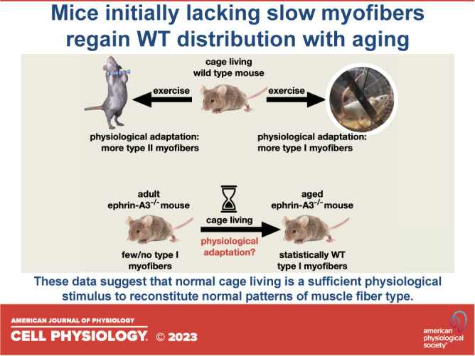
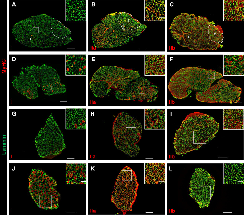
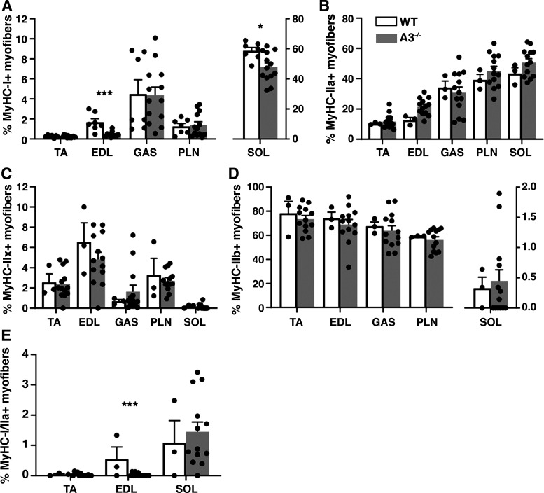
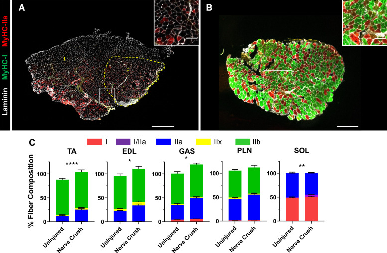
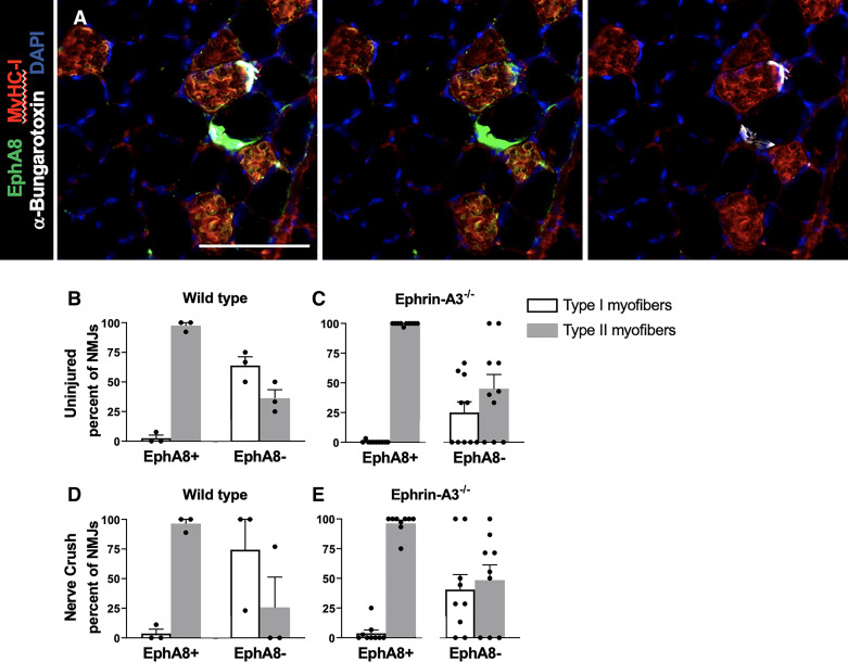
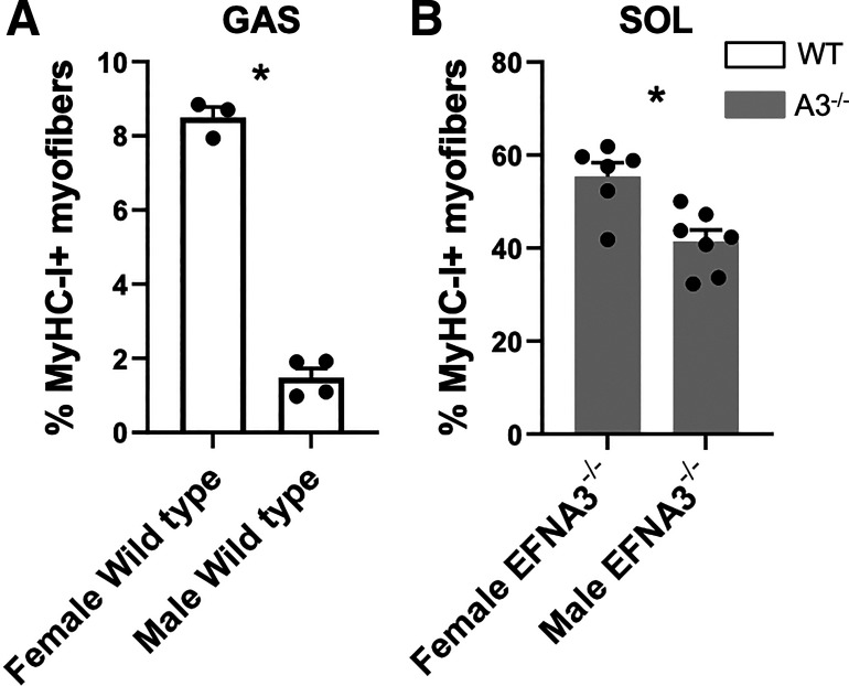
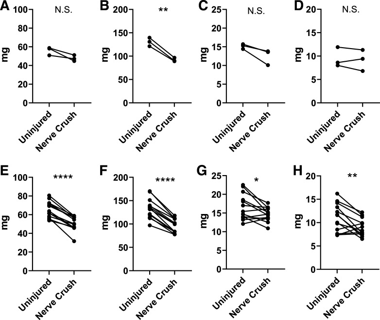
Individual limb muscles have characteristic representation and spatial distribution of muscle fiber types (one slow and up to three fast isoforms) appropriate to their unique anatomical location and function. This distribution can be altered by physiological stimuli such as training (i.e., for increased endurance or force) or pathological conditions such as aging. Our group previously showed that ephrin-A3 is expressed only on slow myofibers, and that adult mice lacking ephrin-A3 have dramatically reduced numbers of slow myofibers due to postnatal innervation of previously slow myofibers by fast motor neurons. In this study, fiber type composition of hindlimb muscles of aged and denervated/reinnervated C57BL/6 and ephrin-A3-/- mice was analyzed to determine whether the loss of slow myofibers persists across the lifespan. Surprisingly, fiber-type composition of ephrin-A3-/- mouse muscles at two years of age was nearly indistinguishable from age-matched C57BL/6 mice. After challenge with nerve crush, the percentage of IIa and I/IIa hybrid myofibers increased significantly in aged ephrin-A3-/- mice. While EphA8, the receptor for ephrin-A3, is present at all neuromuscular junctions (NMJs) on fast fibers in 3-6 mo old C57BL/6 and ephrin-A3-/- mice, this exclusive localization is lost with aging, with EphA8 expression now found on a subset of NMJs on some slow muscle fibers. This return to appropriate fiber-type distribution given time and under use reinforces the role of activity in determining fiber-type representation and suggests that, rather than being a passive baseline, the developmentally and evolutionarily selected fiber type pattern may instead be actively reinforced by daily living.

Keywords: Eph/ephrin; aging; neuromuscular junction; skeletal muscle fiber type.


Conflict of interest statement

C. L. Lorson is the co-founder and chief scientific officer for Shift Pharmaceuticals. None of the other authors has any conflicts of interest, financial or otherwise, to disclose.

Figures

None
Graphical abstract
Figure 1.
Figure 1.
Fiber-type representation in aged ephrin-A3−/− mouse muscles resembles aged wild-type (WT) mouse muscles. AL: immunohistochemistry showing laminin (green) and myosin heavy chains (MyHCs) (red: MyHC-I, left; MyHC-IIa, middle; MyHC-IIb, right) expression in transverse cryosections from uninjured tibialis anterior (TA)-extensor digitorum longus (EDL) (AC), gastrocnemius (GAS) (DF), plantaris (PLN) (GI), and soleus (SOL) muscles (JL) from an aged ephrin-A3−/− mouse. AF: scale bar, 600 µm. GL: scale bar, 300 µm. Inset scale bar, 100 µm.
Figure 2.
Figure 2.
Muscle fiber-type composition of aged ephrin-A3−/− muscles is nearly indistinguishable from aged wild-type (WT) muscles. Percentage of type I (A), IIa (B), IIx (C), IIb (D), and I/IIa (E) hybrid fibers in tibialis anterior (TA), extensor digitorum longus (EDL), gastrocnemius (GAS), plantaris (PLN), and soleus (SOL) muscles. Data are presented as means ± SE. *P < 0.05; ***P < 0.001.
Figure 3.
Figure 3.
I/IIa hybrid and IIa myofibers increase in aged ephrin-A3−/− mice after nerve crush. Immunohistochemistry showing laminin (white), myosin heavy chain (MyHC)-I (green), and MyHC-IIa (red) expression in transverse cryosections from tibialis anterior (TA)-extensor digitorum longus (EDL) (A) and soleus (SOL) muscles from aged ephin-A3−/− mice 4 wk post-nerve crush (B). C: fiber-type distribution of the TA, EDL, gastrocnemius (GAS), plantaris (PLN), and SOL muscles of aged ephrin-A3−/− mice. Data are presented as means ± SE. *P < 0.05; **P < 0.01; ****P < 0.0001. A: scale bar, 600 µm. B: scale bar, 300 µm. Inset scale bar, 100 µm.
Figure 4.
Figure 4.
The majority of neuromuscular junctions (NMJs) on fast fibers in aged gastrocnemius (GAS) muscle express EphA8. A: section of GAS muscle (myosin heavy chain, MyHC-I+ve myofibers, red) showing expression of EphA8 (green) predominantly at fast myofiber NMJs (a-bungarotoxin, white). Scale bar, 100 µm. B and C: EphA8 expression is restricted to fast myofibers in young/adult mice, whereas slow fibers consistently lack EphA8 (33). In both wild type (WT) and ephrin-A3−/− mice, in uninjured muscle the great majority of EphA8+ve NMJs remain associated with fast myofibers (gray bars), but unlike young/adult mice EphA8−ve NMJs were not restricted to slow myofibers (white bars). n = 3 mice, two sections per sample in WT and n = 10 mice, two sections per sample in ephrin-A3−/−. D and E: after challenge by denervation/renervation, EphA8+ve NMJs in both WT and ephrin-A3−/− mice remained consistently associated with fast myofibers (gray bars) but the number of EphA8−ve fast myofibers was, surprisingly, even greater than in uninjured mice. n = 3 mice, two sections per sample in WT and n = 9 mice, two sections per sample in ephrin-A3−/−. Data are presented as means ± SE.
Figure A1.
Figure A1.
Sex-specific differences in the percentage of type I myofibers in aged uninjured mouse muscles. Changes in type I muscle fiber-type percentage in aged wild-type (WT) gastrocnemius (GAS) (A) and aged ephrin-A3−/− soleus (SOL) muscles (B). Data are presented as means ± SE. *P < 0.05.
Figure A2.
Figure A2.
Decreased muscle mass 4 wk post-nerve crush. Mass of tibialis anterior (TA)-extensor digitorum longus (EDL) (A), gastrocnemius (GAS) (B), plantaris (PLN) (C), and soleus (SOL) (D) muscles in aged wild-type (WT) mice. Mass of TA-EDL (E), GAS (F), PLN (G), and SOL (H) muscles in aged ephrin-A3−/− mice. *P < 0.05; **P < 0.01; ****P < 0.0001; N.S., not significant.

References

    1. Schiaffino S, Reggiani C. Fiber types in mammalian skeletal muscles. Physiol Rev 91: 1447–1531, 2011. doi:10.1152/physrev.00031.2010. - DOI - PubMed
    1. Kanning KC, Kaplan A, Henderson CE. Motor neuron diversity in development and disease. Annu Rev Neurosci 33: 409–440, 2010. doi:10.1146/annurev.neuro.051508.135722. - DOI - PubMed
    1. Pette D, Staron RS. Myosin isoforms, muscle fiber types, and transitions. Microsc Res Tech 50: 500–509, 2000. doi:10.1002/1097-0029(20000915)50:6<500::AID-JEMT7>3.0.CO;2-7. - DOI - PubMed
    1. Agbulut O, Li Z, Mouly V, Butler-Browne GS. Analysis of skeletal and cardiac muscle from desmin knock-out and normal mice by high resolution separation of myosin heavy-chain isoforms. Biol Cell 88: 131–135, 1996. doi:10.1016/S0248-4900(97)83528-9. - DOI - PubMed
    1. Hämäläinen N, Pette D. The histochemical profiles of fast fiber Types IIB, ID, and IIA in skeletal muscles of mouse, rat, and rabbit. J Histochem Cytochem 41: 733–743, 1993. doi:10.1177/41.5.8468455. - DOI - PubMed

Publication types

LinkOut - more resources