. 2023 Apr 1;324(4):R470-R479.
doi: 10.1152/ajpregu.00241.2022. Epub 2023 Jan 30.

Adult skeletal muscle peroxisome proliferator-activated receptor γ -related coactivator 1 is involved in maintaining mitochondrial content

Affiliations

Adult skeletal muscle peroxisome proliferator-activated receptor γ -related coactivator 1 is involved in maintaining mitochondrial content

Drue Benefield et al. Am J Physiol Regul Integr Comp Physiol. .

Abstract

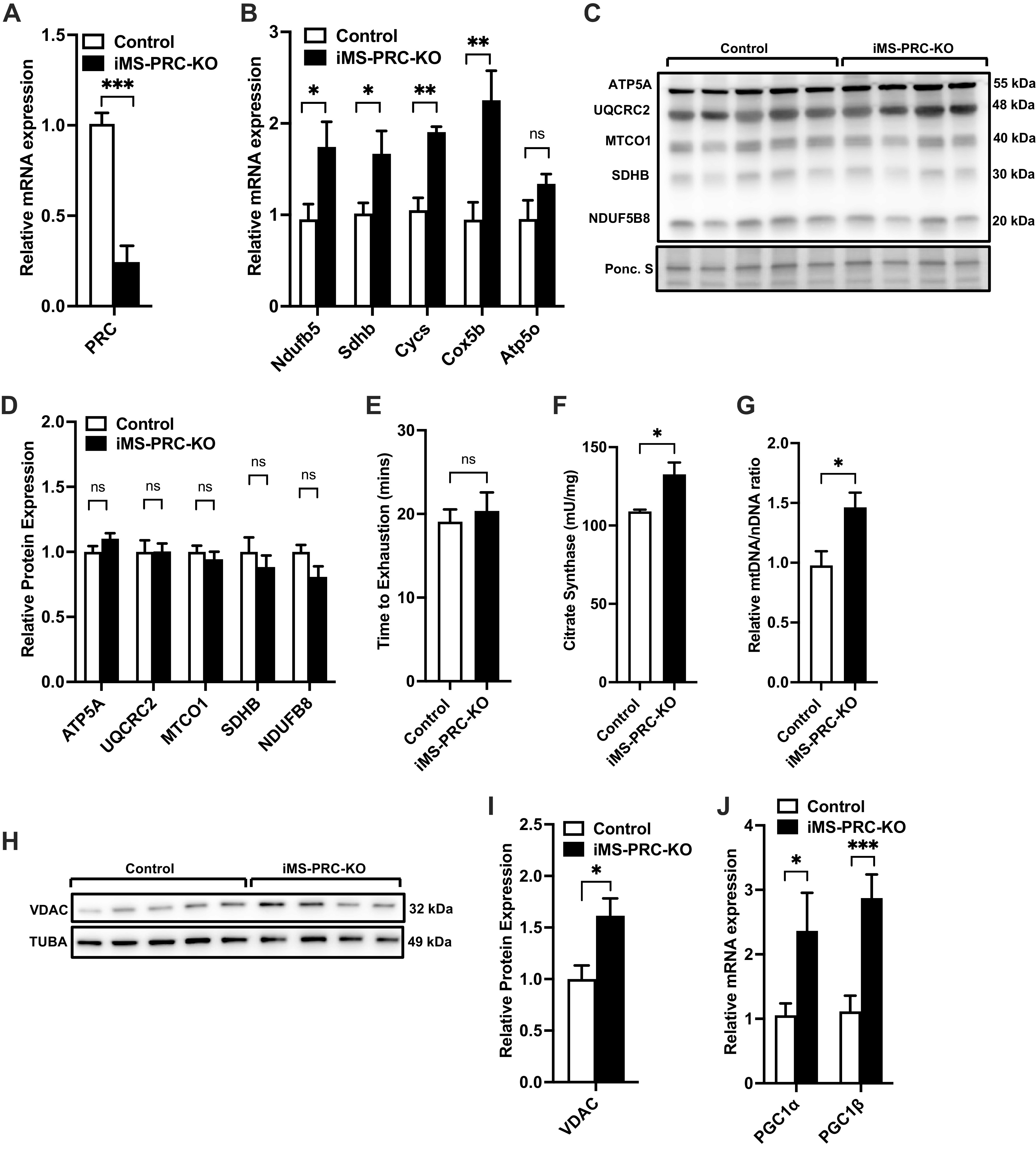
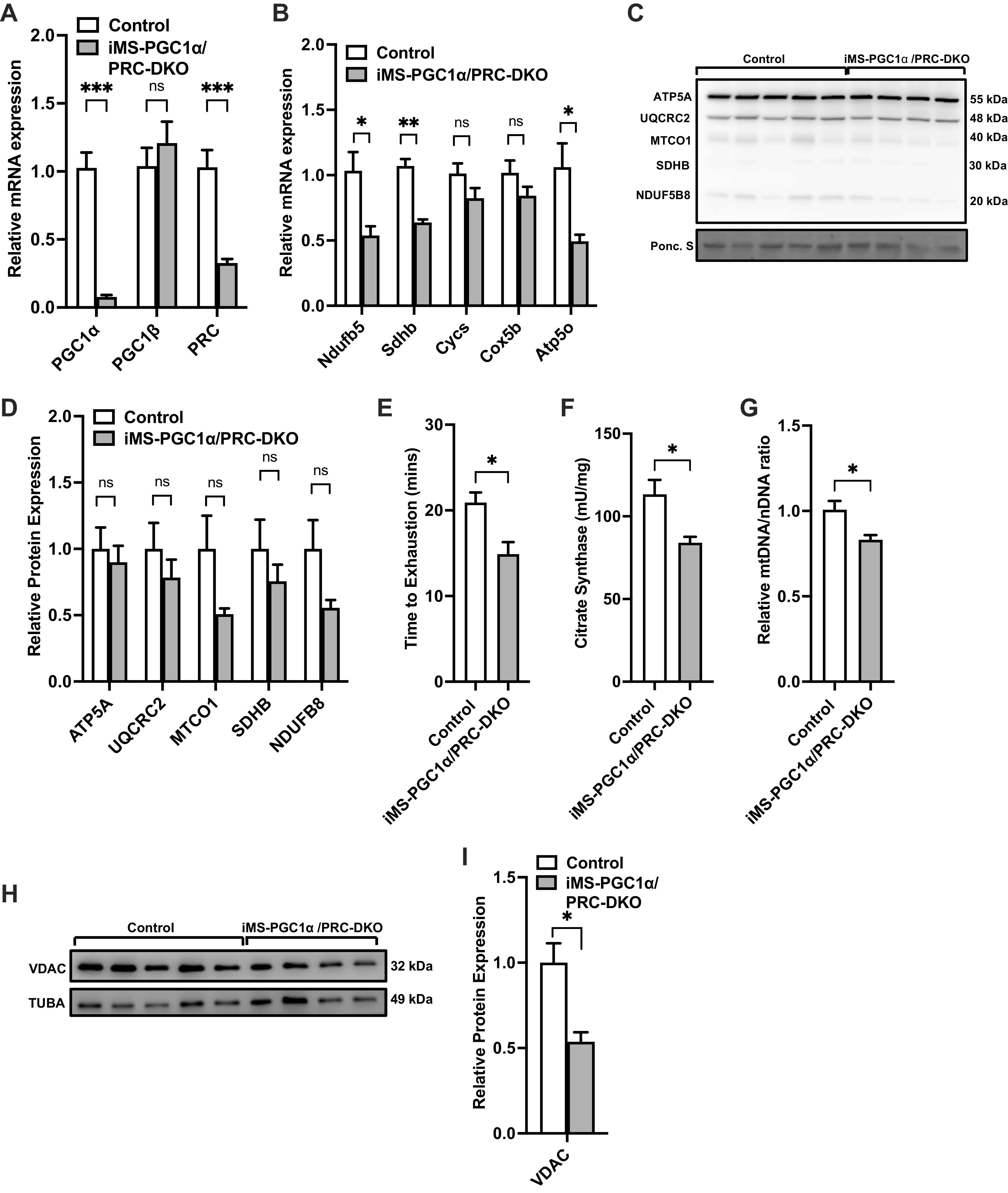
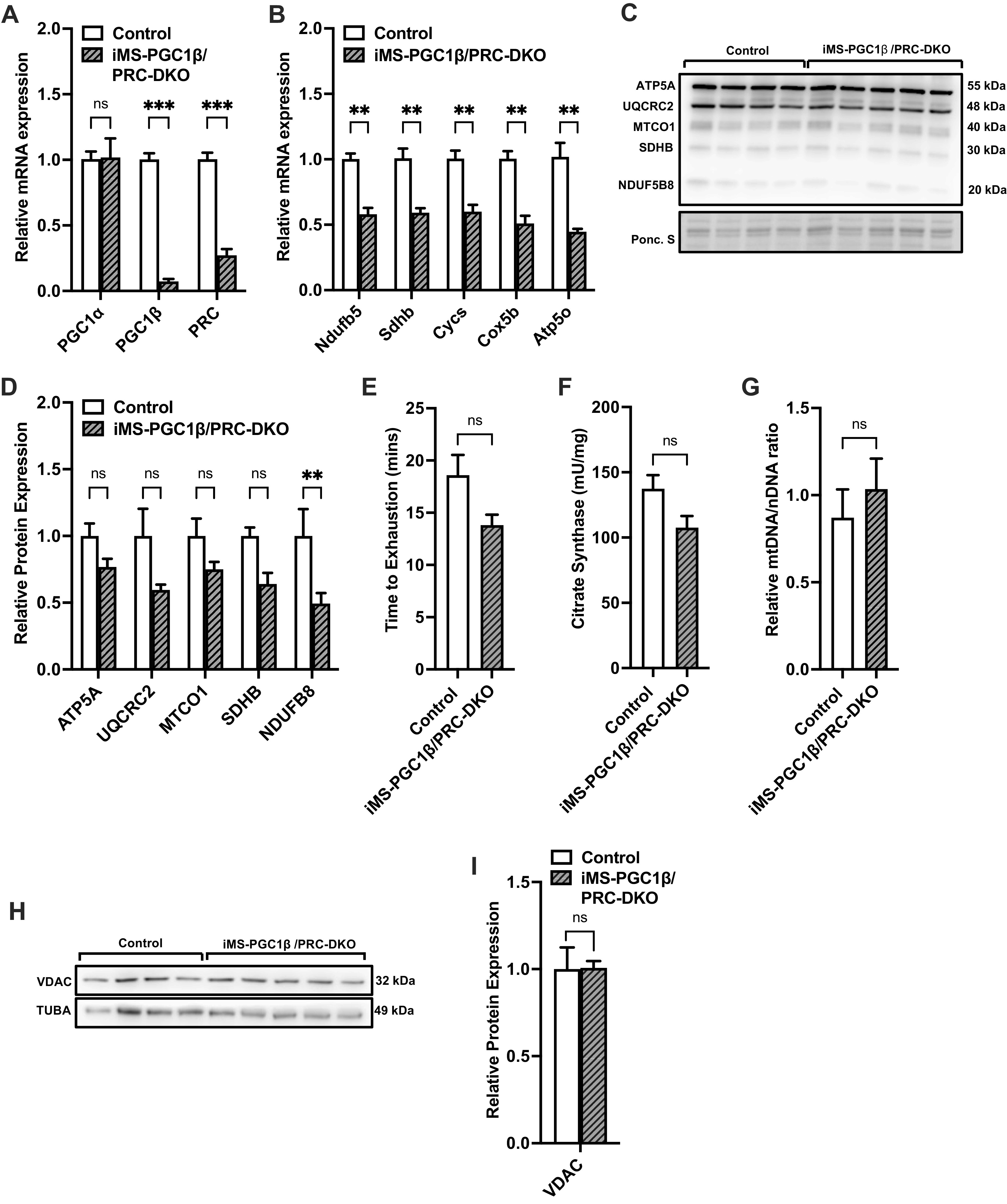
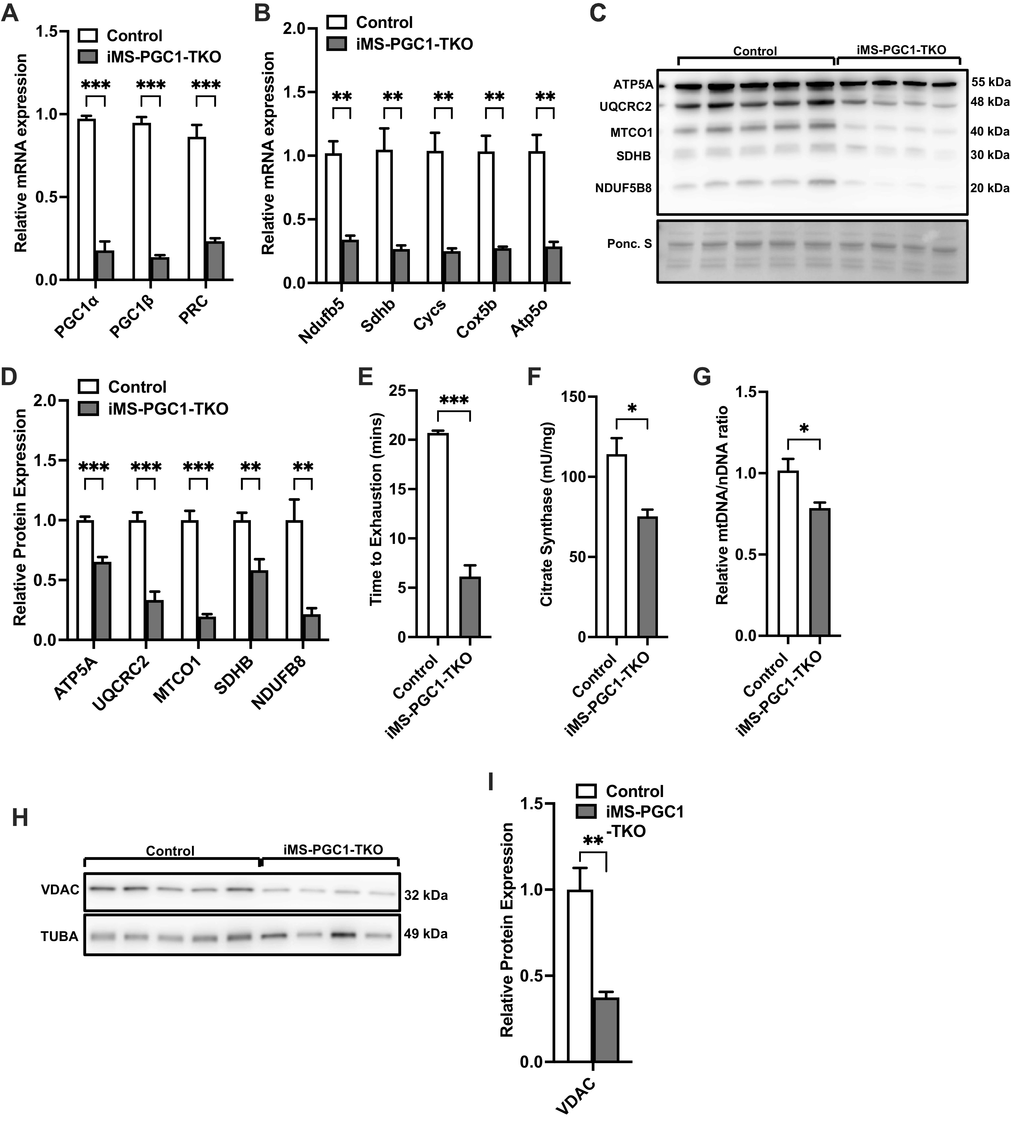
The peroxisome proliferator-activated receptor γ coactivator-1 (PGC-1) family of transcriptional coactivators are regulators of mitochondrial oxidative capacity and content in skeletal muscle. Many of these conclusions are based primarily on gain-of-function studies using muscle-specific overexpression of PGC1s. We have previously reported that genetic deletion of both PGC-1α and PGC-1β in adult skeletal muscle resulted in a significant reduction in oxidative capacity with no effect on mitochondrial content. However, the contribution of PGC-1-related coactivator (PRC), the third PGC-1 family member, in regulating skeletal muscle mitochondria is unknown. Therefore, we generated an inducible skeletal muscle-specific PRC knockout mouse (iMS-PRC-KO) to assess the contribution of PRC in skeletal muscle mitochondrial function. We measured mRNA expression of electron transport chain (ETC) subunits as well as markers of mitochondrial content in the iMS-PRC-KO animals and observed an increase in ETC gene expression and mitochondrial content. Furthermore, the increase in ETC gene expression and mitochondrial content was associated with increased expression of PGC-1α and PGC-1β. We therefore generated an adult-inducible PGC-1 knockout mouse in which all PGC-1 family members are deleted (iMS-PGC-1TKO). The iMS-PGC-1TKO animals exhibited a reduction in ETC mRNA expression and mitochondrial content. These data suggest that in the absence of PRC alone, compensation occurs by increasing PGC-1α and PGC-1β to maintain mitochondrial content. Moreover, the removal of all three PGC-1s in skeletal muscle results in a reduction in both ETC mRNA expression and mitochondrial content. Taken together, these results suggest that PRC plays a role in maintaining baseline mitochondrial content in skeletal muscle.

Keywords: PGC-1-related coactivator; mitochondria; mitochondrial biogenesis; skeletal muscle.


Conflict of interest statement

No conflicts of interest, financial or otherwise, are declared by the authors.

Figures

Figure 1.
Figure 1.
Effect of adult deletion of PRC on skeletal muscle ETC expression and mitochondrial content. PRC mRNA expression (A), nuclear-encoded ETC mRNA expression (B), protein expression of ETC subunits (C), densitometry quantification of ETC subunits protein expression to Ponceau S staining in gastrocnemius muscle (D), time to exhaustion (E), citrate synthase activity (F), mtDNA-to-nDNA (mtDNA/nDNA) ratio (G), VDAC protein expression (H), densitometry quantification of VDAC protein expression normalized to Tuba protein expression (I) and PGC-1α and PGC-1β mRNA expression in gastrocnemius muscle in adult-inducible skeletal muscle-specific PRC knockouts (iMS-PRC-KO) and control littermates (J). Data are presented as means ± SE; n = 4–5 per group; *P < 0.05, **P < 0.01, ***P < 0.001 compared with control as determined by unpaired t test. ETC, electron transport chain; KO, knockout; mtDNA, mitochondrial DNA; nDNA, nuclear DNA; PGC-1, peroxisome proliferator-activated receptor γ coactivator-1; PRC, PGC-1-related coactivator; VDAC, voltage-dependent anion channel.
Figure 2.
Figure 2.
Effect of adult deletion of PRC and PGC-1α on skeletal muscle ETC expression and mitochondrial content. PGC-1α, PGC-1β, and PRC mRNA expression (A); nuclear-encoded ETC mRNA expression (B); protein expression of ETC subunits (C); densitometry quantification of ETC subunits protein expression to Ponceau S staining in gastrocnemius muscle (D); time to exhaustion (E); citrate synthase activity (F); mtDNA-to-nDNA (mtDNA/nDNA) ratio (G); VDAC protein expression (H); densitometry quantification of VDAC protein expression normalized to Tuba protein expression in gastrocnemius muscle in adult-inducible skeletal muscle-specific PGC-1α/PRC knockouts (iMS-PGC-1α/PRC-DKO) and control littermates (I). Data are presented as means ± SE; n = 4–5 per group; *P < 0.05, **P < 0.01, ***P < 0.001 compared with control as determined by unpaired t test. ETC, electron transport chain; KO, knockout; mtDNA, mitochondrial DNA; nDNA, nuclear DNA; PGC-1, peroxisome proliferator-activated receptor γ coactivator-1; PRC, PGC-1-related coactivator; VDAC, voltage-dependent anion channel.
Figure 3.
Figure 3.
Effect of adult deletion of PRC and PGC-1β on skeletal muscle ETC expression and mitochondrial content. PGC-1α, PGC-1β, and PRC mRNA expression (A); nuclear-encoded ETC mRNA expression (B); protein expression of ETC subunits (C); densitometry quantification of ETC subunits protein expression to Ponceau S staining in gastrocnemius muscle (D); time to exhaustion (E); citrate synthase activity (F); mtDNA-to-nDNA (mtDNA/nDNA) ratio (G); VDAC protein expression (H); densitometry quantification of VDAC protein expression normalized to Tuba protein expression in gastrocnemius muscle in adult-inducible skeletal muscle-specific PGC-1β/PRC knockouts (iMS-PGC-1β/PRC-DKO) and control littermates (I). Data are presented as means ± SE; n = 4–5 per group; *P < 0.05, **P < 0.01, ***P < 0.001 compared with control as determined by unpaired t test. ETC, electron transport chain; KO, knockout; mtDNA, mitochondrial DNA; nDNA, nuclear DNA; PGC-1, peroxisome proliferator-activated receptor γ coactivator-1; PRC, PGC-1-related coactivator; VDAC, voltage-dependent anion channel.
Figure 4.
Figure 4.
Effect of adult deletion of all three PGC-1s on skeletal muscle ETC expression and mitochondrial content. PGC-1α, PGC-1β, and PRC mRNA expression (A); nuclear-encoded ETC mRNA expression (B); protein expression of ETC subunits (C); densitometry quantification of ETC subunits protein expression to Ponceau S staining in gastrocnemius muscle (D); time to exhaustion (E); citrate synthase activity (F); mtDNA-to-nDNA (mtDNA/nDNA) ratio (G); VDAC protein expression (H); densitometry quantification of VDAC protein expression normalized to Tuba protein expression in gastrocnemius muscle in adult-inducible skeletal muscle-specific PGC-1α/PGC-1β/PRC knockouts (iMS-PGC-1-TKO) and control littermates (I). Data are presented as means ± SE; n = 4–5 per group; *P < 0.05, **P < 0.01, ***P < 0.001 compared with control as determined by unpaired t test. ETC, electron transport chain; KO, knockout; mtDNA, mitochondrial DNA; nDNA, nuclear DNA; PGC-1, peroxisome proliferator-activated receptor γ coactivator-1; PRC, PGC-1-related coactivator; VDAC, voltage-dependent anion channel.

References

    1. Goodpaster BH, Sparks LM. Metabolic flexibility in health and disease. Cell Metab 25: 1027–1036, 2017. doi:10.1016/j.cmet.2017.04.015. - DOI - PMC - PubMed
    1. Hood DA, Memme JM, Oliveira AN, Triolo M. Maintenance of skeletal muscle mitochondria in health, exercise, and aging. Annu Rev Physiol 81: 19–41, 2019. doi:10.1146/annurev-physiol-020518-114310. - DOI - PubMed
    1. Rowe GC, Safdar A, Arany Z. Running forward: new frontiers in endurance exercise biology. Circulation 129: 798–810, 2014. doi:10.1161/CIRCULATIONAHA.113.001590. - DOI - PMC - PubMed
    1. Heo JW, No MH, Park DH, Kang JH, Seo DY, Han J, Neufer PD, Kwak HB. Effects of exercise on obesity-induced mitochondrial dysfunction in skeletal muscle. Korean J Physiol Pharmacol 21: 567–577, 2017. doi:10.4196/kjpp.2017.21.6.567. - DOI - PMC - PubMed
    1. Hesselink MK, Schrauwen-Hinderling V, Schrauwen P. Skeletal muscle mitochondria as a target to prevent or treat type 2 diabetes mellitus. Nat Rev Endocrinol 12: 633–645, 2016. doi:10.1038/nrendo.2016.104. - DOI - PubMed

Publication types

MeSH terms

Substances