. 2023 Jan 30;28(1):10.
doi: 10.1186/s11658-023-00424-1.

A therapeutic target for CKD: activin A facilitates TGFβ1 profibrotic signaling

Affiliations

A therapeutic target for CKD: activin A facilitates TGFβ1 profibrotic signaling

Asfia Soomro et al. Cell Mol Biol Lett. .

Abstract

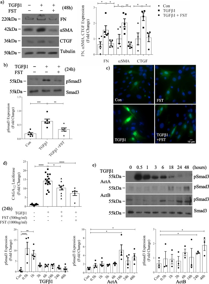
Background: TGFβ1 is a major profibrotic mediator in chronic kidney disease (CKD). Its direct inhibition, however, is limited by adverse effects. Inhibition of activins, also members of the TGFβ superfamily, blocks TGFβ1 profibrotic effects, but the mechanism underlying this and the specific activin(s) involved are unknown.

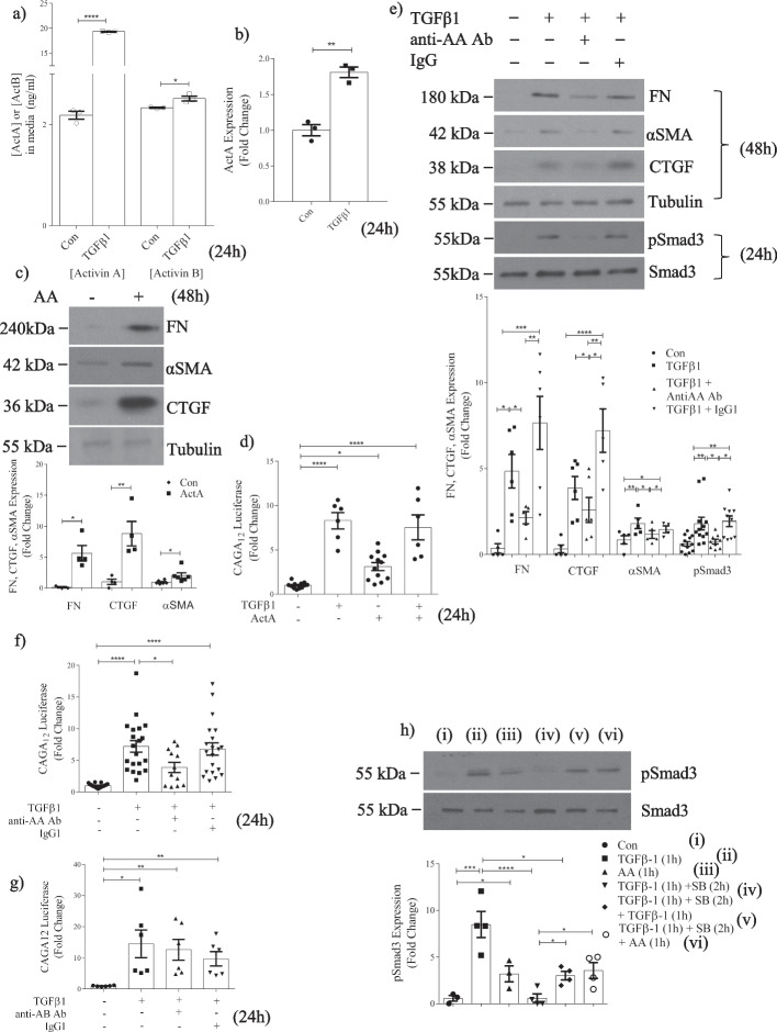
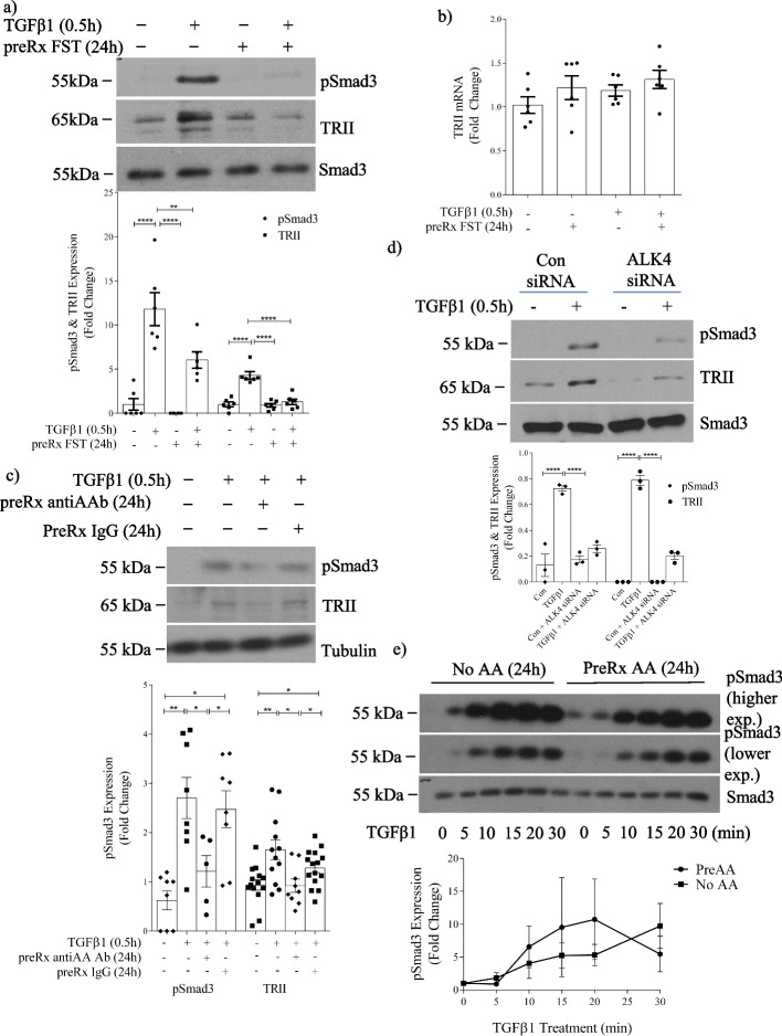
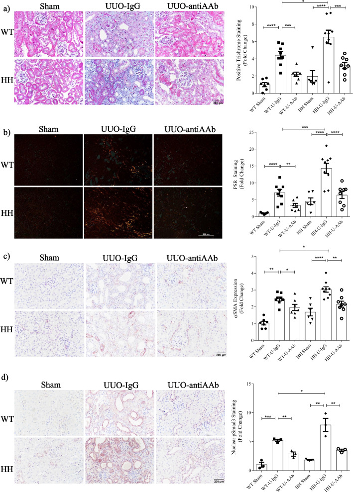
Methods: Cells were treated with TGFβ1 or activins A/B. Activins were inhibited generally with follistatin, or specifically with neutralizing antibodies or type I receptor downregulation. Cytokine levels, signaling and profibrotic responses were assessed with ELISA, immunofluorescence, immunoblotting and promoter luciferase reporters. Wild-type or TGFβ1-overexpressing mice with unilateral ureteral obstruction (UUO) were treated with an activin A neutralizing antibody.

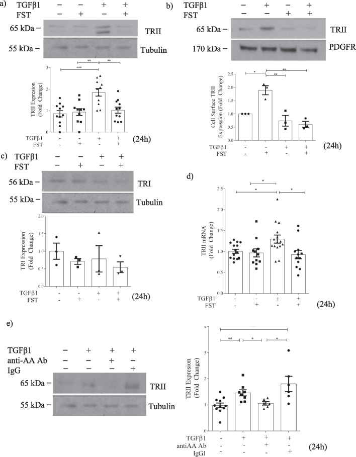
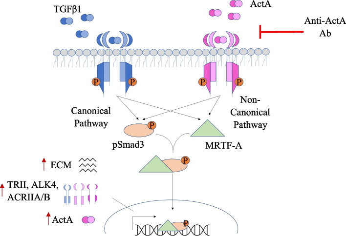
Results: In primary mesangial cells, TGFβ1 induces secretion primarily of activin A, which enables longer-term profibrotic effects by enhancing Smad3 phosphorylation and transcriptional activity. This results from lack of cell refractoriness to activin A, unlike that for TGFβ1, and promotion of TGFβ type II receptor expression. Activin A also supports transcription through regulating non-canonical MRTF-A activation. TGFβ1 additionally induces secretion of activin A, but not B, from tubular cells, and activin A neutralization prevents the TGFβ1 profibrotic response in renal fibroblasts. Fibrosis induced by UUO is inhibited by activin A neutralization in wild-type mice. Worsened fibrosis in TGFβ1-overexpressing mice is associated with increased renal activin A expression and is inhibited to wild-type levels with activin A neutralization.

Conclusions: Activin A facilitates TGFβ1 profibrotic effects through regulation of both canonical (Smad3) and non-canonical (MRTF-A) signaling, suggesting it may be a novel therapeutic target for preventing fibrosis in CKD.

Keywords: Activin A; Extracellular matrix; Kidney fibrosis; TGFβ1.


Conflict of interest statement

All authors declare no competing interests.

Figures

Fig. 1
Fig. 1
Activin inhibition attenuates TGFβ1-induced fibrotic responses and Smad3 activation in MC. Activin inhibition with follistatin (FST) decreases TGFβ1-induced: a FN, αSMA and CTGF upregulation at 48 h (n = 5), b Smad3 phosphorylation (pSmad3) at 24 h (n = 5), c Smad3 nuclear translocation as assessed using eGFP-Smad3 (n = 3; 25–30 cells quantified per treatment group) at 24 h, and d Smad3 transcriptional activity at 24 h (n = 8). e Time course experiments show increases in pSmad3 occur earlier (30–60 min) with TGFβ1 (n = 4) compared with actA (n = 4) or actB (n = 3) (18–48 h). *, **, ***, ****P < 0.05, 0.01, 0.001, 0.0001; one-way ANOVA with Tukey’s multiple comparisons post hoc test
Fig. 2
Fig. 2
Specific activin A inhibition attenuates TGFβ1-induced Smad3 activation and profibrotic responses in MC. a ELISA demonstrates that TGFβ1 (24 h) increases actA and actB secretion (n = 3) to 19.5 ng/ml and 2.5 ng/ml, representing an 8.9- and 1.06-fold induction respectively. b TGFβ1 increases actA in whole cell lysate by 1.8-fold (n = 3). c ActA (20 ng/ml) upregulates FN (n = 3–4), CTGF (n = 4) and αSMA (n = 6) at 48 h. d TGFβ1 and actA both increase Smad3 transcriptional activity; no synergistic effect is seen (n = 6–12). e An actA neutralizing antibody attenuates TGFβ1-induced FN (n = 5–6), αSMA (n = 5), CTGF (n = 5–6), and Smad3 activation (n = 10–12). f ActA neutralization decreases TGFβ1-induced Smad3 transcriptional activity at 24 h (n = 9–15), but this is not decreased by actB neutralization (n = 6) (g). h MC were stimulated with TGFβ1 or actA for 1 h, then treated with their type I receptor inhibitor SB431542 (50 µM). Restimulation with the same ligand shows that cells become refractory to TGFβ1, but not actA (n = 4). *, **, ***, ****P < 0.05, 0.01, 0.001, 0.0001; one-way ANOVA with Tukey’s multiple comparisons post hoc test
Fig. 3
Fig. 3
Activins facilitate TGFβ1 receptor type II upregulation in response to TGFβ1 in MC. a Follistatin (FST) inhibits the TGFβ1-induced increase in TRII expression at 24 h in whole cell lysate (n = 10–11). b The TGFβ1-induced increase in cell surface TRII was diminished by follistatin at 24 h (n = 3). c No change in response to either TGFβ1 or follistatin were seen with TRI (n = 6). d The TGFβ1-induced increase in TRII transcript was also diminished by follistatin at 24 h (n = 13–15). e TGFβ1 induction of TRII expression is reduced with an actA neutralizing antibody (n = 6). *, **, ***, ****P < 0.05, 0.01, 0.001, 0.0001; one-way ANOVA with Tukey’s multiple comparisons post hoc test
Fig. 4
Fig. 4
Activin A facilitates TGFβ1-induced type II receptor expression without altering TGFβ1 receptor sensitivity in MC. a TGFβ1 for 30 min increased TRII expression and Smad3 activation in whole cell lysate, and both were prevented by 24 h pretreatment with follistatin (n = 6). b There was no change in TRII transcript after 24 h of follistatin with or without TGFβ1 for 30 min. c Pretreatment with an actA neutralizing antibody for 24 h inhibited acute (30 min) TGFβ1-induced Smad3 activation and TRII upregulation (n = 6–7). These were similarly inhibited by downregulation of the actA type I receptor ALK4 with siRNA (d) (n = 3). e Pretreatment with actA (20 ng/ml) for 24 h did not affect acute TGFβ1-induced Smad3 activation (n = 6). *, **, ****P < 0.05, 0.01, 0.0001; one-way ANOVA with Tukey’s multiple comparisons post hoc test
Fig. 5
Fig. 5
Activin A regulation of MRTF-A contributes to αSMA induction by TGFβ1 in MC. a ActA (20 ng/ml) and TGFβ1 show an additive effect on αSMA promoter transcriptional activation at 24 h (n = 6). Activin inhibition with b follistatin (n = 7) and c an actA neutralizing antibody decrease TGFβ1-induced αSMA promoter transcriptional activity (n = 14–23). d actB neutralization does not decrease TGFβ1-induced αSMA promoter transcriptional activity (n = 6–9). e actA increases MRTF-A nuclear localization at 18 h (n = 5). TGFβ1-induced nuclear MRTF-A translocation is attenuated with f follistatin (n = 9) and g actA neutralization (n = 5). h ActA-induced αSMA promoter transcriptional activity is inhibited by MRTF-A siRNA (n = 9) and i the Smad3 inhibitor SIS3 (n = 3). *, **, ***, ****P < 0.05, 0.01, 0.001, 0.0001; one-way ANOVA with Tukey’s multiple comparisons post hoc test
Fig. 6
Fig. 6
Activin A neutralization inhibits renal fibrosis in TGFβ1-overexpressing mice. a TGFβ1 transcript is increased in mice genetically engineered to overexpress TGFβ1 (HH) compared with wild-type mice (WT) (n = 6–7, *p ≤ 0.05). b Serum actA is elevated in wild-type and HH mice after UUO. This is decreased by treatment with a neutralizing actA antibody (anti-actA) in HH mice. c Renal actA is increased after UUO, with a greater induction in HH mice. Both are attenuated by actA neutralization. Boxed areas are shown at higher magnification immediately below. ActA increases are seen particularly in tubular epithelial cells. d Renal αSMA, fibronectin (FN), pSmad3 and MRTF-A are increased after UUO and this is augmented in HH kidneys. Expression of all is attenuated by actA neutralization in both WT and HH kidneys. (n = 6–9) *, **, ***, ****P < 0.05, 0.01, 0.001, 0.0001; one-way ANOVA with Tukey’s multiple comparisons post hoc test where there are > 2 groups; t-test for 2 groups
Fig. 7
Fig. 7
Augmented fibrosis in UUO by TGFβ1 overexpression is attenuated by actA neutralization. TGFβ1 overexpression worsened UUO-induced fibrosis as assessed by a Trichrome and b PSR. Both were attenuated by actA neutralization. c TGFβ1 overexpression augmented UUO-induced expression of the fibroblast marker αSMA, which was attenuated by actA inhibition. d Nuclear levels of phosphorylated Smad3 were also augmented by TGFβ1 overexpression after UUO and this was inhibited by actA neutralization (n = 6–9) *, **, ***, ****P < 0.05, 0.01, 0.001, 0.0001; one-way ANOVA with Tukey’s multiple comparisons post hoc tests
Fig. 8
Fig. 8
A schematic representation showing ActA-stimulated Smad3 and MRTF-A signaling converge to enable sustained TGFβ1 profibrotic responses. ActA regulation of TRII and actA receptors, as well as ongoing actA receptor responsiveness to its ligand in contrast to the refractoriness of TGFβ1 receptors to TGFβ1, are important in maintaining profibrotic TGFβ1 signaling. Neutralization of actA attenuates TGFβ1-induced fibrosis

References

    1. Hill NR, Fatoba ST, Oke JL, Hirst JA, O'Callaghan CA, Lasserson DS, et al. Global prevalence of chronic kidney disease—a systematic review and meta-analysis. PLoS ONE. 2016;11(7):e0158765. - PMC - PubMed
    1. Moody WE, Chue CD, Inston NG, Edwards NC, Steeds RP, Ferro CJ, et al. Understanding the effects of chronic kidney disease on cardiovascular risk: are there lessons to be learnt from healthy kidney donors? J Hum Hypertens. 2012;26(3):141–148. - PubMed
    1. Chen TK, Knicely DH, Grams ME. Chronic kidney disease diagnosis and management: a review. JAMA. 2019;322(13):1294–1304. - PMC - PubMed
    1. Heerspink HJL, Stefansson BV, Correa-Rotter R, Chertow GM, Greene T, Hou FF, et al. Dapagliflozin in patients with chronic kidney disease. N Engl J Med. 2020;383(15):1436–1446. - PubMed
    1. Meng XM, Nikolic-Paterson DJ, Lan HY. TGF-beta: the master regulator of fibrosis. Nat Rev Nephrol. 2016;12(6):325–338. - PubMed