. 2023 Jan 30;21(1):61.
doi: 10.1186/s12967-023-03900-6.

Characterization of large extracellular vesicles (L-EV) derived from human regulatory macrophages (Mreg): novel mediators in wound healing and angiogenesis?

Affiliations

Characterization of large extracellular vesicles (L-EV) derived from human regulatory macrophages (Mreg): novel mediators in wound healing and angiogenesis?

Martin Albrecht et al. J Transl Med. .

Abstract

Background: Large extracellular vesicles (L-EV) with a diameter between 1 and 10 µm are released by various cell types. L-EV contain and transport active molecules which are crucially involved in cell to cell communication. We have shown that secretory products of human regulatory macrophages (Mreg) bear pro-angiogenic potential in-vitro and our recent findings show that Mreg cultures also contain numerous large vesicular structures similar to L-EV with so far unknown characteristics and function.

Aim of this study: To characterize the nature of Mreg-derived L-EV (L-EVMreg) and to gain insights into their role in wound healing and angiogenesis.

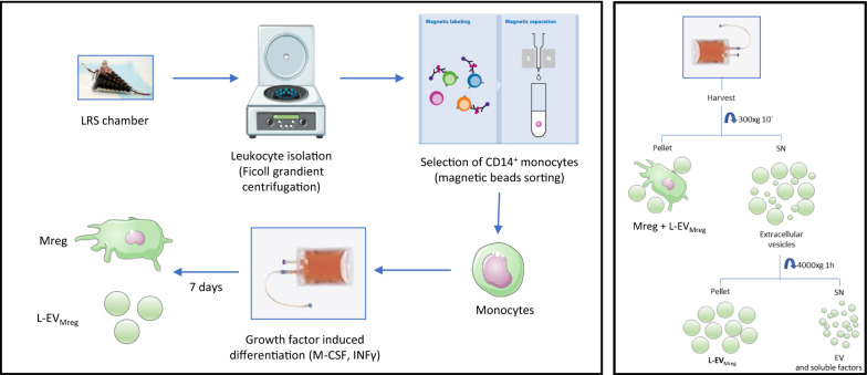
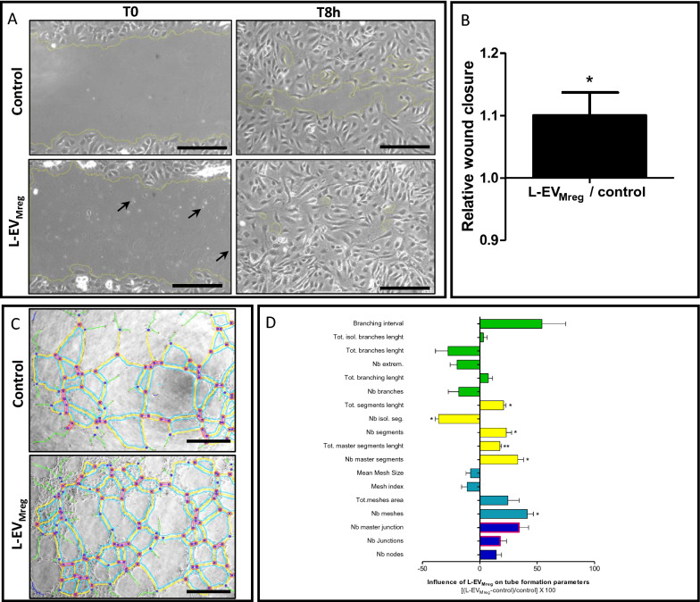
Methods: Mreg were differentiated using blood monocytes from healthy donors (N = 9) and L-EVMreg were isolated from culture supernatants by differential centrifugation. Characterization of L-EVMreg was performed by cell/vesicle analysis, brightfield/transmission electron microscopy (TEM), flow cytometry and proteome profiling arrays. The impact of L-EVMreg on wound healing and angiogenesis was evaluated by means of scratch and in-vitro tube formation assays.

Results: Mreg and L-EVMreg show an average diameter of 13.73 ± 1.33 µm (volume: 1.45 ± 0.44 pl) and 7.47 ± 0.75 µm (volume: 0.22 ± 0.06 pl) respectively. Flow cytometry analyses revealed similarities between Mreg and L-EVMreg regarding their surface marker composition. However, compared to Mreg fewer L-EVMreg were positive for CD31 (P < 0.01), CD206 (P < 0.05), CD103 (P < 0.01) and CD45 (P < 0.05). Proteome profiling suggested that L-EVMreg contain abundant amounts of pro-angiogenic proteins (i.e. interleukin-8, platelet factor 4 and serpin E1). From a functional point of view L-EVMreg positively influenced in-vitro wound healing (P < 0.05) and several pro-angiogenic parameters in tube formation assays (all segment associated parameters, P < 0.05; number of meshes, P < 0.05).

Conclusion: L-EVMreg with regenerative and pro-angiogenic potential can be reproducibly isolated from in-vitro cultured human regulatory macrophages. We propose that L-EVMreg could represent a putative therapeutic option for the treatment of chronic wounds and ischemia-associated diseases.

Keywords: Angiogenesis; Large extracellular vesicles; Macrophages; Wound healing.


Conflict of interest statement

The authors (MA, KZ, FF, RB) are involved in a pending patent concerning L-EVMreg and results of the current study are included in the patent application.

Figures

Fig. 1
Fig. 1
Schematic representation of in-vitro differentiation of human monocytes to Mreg and L-EVMreg (left) and differential centrifugation steps leading to L-EVMreg enriched pellets (right). SN, supernatant; EV, extracellular vesicles
Fig. 2
Fig. 2
Vesicle and cell analysis. A. Representative analysis of a sample containing Mreg and L-EVMreg using the MOXI counter. B. Representative analysis of a sample containing Mreg and L-EVMreg using the Nucleocounter. Note that nucleus lacking vesicles are not detected using the Nucleocounter method. C. Quantitative results from vesicle and cell analysis
Fig. 3
Fig. 3
Morphology of Mreg derived L-EV (L-EVMreg). A. Brighfield microscopy after harvest on day 7. B. Transmission electron microscopy after harvest on day 7. Note that L-EVMreg share several morphological features of Mreg cells such as numerous of vesicles/vacuoles and pseudopodia-like extensions. N nucleus, V vesicle/vacuole, P pseudopodia
Fig. 4
Fig. 4
Cell surface marker characterization of Mreg derived L-EV (L-EVMreg). A. Representative scatter plot (FCS vs. SSC) showing L-EVMreg and Mreg populations at day 7. B. Flow cytometric analysis of surface marker characteristics of L-EVMreg and Mreg. FSC, forward scatter; SSC, sideward scatter; *, P < 0.05; **, P < 0.01
Fig. 5
Fig. 5
Characterization of angiogenic proteins in Mreg derived L-EV (L-EVMreg). A. Representative proteome profiling array membrane incubated with sonicated L-EVMreg showing the relative abundance of 55 proteins involved in angiogenesis. Note that each protein is represented by duplicate spots on the respective membrane. B. Three-dimensional heatmap analysis of A. C. Arrangement of protein spots on the membrane. Proteins with the highest signal intensities are depicted in bold
Fig. 6
Fig. 6
Effects of Mreg derived L-EV (L-EVMreg) on in-vitro wound healing and angiogenesis. A. Photomicrographs of a representative wound healing assay employing human endothelial cells (HUVEC) with addition of 106 L-EVMreg/ml and without addition of L-EVMreg. Note the presence of numerous L-EV in the treatment group (arrows). Scale bars denote 250 µm. B. Statistical analysis of the effect of L-EVMreg addition on relative wound closure (control = 1) after 8 h. The column denotes the mean ± SD of 3 independent experiments; *, P < 0.05 (one sample student t-test vs. 1). C. Representative images of tube formation analysis employing HUVEC cell cultures with and without the addition of L-EVMreg. Scale bars denote 500 µm. D. The influence of L-EVMreg addition on various tube formation parameters. Horizontal columns denote the mean ± SD of 3 independent experiments. *, P < 0.05; **, P < 0.01 (one sample student t-test vs. 0). Green color, branches related parameters; yellow color, segments related parameters; cyan color, meshes related parameters; blue color, nodes related parameters; red color, junctions related parameters. For parameter definitions and details on the angiogenesis analyzer tool please refer to: http://image.bio.methods.free.fr/ImageJ/?Angiogenesis-Analyzer-for-ImageJ&lang=en

References

    1. Nieuwland R, Falcon-Perez JM, Soekmadji C, Boilard E, Carter D, Buzas EI. Essentials of extracellular vesicles: posters on basic and clinical aspects of extracellular vesicles. J Extracell Vesicles. 2018;7:1548234. - PMC - PubMed
    1. Couch Y, Buzas EI, Di Vizio D, Gho YS, Harrison P, Hill AF, Lotvall J, Raposo G, Stahl PD, Thery C, et al. A brief history of nearly EV-erything—the rise and rise of extracellular vesicles. J Extracell Vesicles. 2021;10:e12144. - PMC - PubMed
    1. Wang X, Jiang L, Liu Q. miR-18a-5p derived from mesenchymal stem cells-extracellular vesicles inhibits ovarian cancer cell proliferation, migration, invasion, and chemotherapy resistance. J Transl Med. 2022;20:258. - PMC - PubMed
    1. Sun IO, Bae YU, Lee H, Kim H, Jeon JS, Noh H, Choi JS, Doh KO, Kwon SH. Circulating miRNAs in extracellular vesicles related to treatment response in patients with idiopathic membranous nephropathy. J Transl Med. 2022;20:224. - PMC - PubMed
    1. Paolicelli RC, Bergamini G, Rajendran L. Cell-to-cell communication by extracellular vesicles: focus on microglia. Neuroscience. 2019;405:148–157. - PubMed

Publication types