. 2023 Jan 30;9(1):4.
doi: 10.1186/s40942-022-00439-4.

Surgical classification for large macular hole: based on different surgical techniques results: the CLOSE study group

Affiliations

Surgical classification for large macular hole: based on different surgical techniques results: the CLOSE study group

Flavio A Rezende et al. Int J Retina Vitreous. .

Abstract

Background: The CLOSE study group proposes an updated surgical classification for large macular holes based on a systematic review of new treatments. Recently, many new techniques have been introduced to treat large full-thickness macular holes (FTMH); although the indications are not clear. An updated surgical classification is needed to help surgical decision-making.

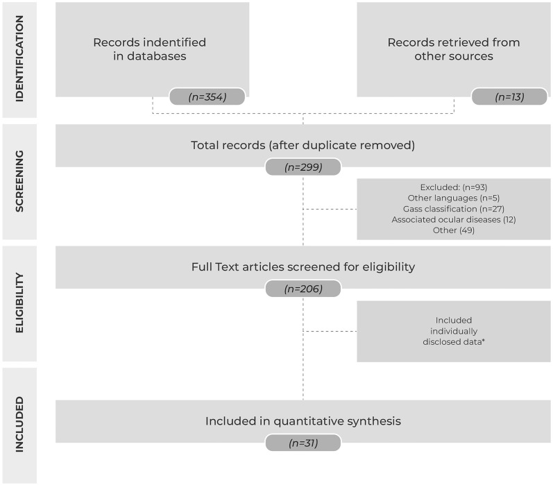
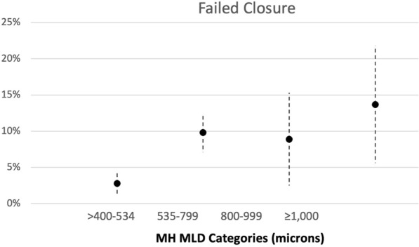
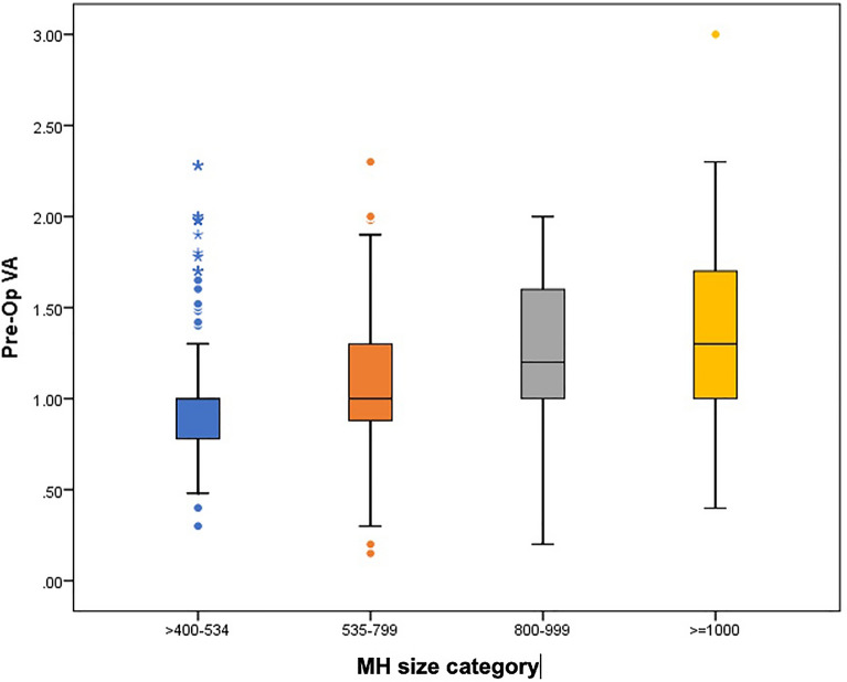
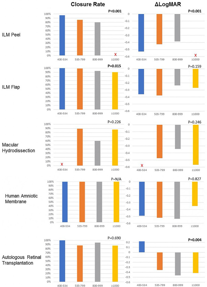
Methods: We gathered published series by the CLOSE Study Group members and from literature search until June 2021. Techniques included: internal limiting membrane peeling (ILM peeling), ILM flaps, macular hydrodissection (macular hydro), human amniotic membrane graft (hAM), and autologous retinal transplantation (ART). Within each technique, chi-square test assessed association between the minimal linear diameter (MLD) (in µm) and closure rate; the postoperative best-corrected visual acuity (BCVA) gains were compared among groups.

Results: Data extraction included 31 published articles: total of 1135 eyes. Eyes were divided into the following groups: ILM peel (n: 683), ILM Flap (n: 233), macular hydrodissection (n: 64), hAM (n: 59), and ART (n: 96). The initial BCVA and size were heterogenous between the groups. ILM peel showed the best results in large FTMH ≤ 535 µm (closure rate 96.8%); adjusted mean BCVA: 0.49 (LogMAR) with a statistical difference among groups. Large FTMH between 535 and 799 µm: ILM flap technique showed better results (closure rate 99.0%); adjusted mean BCVA: 0.67(LogMAR); also with a statistical difference. For large FTMH ≥ 800 µm more invasive techniques are required. Use of hAM, macular hydrodissection and ART showed higher closure rates for this category (100%, 83.3% and 90.5% respectively), and adjusted mean BCVA varied from 0.76 to 0.89. Although there was no statistical difference between those techniques for this group due to the smaller number of cases.

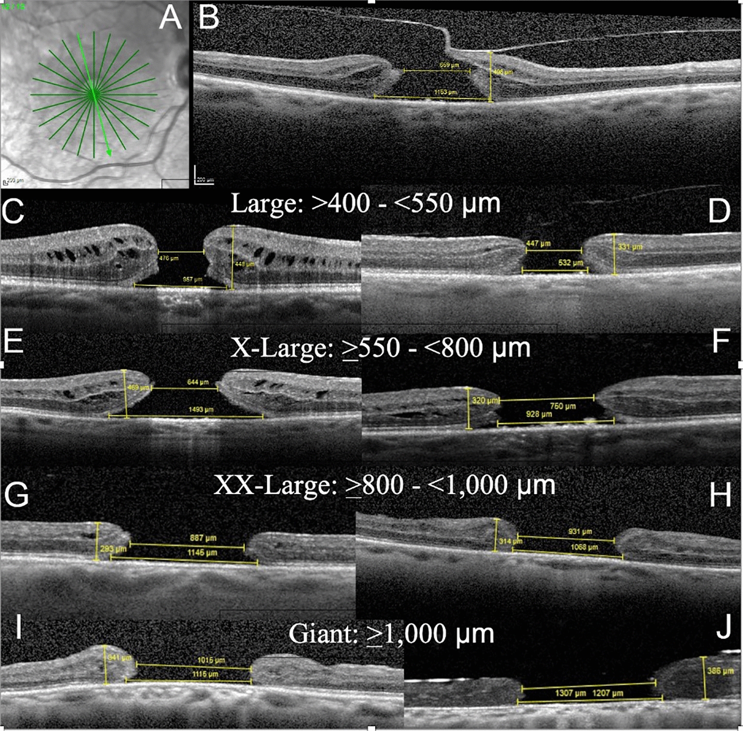
Conclusions: The CLOSE study group demonstrated the potential usefulness of a new surgical classification for large FTMHs and propose OCT biomarkers for use in clinical practice and future research. This new classification demonstrated that Large (400-550 µm) and X-Large (550-800 µm) holes can be treated highly successfully with ILM peel and ILM flap techniques, respectively. Further studies are necessary for the larger FTMHs (XX-Large and Giant), using the CLOSE classification, in order to determine which technique is better suited for each hole size and characteristics.

Keywords: Close study group; Large macular holes; Surgical macular hole classification.


Conflict of interest statement

The authors declare that they have no competing interests.

Figures

Fig. 1
Fig. 1
Flow diagram of the search process and study selection of articles (n = number of records in each category)
Fig. 2
Fig. 2
Correlation of macular hole (MH) categories (divided into 4 groups) based on the preoperative minimum linear diameter (MLD) and percentage of postoperative failure to close
Fig. 3
Fig. 3
Association between the preoperative macular hole (MH) minimum linear diameter (MLD) size measured by optical coherence tomography (in µm) and the preoperative logarithm of the minimum angle of resolution visual acuity (pre-op VA)
Fig. 4
Fig. 4
The graphs show the outcomes for each technique. Outcomes are categorized by the preoperative minimum linear diameter (MLD) measured on optical coherence tomography. The graphs on the left show the percentages of macular hole (MH) closure for each MLD size group. The graphs on the right show the improvement in the logarithm of the minimum angle of resolution (logMAR) (ΔlogMAR) visual acuity for MHs in each MLD size group. The X indicates MLD size groups with an insufficient number of eyes to be included in the analysis. ILM = internal limiting membrane
Fig. 5
Fig. 5
Spectral-domain optical coherence tomography (OCT) radial scans of full-thickness macular hole (FTMH) over 400 µm in minimum linear diameter (MLD) show each hole size group. The measurements of the MLD, base linear diameter (BLD), and hole height (at the highest point around the hole) are shown. The macular hole index (MHI) is calculated by the ratio of height to BLD. The edge configuration is described as having a fluid cuff or being flat and the presence/absence of edematous cysts. Vitreomacular traction (VMT) and epiretinal membranes (ERMs) also are described. With increasing hole size, the fluid cuff and edematous cysts tend to disappear. A Radial scans centered on the FTMH are used to detect the largest MLD and presence of VMT. B Only one of the radial scans from A shows focal VMT in this primary X-large (XL) hole with a MLD of 659 µm, a BLD of 1153 µm, height of 496 µm, MHI of 0.43 with a fluid cuff and edematous cysts, and no ERM. C A primary large FTMH with a MLD of 476 µm, BLD of 957 µm, height of 448, MHI of 0.47, with a cuff and edematous cysts, no VMT, and no ERM. D A primary large FTMH with a MLD of 447 µm, BLD of 532 µm, height of 331 µm, MHI of 0.62, no cuff, no cysts, no VMT, and no ERM. E A primary XL hole with a MLD of 644 µm, BLD of 1493, height of 469 µm, MHI of 0.31, with cuff and cysts, no VMT, and no ERM. F A refractory XL hole with a MLD of 750 µm, BLD of 928 µm, height of 320 µm, MHI of 0.35, no cuff, no cysts, no VMT, and no ERM. G A refractory XXL hole with a MLD of 887 µm, BLD of 1145 µm, height of 293 µm, MHI of 0.26, with a cuff, no cysts, no VMT, and no ERM. H A refractory XXL hole with a MLD of 931 µm, BLD of 1068 µm, height of 314, MHI of 0.29, no cuff, no cysts, no VMT, and no ERM. I A refractory giant hole with a MLD of 1015 µm, BLD of 1115 µm, height of 341 µm, MHI of 0.31, no cuff, no cysts, and no VMT or ERM. J A refractory giant hole under silicone oil tamponade with a MLD of 1207 µm, BLD of 1307 µm, height of 386 µm, and no cuff, cysts, VMT, or ERM

References

    1. Gass JDM. Reappraisal of biomicroscopic classification of stages of development of a macular hole. Am J Ophthalmol. 1995;119:752–759. doi: 10.1016/S0002-9394(14)72781-3. - DOI - PubMed
    1. Kelly NE, Wendel RT. Vitreous surgery for idiopathic macular holes: results of a pilot study. Arch Ophthalmol. 1991;109:654–659. doi: 10.1001/archopht.1991.01080050068031. - DOI - PubMed
    1. Rodrigues EB, Meyer CH, Kroll P. Chromovitrectomy: A new field in vitreoretinal surgery. Graefe Arch Clin Exp Ophthalmol. 2005;243:291–293. doi: 10.1007/s00417-004-0992-x. - DOI - PubMed
    1. Duker JS, Kaiser PK, Binder S, et al. The international vitreomacular traction study group classification of vitreomacular adhesion, traction, and macular hole. Ophthalmology. 2013;120:2611–2619. doi: 10.1016/j.ophtha.2013.07.042. - DOI - PubMed
    1. Liu L, Enkh-Amgalan I, Wang N-K, et al. Results of macular hole surgery. Retina. 2018;38:900–906. doi: 10.1097/IAE.0000000000001647. - DOI - PubMed

LinkOut - more resources