Case Reports
. 2023 Jan;19(1):70-73.
doi: 10.5152/iao.2023.22716.

Cholesteatoma Masquerading as Recurrent Langerhans Cell Histiocytosis

Affiliations
Case Reports

Cholesteatoma Masquerading as Recurrent Langerhans Cell Histiocytosis

Adrien Gendre et al. J Int Adv Otol. 2023 Jan.

Abstract

Langerhans cell histiocytosis is a rare condition affecting the temporal bone in up to 60% of cases. Symptoms are non-specific and the differential diagnosis includes infection, benign lesions such as cholesteatoma, and malignant lesions of the skull base. Here, we report the case of a 14-yearold child referred with chronic ear discharge, and background of multifocal Langerhans cell histiocytosis 9 years prior. Recurrence of Langerhans cell histiocytosis was initially suspected and systemic treatment was considered. Further imaging workup and surgical exploration of the mastoid showed a secondary acquired cholesteatoma arising from a dehiscent posterior ear canal wall. Surgical removal of the cholesteatoma was performed with a canal wall down procedure. We review the presentation and management of temporal bone Langerhans cell histiocytosis. We recommend that cholesteatoma should be considered in case of recurrence of otological symptoms in patients with a background of Langerhans cell histiocytosis.


Figures

Figure 1.
Figure 1.
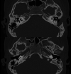
Axial high-resolution CT scan of the temporal bone (initial presentation). There is extensive bony destruction in the left lateral skull base. The otic capsule is intact. CT, computed tomography.
Figure 2.
Figure 2.
Coronal MRI of the skull base, T1 with contrast (gadolinium) (initial presentation). It shows a soft tissue mass in the lateral skull base of the left. It demonstrates T1 hyperintensity enhanced with contrast. MRI, magnetic resonance imaging.
Figure 3.
Figure 3.
Axial high-resolution CT scan of the temporal bone. The middle ear and tympanic membrane appear to be intact. There is a defect in the posterior wall of the external ear canal, the mastoid is filled with a soft tissue mass. Of note, the temporal bone lateral to the otic capsule has mostly re-appeared with growth since the initial CT scan at the time of the primary LCH diagnosis (Figure 1). CT, computed tomography; LCH, Langerhans cell histiocytosis.
Figure 4.
Figure 4.
Coronal MRI of the skull base, T2-weighted. The soft tissue mass in the left mastoid is non-specific with heterogeneous T2 hyper- and hypointensity. MRI, magnetic resonance imaging.

References

    1. Devaney KO, Putzi MJ, Ferlito A, Rinaldo A. Head and neck Langerhans cell histiocytosis. Ann Otol Rhinol Laryngol. 1997;106(6):526 532. (10.1177/000348949710600616) - DOI - PubMed
    1. Guo Y, Ning F, Wang G.et al. Retrospective study of Langerhans cell histiocytosis in ear, nose and neck. Am J Otolaryngol. 2020;41(2):102369. (10.1016/j.amjoto.2019.102369) - DOI - PubMed
    1. Mayer S, Raggio BS, Master A, Lygizos N. Langerhans cell histiocytosis of the temporal bone. Ochsner J. 2020;20(3):315 318. (10.31486/toj.19.0032) - DOI - PMC - PubMed
    1. Berres ML, Merad M, Allen CE. Progress in understanding the pathogenesis of Langerhans cell histiocytosis: back to histiocytosis. Br J Haematol. 2015;169(1):3 13. - PMC - PubMed
    1. Thacker NH, Abla O. Pediatric Langerhans cell histiocytosis: state of the science and future directions. Clin Adv Hematol Oncol. 2019;17(2):122 131. - PubMed

Publication types