. 2022 Nov 29;3(1):63-73.
doi: 10.1021/acsphyschemau.2c00042. eCollection 2023 Jan 25.

Photoinduced Electron Transfer from the Tryptophan Triplet State in Zn-Azurin

Affiliations

Photoinduced Electron Transfer from the Tryptophan Triplet State in Zn-Azurin

Joel J Rivera et al. ACS Phys Chem Au. .

Abstract

Tryptophan is one of few residues that participates in biological electron transfer reactions. Upon substitution of the native Cu2+ center with Zn2+ in the blue-copper protein azurin, a long-lived tryptophan neutral radical can be photogenerated. We report the following quantum yield values for Zn-substituted azurin in the presence of the electron acceptor Cu(II)-azurin: formation of the tryptophan neutral radical (Φrad), electron transfer (ΦET), fluorescence (Φfluo), and phosphorescence (Φphos), as well as the efficiency of proton transfer of the cation radical (ΦPT). Increasing the concentration of the electron acceptor increased Φrad and ΦET values and decreased Φphos without affecting Φfluo. At all concentrations of the acceptor, the value of ΦPT was nearly unity. These observations indicate that the phosphorescent triplet state is the parent state of electron transfer and that nearly all electron transfer events lead to proton loss. Similar results regarding the parent state were obtained with a different electron acceptor, [Co(NH3)5Cl]2+; however, Stern-Volmer graphs revealed that [Co(NH3)5Cl]2+ was a more effective phosphorescence quencher (K SV = 230 000 M-1) compared to Cu(II)-azurin (K SV = 88 000 M-1). Competition experiments in the presence of both [Co(NH3)5Cl]2+ and Cu(II)-azurin suggested that [Co(NH3)5Cl]2+ is the preferred electron acceptor. Implications of these results in terms of quenching mechanisms are discussed.


Conflict of interest statement

The authors declare no competing financial interest.

Figures

Figure 1
Figure 1
Absorption (red) and normalized emission (blue) spectra of deoxygenated ZnAzW48 in 20 mM KPi, pH = 7.0. The normalized emission spectrum of NATA in air is shown as the green dashed curve. The inset shows the phosphorescence region of deoxygenated ZnAzW48 (solid) and ZnAzW48 in air (dashed).
Figure 2
Figure 2
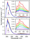
Normalized emission spectra of 25 ± 2 μM ZnAzW48 with increasing concentrations of electron acceptor CuAzW48 (top) and 26 ± 1 μM ZnAzW48 with increasing concentrations of [Co(NH3)5Cl]2+ (bottom). The ratios [CuAz]/[ZnAz] for these representative trials were 0, 0.26, 0.51, 0.80, 1.1, 1.5, 2.0, and 3.2, and [Co(III)]/[ZnAz] were 0, 0.10, 0.17, 0.24, 0.46, and 1.9. Insets show the expanded region of the phosphorescence.
Figure 3
Figure 3
Stern–Volmer plot of ZnAzW48 showing the relative phosphorescence (Φphos0/Φphos, open markers) and relative fluorescence (Φfluo0/Φfluo, filled markers) quantum yields with acceptors CuAzW48 (blue squares) and [Co(NH3)5Cl]2+ (red circles) at T = 22 °C. The phosphorescence data were fit to the linear Stern–Volmer equation (solid line), and the resulting values of KSV (slope, units M–1) and y-intercepts are indicated. Data are shown up to [CuAz]/[ZnAz] =1.5 and [Co(III)]/[ZnAz] = 0.46.
Figure 4
Figure 4
Top panel: Representative absorption spectra during the photolysis of ZnAzW48 in the presence of CuAzW48 with [CuAz]/[ZnAz] = 0.80 for 1 to 40 min. The black curve is the prephotolysis spectrum. Inset shows the region of the isosbestic point. Bottom panel: Difference spectra calculated by subtracting the prephotolysis spectrum from each spectrum on the top panel. The difference spectra have been corrected for the 628 nm bleach. The inset shows the increase in concentration of W48• in ZnAzW48 and decrease in concentration (bleach) of the Cu(II) metal center in CuAzW48.
Figure 5
Figure 5
Representative difference spectra for the photolysis of ZnAzW48 in the presence of [Co(NH3)5Cl]2+ with [Co(III)]/[ZnAz] = 1.9 for 3 to 110 min. The inset shows the increase in concentration of W48•.
Figure 6
Figure 6
Growth of the 515 nm absorbance (top, open symbols) and decay of the 628 nm absorbance (bottom, filled symbols) during photolysis of ZnAzW48 in the presence of CuAzW48 or both [Co(NH3)5Cl]2+ and CuAzW48 electron acceptors. The concentrations are as follows: Black squares [ZnAzW48] = 23 μM and [CuAzW48] = 29 μM; green circles [ZnAzW48] = 24 μM, [CuAzW48] = 29 μM, and [Co(NH3)5Cl]2+ = 4 μM; red triangles [ZnAzW48] = 26 μM, [CuAzW48] = 31 μM, and [Co(NH3)5Cl]2+ = 7 μM; blue stars [ZnAzW48] = 28 μM, [CuAzW48] = 35 μM, and [Co(NH3)5Cl]2+ = 34 μM.
Figure 7
Figure 7
Left axis: Relative quantum yields for fluorescence (Φfluofluo0, black filled circle) and phosphorescence (Φphosphos0, black open circle) for ZnAzW48 in the presence of CuAzW48 (top panel) and [Co(NH3)5Cl]2+ (bottom panel). Right axis (blue and red curves): Quantum yields for radical formation (Φrad, blue filled circle) and ET (ΦET, red filled circle). Lines connect the points to help guide the eye.
Figure 8
Figure 8
Relevant relaxation pathways and measured quantum yields for photoexcited W48 in ZnAzW48 in the presence (right path) and absence (left path) of electron acceptor; all values are from Table 1. In the presence of the CuAzW48 electron acceptor [CuAz]/[ZnAz] = 2.4 ± 0.5), the triplet state relaxes via ET to the Cu(II) metal center and the value of Φphos is negligible at 0.003. In the absence of an electron acceptor, ET from T1 is not possible and Φphos is high at 0.018. In contrast to Φphos, the value of Φfluo is not significantly affected by the presence of an electron acceptor.

Similar articles

Cited by

References

    1. Page C. C.; Moser C. C.; Chen X.; Dutton P. L. Natural engineering principles of electron tunnelling in biological oxidation-reduction. Nature. 1999, 402 (6757), 47–52. 10.1038/46972. - DOI - PubMed
    1. Gray H. B.; Winkler J. R. Electron tunneling through proteins. Q. Rev. Biophys. 2003, 36 (3), 341–372. 10.1017/S0033583503003913. - DOI - PubMed
    1. Onuchic J. N.; Beratan D. N.; Winkler J. R.; Gray H. B. Pathway analysis of protein electron-transfer reactions. Annu. Rev. Biophys. Biomol. Struct. 1992, 21 (1), 349–377. 10.1146/annurev.bb.21.060192.002025. - DOI - PubMed
    1. Stubbe J.; van der Donk W. A. Protein raicals in enzyme catalysis. Chem. Rev. 1998, 98 (2), 705–762. 10.1021/cr9400875. - DOI - PubMed
    1. Eklund H.; Uhlin U.; Färnegårdh M.; Logan D. T.; Nordlund P. Structure and function of the radical enzyme ribonucleotide reductase. Prog. Biophys. Mol. Biol. 2001, 77 (3), 177–268. 10.1016/S0079-6107(01)00014-1. - DOI - PubMed