. 2023 Feb 14;17(3):2089-2100.
doi: 10.1021/acsnano.2c07677. Epub 2023 Jan 31.

Strongly Confined CsPbBr3 Quantum Dots as Quantum Emitters and Building Blocks for Rhombic Superlattices

Affiliations

Strongly Confined CsPbBr3 Quantum Dots as Quantum Emitters and Building Blocks for Rhombic Superlattices

Simon C Boehme et al. ACS Nano. .

Abstract

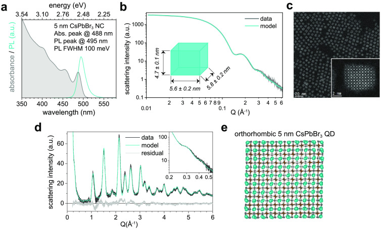
The success of the colloidal semiconductor quantum dots (QDs) field is rooted in the precise synthetic control of QD size, shape, and composition, enabling electronically well-defined functional nanomaterials that foster fundamental science and motivate diverse fields of applications. While the exploitation of the strong confinement regime has been driving commercial and scientific interest in InP or CdSe QDs, such a regime has still not been thoroughly explored and exploited for lead-halide perovskite QDs, mainly due to a so far insufficient chemical stability and size monodispersity of perovskite QDs smaller than about 7 nm. Here, we demonstrate chemically stable strongly confined 5 nm CsPbBr3 colloidal QDs via a postsynthetic treatment employing didodecyldimethylammonium bromide ligands. The achieved high size monodispersity (7.5% ± 2.0%) and shape-uniformity enables the self-assembly of QD superlattices with exceptional long-range order, uniform thickness, an unusual rhombic packing with an obtuse angle of 104°, and narrow-band cyan emission. The enhanced chemical stability indicates the promise of strongly confined perovskite QDs for solution-processed single-photon sources, with single QDs showcasing a high single-photon purity of 73% and minimal blinking (78% "on" fraction), both at room temperature.

Keywords: colloidal nanocrystals; excitons; perovskites; quantum confinement; self-assembly.


Conflict of interest statement

The authors declare no competing financial interest.

Figures

Figure 1
Figure 1
Strongly confined DDAB-capped 5 nm CsPbBr3QDs with narrow-band PL and well-resolved absorption features. (a) Absorbance (gray) and PL (cyan) spectra. (b) Fitting the experimental SAXS data of a QD dispersion (gray line) via an analytical model (cyan line) allows for particle shape determination (see cyan particle model in the inset), yielding an oblate shape with edge lengths of 4.7 ± 0.1 nm, 5.6 ± 0.2 nm, and 5.8 ± 0.2 nm, respectively. (c) High-resolution STEM image, with a single QD shown in the inset. (d) The Porod small-angle region and the WAXTS experimental data (black line) simultaneously fitted via the DSE (cyan line, residuals: gray line), utilizing atomistic models of QDs with an orthorhombic crystal structure (see main text and Supporting Information for details). (e) Atomistic model of a size-averaged QD, obtained from the fit in (d) and a subsequent geometry relaxation at the DFT/PBE level of theory; Cs, Pb, and Br atoms are depicted in cyan, brown, and gray, respectively.
Figure 2
Figure 2
Self-assembly of rhombic QD SLs from cuboidal 5 nm CsPbBr3 QDs. (a) Optical microscopy image of 3D QD SLs under UV excitation; the upper inset shows a magnified view of a representative SL to better visualize the nonorthogonal in-plane angle of about 104°; the lower inset shows an angle histogram of all SLs in the frame. (b) SEM images and (c) AFM image of representative SLs. (d) TEM image of a 2D QD SL and (e) its associated autocorrelation function (ACF), both featuring QD packing with an in-plane angle of about 104°. (f) Larger-area TEM image; inset: suggested structural model based on an overlay of the axes of QD lattice (gray) and SL (black), inferred from the images in (f) and (g), respectively, with a relative tilt of about 7 ± 2°. (g) WAED at the same sample position and orientation as in (f) suggests that the atomic order of bulk CsPbBr3 is largely preserved, as demonstrated by the (pseudo)cubic indexing of the WAED reflections. (h) Suggested structural model of the rhombic QD SL, based on the size and shape of the oblate QDs (from SAXS, Figure 1b) and the QD packing (from TEM and WAED); the average inter-QD facet-to-facet separation of 2.9 ± 0.4 nm is equal in all three spatial directions (a = b = c = 8.2 nm); the QD packing with rhombic repeating units (black dashed rhombus) exhibits a C-centered orthorhombic symmetry (black solid rectangle). (i) 2D GISAXS pattern (with false colors representing the diffracted intensity) demonstrating high long-range in-plane and out-of-plane order; gray and red markers indicate diffraction patterns resulting from reflected and transmitted channel, respectively; upper panel: a Gaussian fit to the orthorhombic 201SL peak (L = 1; indicated with an arrow in the lower panel) yields a fwhm of 0.0059 Å–1 corresponding to a lower limit for the coherent SL domain size of ca. 106 nm. (j) 1D in-plane scattering intensity obtained from horizontal cuts at L = 0 (solid red line) and L = 1 diffraction order (transmitted light only, solid orange line). Comparison with the in-plane cuboidal QD shape form factor (gray dashed line) explains the intensity variation of both pronounced (indicated by solid vertical lines) and suppressed peaks (dashed vertical lines), particularly at about ±0.12 Å–1.
Figure 3
Figure 3
Strongly confined single 5 nm QDs as single-photon sources. (a) Room-temperature PL spectrum of a single QD after pulsed excitation (405 nm, 10 MHz); inset: time trace over 100 s, showing spectrally stable emission and several blinking events. (b) Second-order correlation function g(2)(t) with g(2)(0) = 0.27, indicative of the emission of single photons from single QDs at room temperature. (c) Upper left panel: intensity–time trace showing the blinking of a single QD at room temperature, obtained via either 1 ms time binning (gray trace) or via a bias-free Bayesian change-point analysis algorithm (CPA, black trace) adapted from Palstra et al.;upper right panel: a 78% ON state fraction is derived from the count histogram with a threshold indicated by the gray dashed line; lower panel: a magnified view of the time span indicated by the gray dashed box in the upper panel. (d) PL spectrum of a single QD at 4 K; inset: time trace over 60 s. (e) Time-resolved PL of a single QD at 4 K (blue circles) and 300 K (gray circles), respectively; the initial decays are well fitted by single-exponential decays (red lines) with time constants of ∼0.4 ns and ∼2.6 ns, respectively. (f) Statistics of the PL lifetime and peak energy of several single QDs at 4 K (blue circles), with the mean and standard deviation shown in red.
Figure 4
Figure 4
Narrow emission in strongly confined QD SLs. (a) Temperature-dependent PL spectra of a SL of 5 nm QDs with rhombic shape. (b) PL fwhm as a function of temperature. (c) Time-resolved PL of a QD SL at 4 K (blue circles) and double-exponential fit (gray line), revealing a short lifetime of ∼0.4 ns. (d–i) PL microscopy images of a single SL at 4 K (d–f) and 295 K (g–i), revealing a high spatial uniformity in the PL intensity (d,g), 1/e PL lifetime (τ1/e) (e,h), and center-of-mass energy ECOM (f,i), respectively.

References

    1. Protesescu L.; Yakunin S.; Bodnarchuk M. I.; Krieg F.; Caputo R.; Hendon C. H.; Yang R. X.; Walsh A.; Kovalenko M. V. Nanocrystals of cesium lead halide perovskites (CsPbX3, X = Cl, Br, and I): novel optoelectronic materials showing bright emission with wide color gamut. Nano Lett. 2015, 15 (6), 3692–3696. 10.1021/nl5048779. - DOI - PMC - PubMed
    1. Sercel P. C.; Lyons J. L.; Wickramaratne D.; Vaxenburg R.; Bernstein N.; Efros A. L. Exciton fine structure in perovskite nanocrystals. Nano Lett. 2019, 19 (6), 4068–4077. 10.1021/acs.nanolett.9b01467. - DOI - PubMed
    1. Nguyen T. P. T.; Blundell S. A.; Guet C. One-photon absorption by inorganic perovskite nanocrystals: a theoretical study. Phys. Rev. B 2020, 101 (19), 195414. 10.1103/PhysRevB.101.195414. - DOI
    1. Krieg F.; Sercel P. C.; Burian M.; Andrusiv H.; Bodnarchuk M. I.; Stöferle T.; Mahrt R. F.; Naumenko D.; Amenitsch H.; Rainò G.; et al. Monodisperse long-chain sulfobetaine-capped CsPbBr3 nanocrystals and their superfluorescent assemblies. ACS Cent. Sci. 2021, 7 (1), 135–144. 10.1021/acscentsci.0c01153. - DOI - PMC - PubMed
    1. Kovalenko M. V.; Protesescu L.; Bodnarchuk M. I. Properties and potential optoelectronic applications of lead halide perovskite nanocrystals. Science 2017, 358 (6364), 745–750. 10.1126/science.aam7093. - DOI - PubMed