. 2023 Apr 3;133(7):e164223.
doi: 10.1172/JCI164223.

Neuron-oligodendrocyte potassium shuttling at nodes of Ranvier protects against inflammatory demyelination

Affiliations

Neuron-oligodendrocyte potassium shuttling at nodes of Ranvier protects against inflammatory demyelination

Hannah Kapell et al. J Clin Invest. .

Abstract

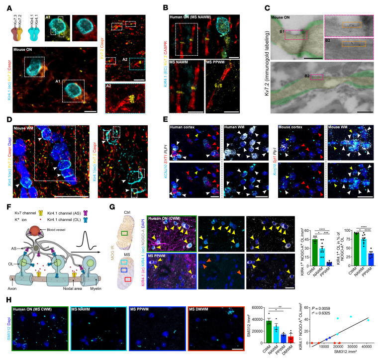
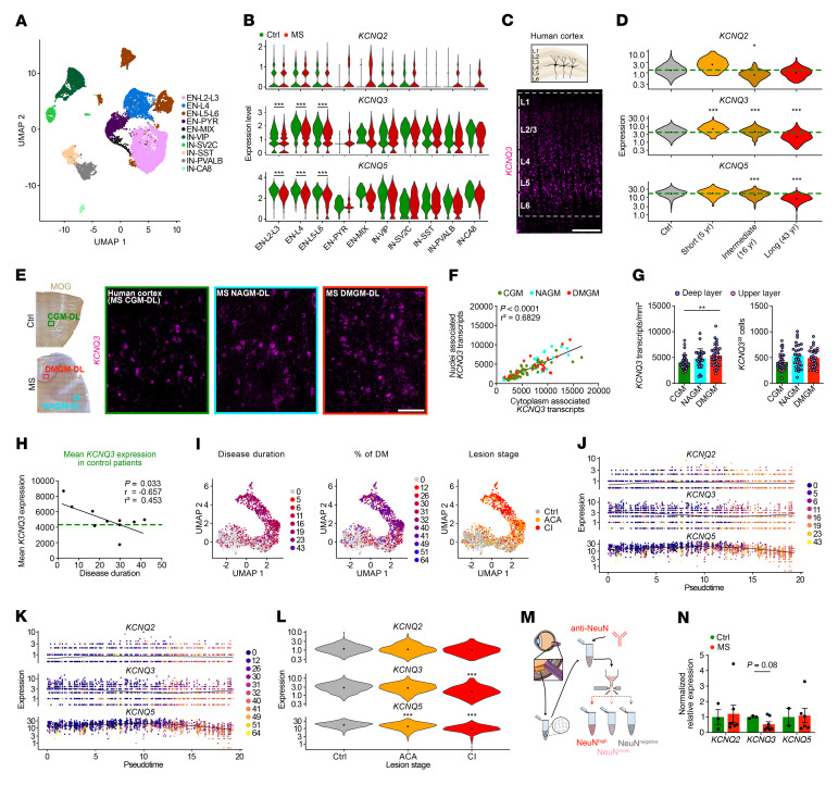
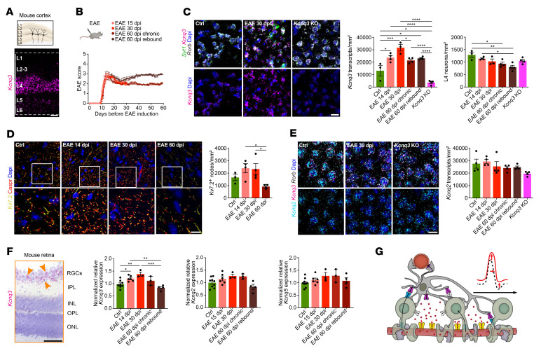
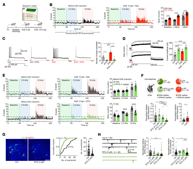
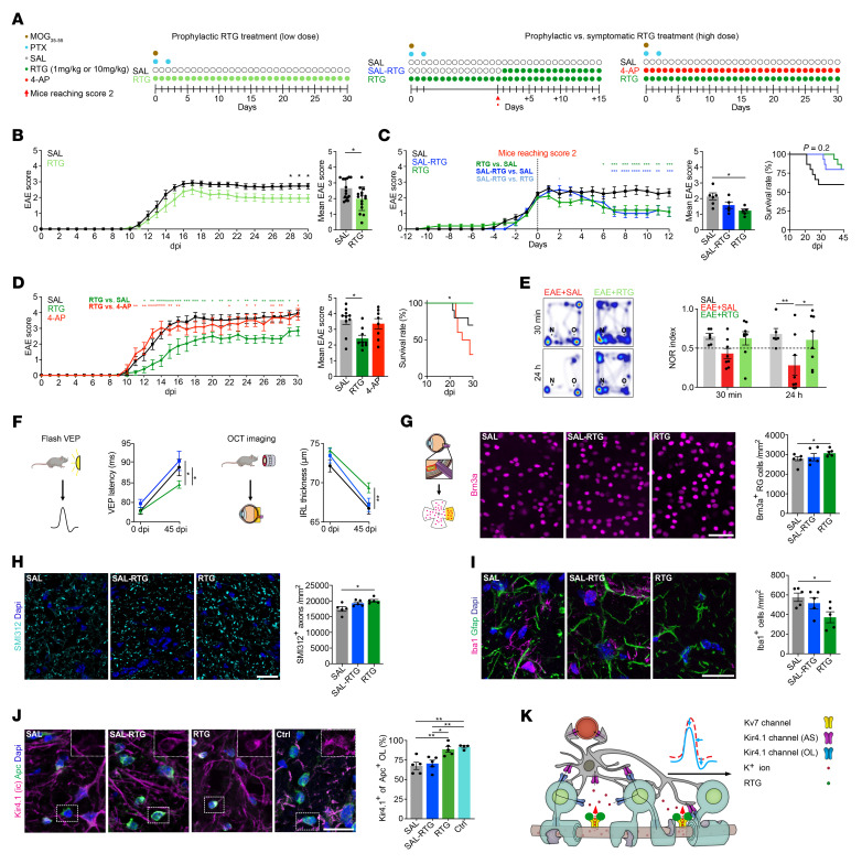
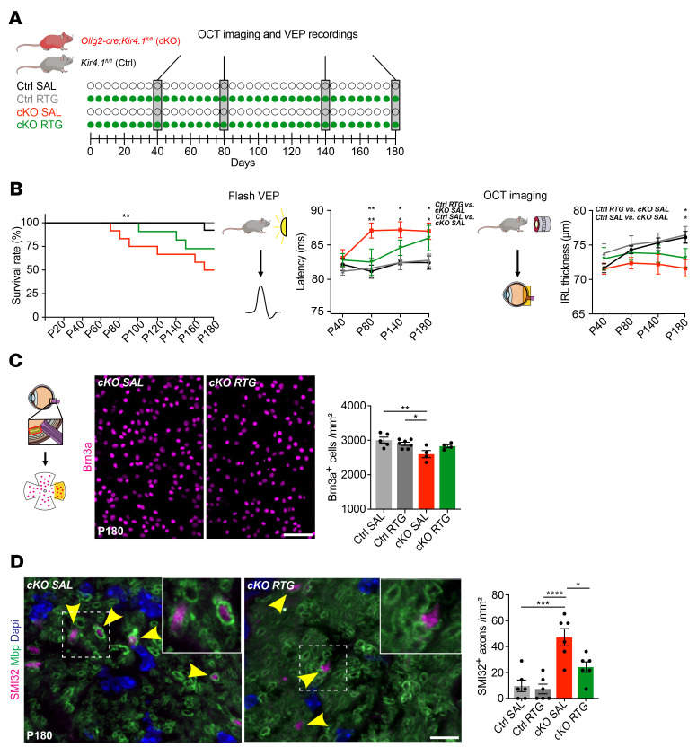
Multiple sclerosis (MS) is a progressive inflammatory demyelinating disease of the CNS. Increasing evidence suggests that vulnerable neurons in MS exhibit fatal metabolic exhaustion over time, a phenomenon hypothesized to be caused by chronic hyperexcitability. Axonal Kv7 (outward-rectifying) and oligodendroglial Kir4.1 (inward-rectifying) potassium channels have important roles in regulating neuronal excitability at and around the nodes of Ranvier. Here, we studied the spatial and functional relationship between neuronal Kv7 and oligodendroglial Kir4.1 channels and assessed the transcriptional and functional signatures of cortical and retinal projection neurons under physiological and inflammatory demyelinating conditions. We found that both channels became dysregulated in MS and experimental autoimmune encephalomyelitis (EAE), with Kir4.1 channels being chronically downregulated and Kv7 channel subunits being transiently upregulated during inflammatory demyelination. Further, we observed that pharmacological Kv7 channel opening with retigabine reduced neuronal hyperexcitability in human and EAE neurons, improved clinical EAE signs, and rescued neuronal pathology in oligodendrocyte-Kir4.1-deficient (OL-Kir4.1-deficient) mice. In summary, our findings indicate that neuron-OL compensatory interactions promoted resilience through Kv7 and Kir4.1 channels and identify pharmacological activation of nodal Kv7 channels as a neuroprotective strategy against inflammatory demyelination.

Keywords: Inflammation; Multiple sclerosis; Neurodegeneration; Neuroscience; Potassium channels.


Figures

Figure 1
Figure 1. Investigation of Kir4.1 and Kv7 channels in neuroglial cell types under homeostatic and inflammatory demyelinating conditions.
(A and B) Triple staining (Caspr, Kir4.1 [extracellular epitope, EC], Kv7.2) revealed specific nodal expression of Kv7.2 (flanked by Caspr IR) adjacent to OL-Kir4.1 channel IR in mouse (A) and human (B) ON. A2 close-up (stimulated emission depletion [STED] image) shows the approximately 190 nm periodic organization of Kv7.2. (C) Kv7.2 immunogold EM labeling shows the presence of gold particles in nodal areas (yellow) between myelin sheets (green) in control mouse ON. (D) Triple staining (compare with A) confirmed the juxtapositioning of OL-Kir4.1 and nodal Kv7.2 channels (white arrows) in other mouse WM tracts (corpus callosum). (E) Perineuronal mouse Kcnj10 and human KCNJ10 expression (ISH) was visualized in mouse and human cortex with OLs coexpressing Plp1+ (mouse) or PLP1+ (human) and Kcnj10+ or KCNJ10+ (white arrowheads) next to Syt1+, SYT1+, Kcnj10, and KCNJ10 neurons (red arrowheads). Yellow arrowheads indicate Kcnj10+, KCNJ10+, Plp1, and PLP1 astrocytes. (F) Cartoon illustrates neuron-OL for the K+ shuttling mechanism: neuronal Kv7 channels mediate axonal K+ efflux, and OL-Kir4.1 channels mediate extracellular K+ uptake and siphoning through interaction with astrocyte (AS) Kir4.1 channels. (G) In human MS ON, KIR4.1 channel IR (antibodies against intracellular [specific for OL-KIR4.1 and AS-KIR4.1] and extracellular [specific for OL-KIR4.1] epitopes) was preserved on astrocyte fibers in lesions. OL-KIR4.1 channel IR (yellow arrows) was reduced in MS NAWM areas (n = 9) and lost in PPWM (n = 6) relative to CWM (n = 4) based on MOG IR. (H) SMI312+ axon density was gradually lost in MS ON tissues toward the lesion rim and correlated with OL-KIR4.1 channel loss. Scale bars: 5 μm (A and B); 0.5 μm (C); 20 μm (D and E, and G); 100 μm (H). Original magnification, ×100 (enlarged insets in A and B) and ×63 (enlarged insets in D). *P < 0.05, **P < 0.01, ***P < 0.001, and ****P < 0.0001, by 1-way ANOVA (G and H, left) and simple linear regression (H, right).
Figure 2
Figure 2. KCNQ3 dysregulation in cortical and retinal MS tissues.
(A) Uniform manifold approximation and projection (UMAP) plot visualizes clustering of human excitatory (EN) and inhibitory (IN) cortical neurons based on a published snRNA-Seq data set (12). (B) Normalized KCNQ2/-3/-5 expression in control (Ctrl) and MS human cortical neurons. (C) Spatial KCNQ3 expression (ISH) in the human cortex. (D) Violin plots visualize average KCNQ2/-3/-5 expression levels (snRNA-Seq, A) in control neurons (n = 5; green dashed line) and representative MS samples from patients with various disease durations. (E) KCNQ3 ISH in human CGM and MS NAGM and DMGM lesion areas based on MOG IR. DL, deep layers. (F) Correlation of nucleus- and cytoplasm-associated KCNQ3 transcript counts within the same cell in human cortical tissues (ISH) from CGM (n = 35 areas, n = 5 patients), NAGM (n = 27 areas, n = 8 patients) and DMGM (n = 34 areas, n = 8 patients). (G) KCNQ3 upregulation in DMGM (ISH) independent of neuronal density. (H) Gradual loss of mean KCNQ3 expression in MS GM tissues (ISH) from patients with a prolonged MS disease duration approached CGM expression levels (n = 5, green dashed line). (I) Unsupervised trajectory inference of upper L2/-3 neuron branch and nuclei distribution along the trajectory (compare with Supplemental Figure 3, E and F) based on MS disease duration, demyelination (DM) extent, and lesion stage. (J and K) Pseudotime-dependent KCNQ2/-3/-5 expression in relation to disease duration and demyelination based on MOG IR. (L) Neuronal KCNQ2/-3/-5 expression grouped by lesion stage. (M) Sorting of retinal nuclei based on NeuN IR. (N) Normalized KCNQ2/-3/-5 expression by qPCR in human RGC nuclei (controls, n = 6; MS, n = 7). Scale bars: 500 μm (C); 100 μm (E). *P < 0.05, **P < 0.01, ***P < 0.001, by Wilcoxon rank-sum test with Bonferroni’s correction (B); generalized linear model by Wald test with Benjamini and Hochberg correction (D and L); simple linear regression (F and H); Kruskal-Wallis test (G); and mixed-effects model with Geisser-Greenhouse correction and Šidák’s multiple-comparison test (N). ACA, acute chronic active; CI, chronic inactive.
Figure 3
Figure 3. Dysregulation of Kv7 subunits in cortical and retinal EAE tissues.
(A) Spatial Kcnq3 expression (ISH) in the mouse cortex. (B) Overview plot visualizes different EAE groups including endpoints at 15, 30, and 60 dpi; note chronic EAE groups (endpoint at 60 dpi) were divided into 2 groups separating animals with or without clinical worsening (rebound). (C) Kcnq3 expression (ISH) in Syt1+ Rorb+ L4 mouse neurons at 14, 30, and 60 dpi in EAE (each, n = 4) and control (n = 3) mice; Kcnq3-KO mouse tissue showed a strong reduction in Kcnq3 expression (n = 4). (D) Density of Kv7.2+ nodes (framed by Caspr+ IR) based on IR in L4 cortical areas at 14, 30, and 60 dpi in EAE (each, n = 4) and control (n = 3) tissues. (E) Kcnq2 expression based on ISH in L4 mouse neurons at 14, 30, and 60 dpi in EAE, control, and Kcnq3-KO (each, n = 4) mice. (F) Kcnq3 expression (ISH) of mouse retinal specimens comprising the inner plexiform layer (IPL), inner nuclear layer (INL), outer plexiform layer (OPL), and outer nuclear layer (ONL) revealed specific Kcnq3 expression in RGCs (yellow arrows). Plots show normalized Kcnq2/-3/-5 expression (qPCR) in sorted mouse NeuNhi RGC nuclei (control, n = 9; 15 dpi, n = 6; 30 dpi, n = 3; 60 dpi chronic, n = 3; 60 dpi rebound, n = 4). (G) Cartoon illustrates dysregulated neuron-OL K+ shuttling during inflammatory demyelination resulting in neuronal hyperexcitability, axonal swelling, and impaired neuronal function in addition to OL-Kir4.1 loss (colorless channels with dashed borders) and transient upregulation of nodal Kv7 channels. Scale bars: 100 μm (A and F); 20 μm (CE). Original magnification, ×63 (enlarged insets in D). *P < 0.05, **P < 0.01, ***P < 0.001, and ****P < 0.0001, by 1-way ANOVA (CF).
Figure 4
Figure 4. Altered neuronal excitability and network activity in EAE.
(A) Cartoon illustrates in vivo recordings from mouse A1. (B) Z score analysis of mouse auditory neurons before EAE induction demonstrated a tonotopic organization of the auditory cortex with a neuronal response to 10 kHz (pink insets) but not 2.5 kHz (blue insets) tones relative to baseline (green insets). EAE induction augmented overall neuronal activity and disrupted tonotopic organization (increased neuronal response to 2.5 kHz) (each, n = 34). (C and D) RTG (30 μM) reduced neuronal excitability (C) and increased M-currents (D) in control (C, untreated, n = 6; RTG, n = 7; D, untreated, n = 10; RTG, n = 9) and 12 dpi EAE (each n = 6) mouse brain sections. (E) Continuous RTG treatment (1 mg/kg) starting at baseline prevented an EAE-associated increase in neuronal excitability at 12 dpi (lower panel, right) resulting in similar z scores before EAE induction (left panels) (each, n = 34). (F) RTG (0.3 μM, n = 15; 1 μM, n = 15; 3 μM, n = 30) reduced normalized relative iEN firing in a dose-dependent manner compared with untreated iENs (n = 41). Kv channel blocker 4-AP increased spontaneous firing (each, n = 29). (G) GCaMP7s reporter iENs showed reduced spontaneous calcium transients in response to RTG (3 μM; each, n = 74). (H) Representative EPSC traces revealed reduced normalized relative EPSC frequency and amplitudes (0.3 μM, n = 15; 1 μM, n = 14 [amplitude], 15 [frequency]; 3 μM, n = 48) of RTG-treated iENs in a dose-dependent manner compared with controls (frequency. n = 56; amplitude. n = 55). Scale bar: 80 μm (G). *P < 0.05, **P < 0.01, ***P < 0.001, and ****P < 0.0001, by 2-way ANOVA (B); 1-way ANOVA (C and D); multiple unpaired t tests (E); Kruskal-Wallis (left) and Mann-Whitney U (right) tests (F); 2-way ANOVA (left) and Mann-Whitney U (right) (G); and Kruskal-Wallis test (H).
Figure 5
Figure 5. Neuroprotective effects of RTG on structural and functional levels.
(A) Illustration shows different RTG treatment regimens. (B) Prophylactic low-dose RTG treatment (1 mg/kg) attenuated motor deficits in chronic EAE (each n = 15). (C) Prophylactic and symptomatic (starting at an EAE score ≥2) high-dose RTG treatment (10 mg/kg; both, n = 5) attenuated EAE courses compared with SAL-treated controls (n = 6). Note that only prophylactic RTG treatment increased survival. (D) High-dose RTG treatment (n = 9) attenuated early EAE progression and increased survival relative to controls (n = 10). 4-AP treatment increased mortality (n = 8). (E) Memory function decline in SAL-treated EAE was prevented by RTG treatment (each, n = 8) compared with non-EAE mice (n = 6) in NOR testing 24 hours after habituation (dashed line indicates threshold for memory impairment). (F) OCT imaging (45 dpi EAE) showed a thinning of IRLs in SAL-treated mice (n = 18) that was prevented by prophylactic (n = 22) but not symptomatic (n = 9) RTG treatment. VEP latency delay (45 dpi EAE) was improved by prophylactic (n = 11) but not symptomatic (n = 5) RTG treatment compared with SAL treatment (n = 7). (GI) Only prophylactic but not symptomatic RTG treatment prevented loss of Brn3a+ RGCs (G) and SMI312+ ON axons (H) and decreased Iba1+ cell infiltration (I) in EAE mice at 45 dpi (each n = 5). (J) Only prophylactic but not symptomatic RTG treatment prevented OL-Kir4.1 channel loss at 45 dpi in EAE (each n = 5), maintaining levels similar to those observed in non-EAE controls (n = 4). (K) Cartoon illustrates neuron-OL for the K+ shuttling mechanism during inflammatory demyelination. Enhanced (transient) neuronal Kv7 channel function augments axonal K+ efflux, counteracting neuronal hyperexcitability and contributing to neuroprotection and preservation of OL-Kir4.1 function. Scale bars: 50 μm (G); 20 μm (HJ). *P < 0.05 and **P < 0.01, by mixed-effects analysis with multiple comparisons (left) and, Mann-Whitney U test (right) (B); 2-way ANOVA (left), Kruskal-Wallis (middle), Mantel-Cox (right) (C and D); 2-way ANOVA (E and F); 1-way ANOVA (GJ).
Figure 6
Figure 6. Effects of chronic RTG treatment in OL-Kcnj10–deficient mice.
(A) Scheme of continuous RTG versus SAL treatment in OL-Kcnj10–KO mice versus controls. (B) Chronic RTG treatment increased survival in both control (SAL, n = 13; RTG, n = 12) and OL-Kcnj10–KO (SAL, n = 12; RTG, n = 11) animals at P180. Delayed VEP latencies in SAL-treated but not RTG-treated (both n = 5) OL-Kcnj10–KO mice versus SAL-treated (n = 9) and RTG-treated (n = 8) mice at P80. Note the delayed VEP latencies with aging in both SAL- and RTG-treated KO groups at P140 and P180. IRLs showed physiological growth during aging in both SAL- and RTG-treated (both, n = 8) control mice and RTG-treated but not SAL-treated (both n = 5) OL-Kcnj10–KO animals until P140. At P180, IRL thinning occurred only in SAL-treated but not RTG-treated KO mice. (C) Chronic RTG treatment (n = 4) prevented loss of Brn3a+ RGCs in SAL-treated (n = 5) OL-Kcnj10–KO mice compared with SAL-treated (n = 5) and RTG-treated (n = 7) controls. (D) RTG treatment prevented increased counts of dystrophic/damaged SMI32+ axons in the ON, as seen in SAL-treated OL-Kcnj10–KO mice compared with controls (n = 6 for each group). Yellow arrowheads indicate SMI32+ (dystrophic) axons sheathed by Mbp+ myelin. Scale bars: 20 μm (C); 10 μm (D). Original magnification, ×63 (enlarged insets in D). *P < 0.05, **P < 0.01, ***P < 0.001, and ****P < 0.0001, by Mantel-Cox (left), 2-way ANOVA (right, VEP and OCT) (B); 1-way ANOVA (C and D).

Comment in

References

    1. Reich DS, et al. Multiple Sclerosis. N Engl J Med. 2018;378(2):169–180. doi: 10.1056/NEJMra1401483. - DOI - PMC - PubMed
    1. Friese MA, et al. Mechanisms of neurodegeneration and axonal dysfunction in multiple sclerosis. Nat Rev Neurol. 2014;10(4):225–238. doi: 10.1038/nrneurol.2014.37. - DOI - PubMed
    1. Schirmer L, et al. Axonal loss and neurofilament phosphorylation changes accompany lesion development and clinical progression in multiple sclerosis. Brain Pathol. 2011;21(4):428–440. doi: 10.1111/j.1750-3639.2010.00466.x. - DOI - PMC - PubMed
    1. Peterson JW, et al. Transected neurites, apoptotic neurons, and reduced inflammation in cortical multiple sclerosis lesions. Ann Neurol. 2001;50(3):389–400. doi: 10.1002/ana.1123. - DOI - PubMed
    1. Caramia MD, et al. Brain excitability changes in the relapsing and remitting phases of multiple sclerosis: a study with transcranial magnetic stimulation. Clin Neurophysiol. 2004;115(4):956–965. doi: 10.1016/j.clinph.2003.11.024. - DOI - PubMed

Publication types

MeSH terms