. 2023 Feb 9;14(5):1272-1278.
doi: 10.1021/acs.jpclett.2c03590. Epub 2023 Jan 31.

Cellular Uptake of Bevacizumab in Cervical and Breast Cancer Cells Revealed by Single-Molecule Spectroscopy

Affiliations

Cellular Uptake of Bevacizumab in Cervical and Breast Cancer Cells Revealed by Single-Molecule Spectroscopy

Aneta Karpinska et al. J Phys Chem Lett. .

Abstract

Bevacizumab is a biological drug that is now extensively studied in clinics against various types of cancer. Although bevacizumab's action is preferably extracellular, there are reports suggesting its internalization into cancer cells, consequently decreasing its therapeutic potential. Here we are solving this issue by applying fluorescence correlation spectroscopy in living cells. We tracked single molecules of fluorescent bevacizumab in MDA-MB-231 and HeLa cells and proved that mobility measurements bring significant added value to standard imaging techniques. We confirmed the presence of the drug in intracellular vesicles. Additionally, we explicitly excluded the presence of a free cytosolic fraction of bevacizumab in both studied cell types. Extracellular and intracellular concentrations of the drug were measured, giving a partition coefficient on the order of 10-5, comparable with the spontaneous uptake of biologically inert nanoparticles. Our work presents how techniques and models developed for physics can answer biological questions.


Conflict of interest statement

The authors declare no competing financial interest.

Figures
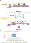
Figure 1
Figure 1
(A) Mechanism of bevacizumab action. The bevacizumab target is the VEGF. In the absence of the drug, the VEGF is bound to its specific receptors, initiating angiogenesis. When the bevacizumab is present, binding between VEGF and the receptor is inhibited as the bevacizumab binds to the VEGF. After the VEGF is attached to the drug, the VEGF cannot bind to the receptor. (B) Possible fractions of bevacizumab during the internalization studies. (a) The drug is not taken up by the cells and freely diffuses in the culture medium. (b) Bevacizumab is internalized into the cell and freely diffuses in the cytosol. (c) Bevacizumab is internalized into the cell but remains closed in the vesicles.
Figure 2
Figure 2
FLIM of HeLa and MDA-MB-231 cells incubated with bevacizumab for 1 h, 24 h, and 2 days. The red signal represents the tested drug, while the blue color corresponds to cell autofluorescence. In the case of HeLa cells after incubation with fluorescent bevacizumab for 24 h, the arrows indicate the elongated stained structures. Two independent experiments were conducted for each cell line within each incubation time.
Figure 3
Figure 3
Quantitative analysis of FLIM images. (A) FLIM image of HeLa cells incubated with bevacizumab for 1 h, with colors representing fluorescence lifetimes: blue for autofluorescence (fl < 2.4 ns) and red for bevacizumab (fl > 2.6 ns). (B) Red channel extracted from image A, with the linear ROI marked. (C) Blue channel extracted from image A, with the linear ROI marked. (D) Fluorescence intensity profiles of the ROIs. (E) FLIM image of HeLa cells incubated with bevacizumab for 24 h, in which the linear ROI was marked with coloring corresponding to the labels in panel F. (F) Fluorescence intensity profile of the red channel of image E. Regions of extracellular, cytosolic, and vesicle areas are marked. (G) Averaged values of fluorescence intensity determined from FLIM images. Four images were captured for each cell line and incubation time. Averaged values came from three measurements taken from each image. The number of analyzed data points was >300. For the analysis, we used ImageJ version 1.53a. Three asterisks denote a statistically significant difference (p < 0.0005).
Figure 4
Figure 4
Comparison of expectations with actual experiments. (A) Scheme showing an example of FCS curves obtained inside a cell filled with the free drug and vesicles. The shape of the FCS curves varies depending on the position of the confocal focus (location of free drug vs location of vesicle). (B) Scheme presenting quantitative and qualitative results of the bevacizumab internalization study. The lack of the drug within the cytosol shown in FLIM was confirmed by the FCS measurements, the lack of an autocorrelation function. The presence of stained (drug-filled) vesicles was quantitatively proven by obtaining the FCS curves presenting the movement of the drug closed in the vesicle and the movement of the vesicle. The data were fitted using eq S5.
Figure 5
Figure 5
Partition coefficients as a parameter defining the effectiveness of bevacizumab internalization. (A) Scheme explaining the way of partition coefficient calculation. Parallel, intracellular, and extracellular drug concentrations were calculated (2a and 2b) using SymphoTime images analysis (1a) and FCS measurements (1b), respectively. Then, the partition coefficient was expressed as a ratio of the drug concentration inside the cells to the extracellular concentration (3). (B) Box plots of obtained partition coefficients for bevacizumab (HeLa after incubation for 24 h and MDA-MB-231 after 24 and 48 h) and TRITC-dextran 155 kDa (HeLa after incubation for 24 h with 500 nM and 50 nM) as a control. The results are averaged from eight independent repetitions. No significant differences between results from different cell lines were detected (t test; p > 0.05).

References

    1. U.S. National Library of Medicine (accessed May 2022).
    1. Deissler H. L.; Lang G. K.; Lang G. E. Internalization of Bevacizumab by Retinal Endothelial Cells and Its Intracellular Fate: Evidence for an Involvement of the Neonatal Fc Receptor. Exp. Eye Res. 2016, 143, 49–59. 10.1016/j.exer.2015.10.007. - DOI - PubMed
    1. Borchers L.; Roider J.; Klettner A. Differences in Uptake and Intracellular Fate between Bevacizumab and Aflibercept after Repetitive Long-Term Treatment in the Retinal Pigment Epithelium. Ophthalmic Res. 2021, 64 (3), 369–388. 10.1159/000511960. - DOI - PubMed
    1. Müller-Greven G.; Carlin C. R.; Burgett M. E.; Ahluwalia M. S.; Lauko A.; Nowacki A. S.; Herting C. J.; Qadan M. A.; Bredel M.; Toms S. A.; Lathia J. D.; Hambardzumyan D.; Sarkaria J. N.; Hamerlik P.; Gladson C. L. Macropinocytosis of Bevacizumab by Glioblastoma Cells in the Perivascular Niche Affects Their Survival. Clin. Cancer Res. 2017, 23 (22), 7059–7071. 10.1158/1078-0432.CCR-17-0249. - DOI - PMC - PubMed
    1. Choi D.-K.; Bae J.; Shin S.-M.; Shin J.-Y.; Kim S.; Kim Y.-S. A General Strategy for Generating Intact, Full-Length IgG Antibodies That Penetrate into the Cytosol of Living Cells. MAbs 2014, 6 (6), 1402–1414. 10.4161/mabs.36389. - DOI - PMC - PubMed