. 2023 Jan 31;16(770):eabo4314.
doi: 10.1126/scisignal.abo4314. Epub 2023 Jan 31.

The dual-function chemokine receptor CCR2 drives migration and chemokine scavenging through distinct mechanisms

Affiliations

The dual-function chemokine receptor CCR2 drives migration and chemokine scavenging through distinct mechanisms

Thomas M Shroka et al. Sci Signal. .

Abstract

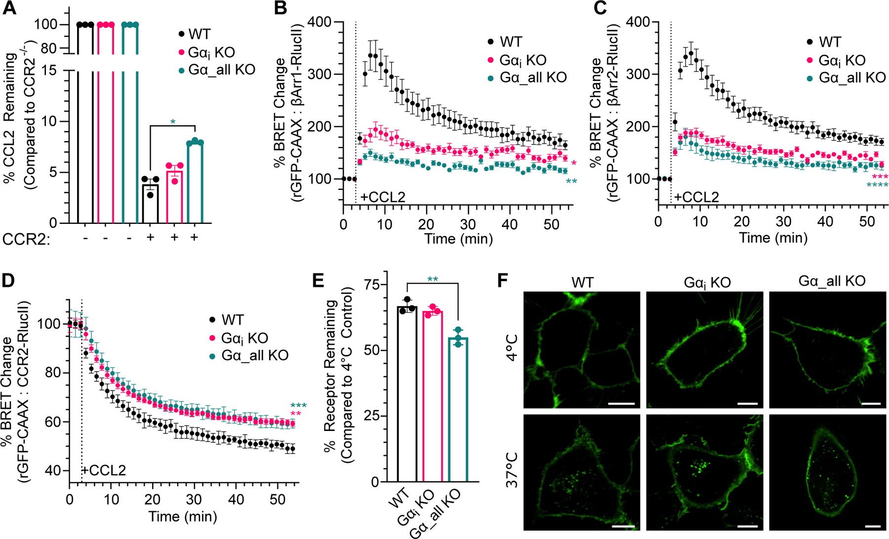
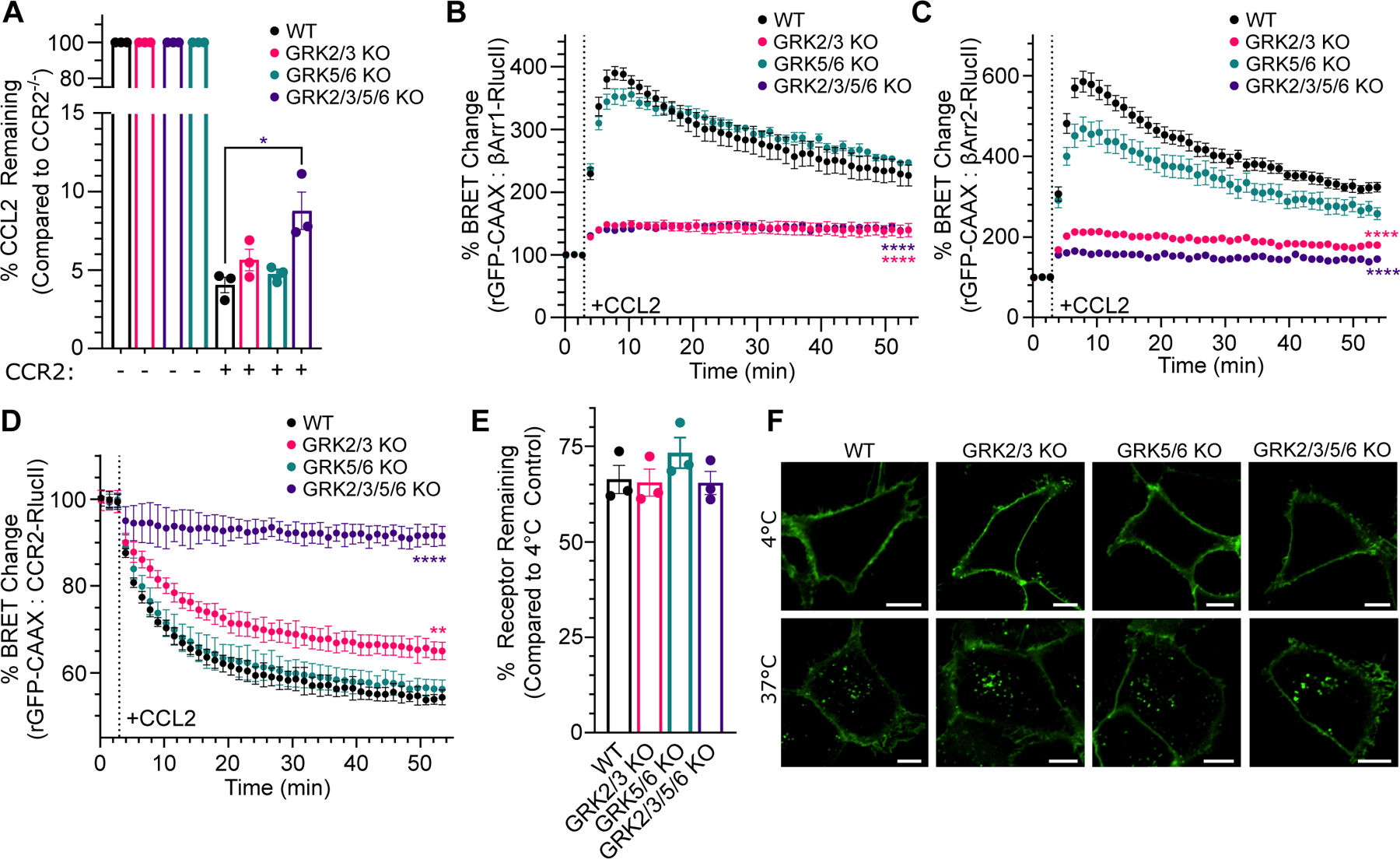
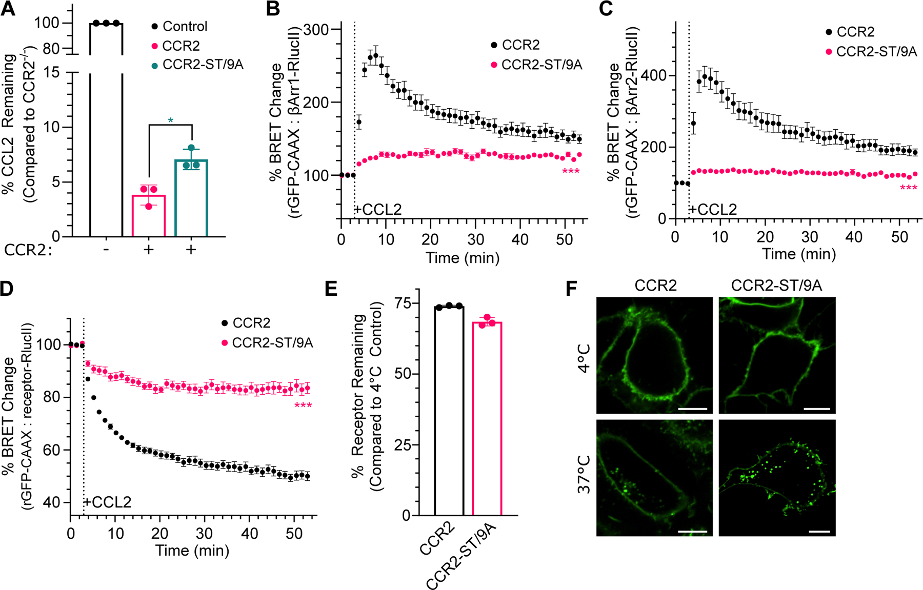
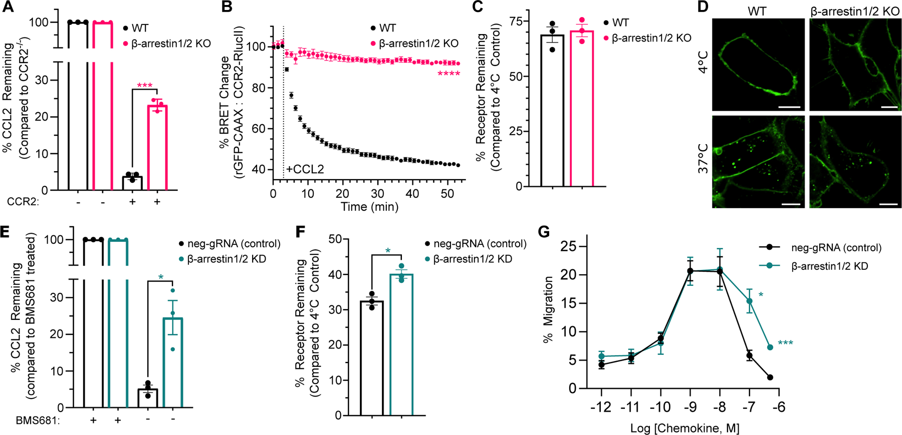
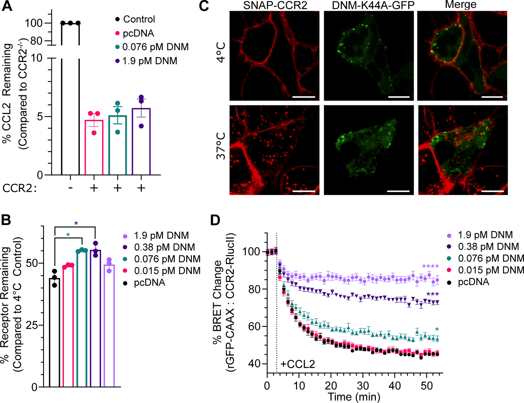
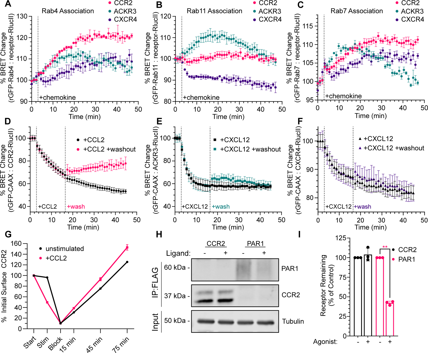
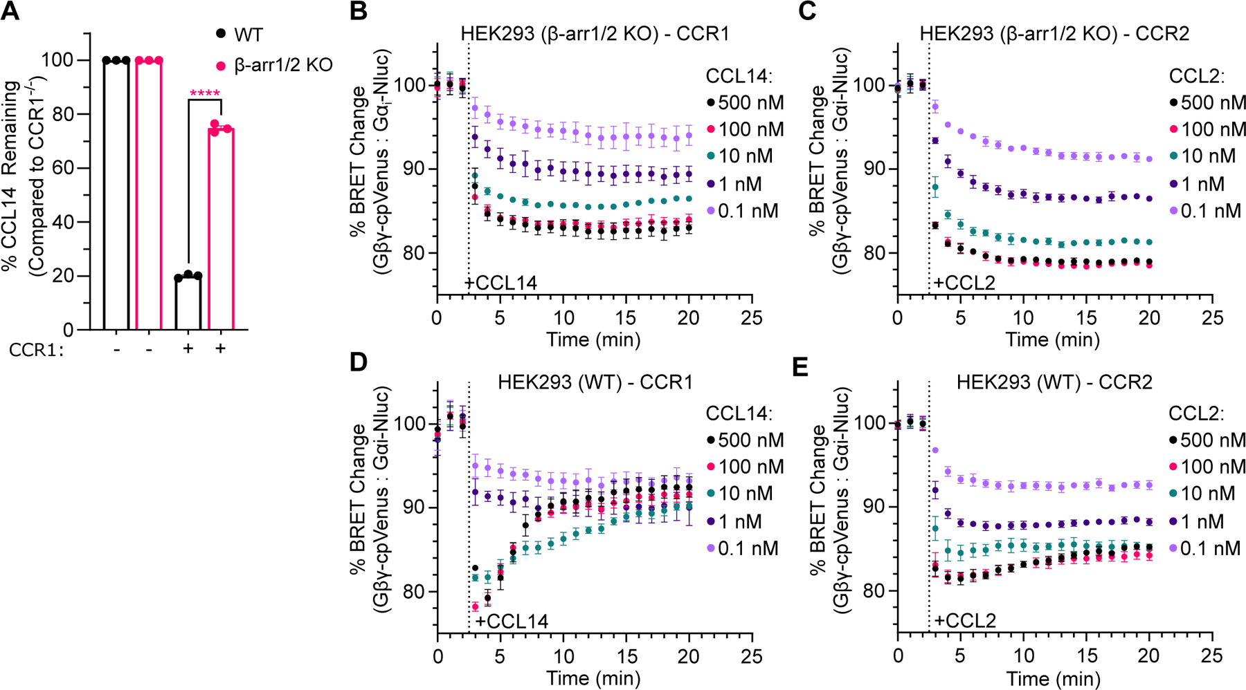
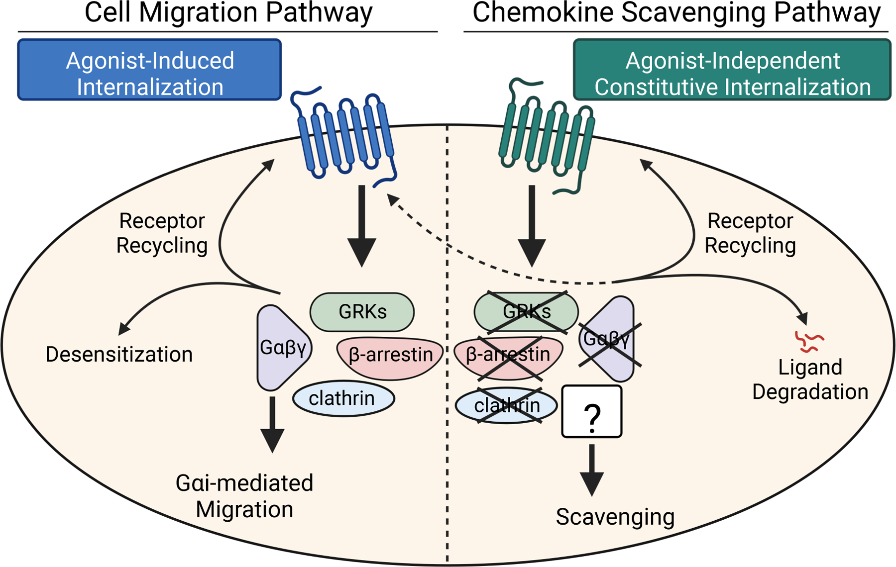
C-C chemokine receptor 2 (CCR2) is a dual-function receptor. Similar to other G protein-coupled chemokine receptors, it promotes monocyte infiltration into tissues in response to the chemokine CCL2, and, like atypical chemokine receptors (ACKRs), it scavenges chemokine from the extracellular environment. CCR2 therefore mediates CCL2-dependent signaling as a G protein-coupled receptor (GPCR) and also limits CCL2 signaling as a scavenger receptor. We investigated the mechanisms underlying CCR2 scavenging, including the involvement of intracellular proteins typically associated with GPCR signaling and internalization. Using CRISPR knockout cell lines, we showed that CCR2 scavenged by constitutively internalizing to remove CCL2 from the extracellular space and recycling back to the cell surface for further rounds of ligand sequestration. This process occurred independently of G proteins, GPCR kinases (GRKs), β-arrestins, and clathrin, which is distinct from other "professional" chemokine scavenger receptors that couple to GRKs, β-arrestins, or both. These findings set the stage for understanding the molecular regulators that determine CCR2 scavenging and may have implications for drug development targeting this therapeutically important receptor.


Conflict of interest statement

Competing interests: Dr. Handel is a co-founder of Lassogen Inc and serves on the Scientific Advisory Boards of Artica, Abilita Bio, and Abalone Bio. The terms of these arrangement have been reviewed and approved by the University of California, San Diego in accordance with its conflict of interest policies. The other authors declare that they have no competing interests.

Figures

Fig. 1.
Fig. 1.. Scavenging of CCR2 is G protein-independent.
(A) WT, Gαi KO and Gα_all KO HEK293 cells stably expressing CCR2 and respective non-expressing cells were cultured in media containing 5 nM CCL2 for 16 h. The remaining levels of CCL2 were measured by ELISA and interpolated from CCL2 standards, and are presented as percentages of remaining CCL2 relative to non-CCR2 expressing cells. (B and C) Cells transfected with β-arrestin1- or β-arrestin2-RlucII and rGFP-CAAX were stimulated with 100 nM CCL2 or left untreated. β-arrestin recruitment was assessed by ebBRET. Data are presented as percentages of BRET values for untreated controls. (D) CCR2 internalization assessed by BRET. Cells transfected with CCR2-RlucII and rGFP-CAAX were stimulated with 100 nM CCL2 or left untreated. The BRET ratio changes upon agonist treatment are expressed as percentages of the BRET ratio observed in untreated controls. (E) Constitutive internalization of WT, Gαi KO and Gα_all KO HEK293 cells stably expressing CCR2 were assessed by pre-label flow cytometry. Data are presented as the percentage of surface receptor remaining compared to non-internalized control. (F) Constitutive internalization was visualized by fluorescence confocal microscopy in WT, Gαi KO and Gα_all KO HEK293 cells expressing SNAP-CCR2 that was labeled with cell impermeable SNAP-Surface Alexa Fluor 488 at 4°C for 1 h. The cells were held at 4°C for 45 min (top panel) or transferred to 37°C for 45 min (bottom panel) before being imaged. Images are representative of three independent experiments. Scale bars, 10 µm. Data are expressed as the means ± SEM of n ≥ 3 independent experiments. *P < 0.05, **P < 0.01, ***P < 0.001, ****P < 0.0001 compared to controls by one-way analysis of variance (ANOVA) with Dunnet’s multiple comparison test.
Fig. 2.
Fig. 2.. CCR2 scavenging of CCL2 is not dependent on GRKs.
(A) WT HEK293A, HEK293A GRK2/3 KO, HEK293A GRK5/6 KO and HEK293A GRK2/3/5/6 KO cells stably expressing CCR2 and the corresponding CCR2 non-expressing cells were cultured in media containing 5 nM CCL2 for 16 h. The remaining levels of CCL2 were interpolated from CCL2 standards and are presented as percentages of remaining CCL2 relative to CCR2 non-expressing cells. (B and C) Cells transfected with β-arrestin1- or β-arrestin2-RlucII and rGFP-CAAX were stimulated with 100 nM CCL2 or left untreated. β-arrestin recruitment was assessed by ebBRET. Data are presented as percentages of BRET values for untreated controls. (D) CCR2 internalization assessed by BRET. Cells transfected with CCR2-RlucII and rGFP-CAAX were stimulated with 100 nM CCL2 or left untreated. The BRET ratio changes upon agonist treatment are expressed as percentages of the BRET ratio observed in the untreated controls. (E) Constitutive internalization of WT HEK293A, HEK293A GRK2/3 KO, HEK293A GRK5/6 KO and HEK293A GRK2/3/5/6 KO stably expressing CCR2 was assessed by pre-label flow cytometry. Data are presented as percentages of surface receptor remaining as compared to non-internalized control. (F) Constitutive internalization was visualized by fluorescence confocal microscopy in WT and corresponding GRK KO HEK293A cells expressing SNAP-CCR2 that was labeled with cell impermeable SNAP-Surface Alexa Fluor 488 at 4°C for 1 h. The cells were then held at 4°C for 45 min (top panel) or transferred to 37°C for 45 min (bottom panel) before being imaged. Images are representative of three independent experiments. Scale bars, 10 µm. Data are expressed as the means ± SEM of n ≥ 3 independent experiments. *P < 0.05, **P < 0.01, ***P < 0.001, ****P < 0.0001 compared to controls by one-way analysis of variance (ANOVA) with Dunnet’s multiple comparison test.
Fig. 3.
Fig. 3.. CCR2 scavenging of CCL2 occurs independently of receptor phosphorylation.
(A) HEK293 cells stably expressing CCR2 or CCR2 with nine C-terminal serine/threonine residues mutated to alanine (CCR2-ST/9A) and non-expressing cells were cultured in media containing 5 nM CCL2 for 16 h. The remaining levels of CCL2 were interpolated from CCL2 standards and are presented as percentages of the respective non-CCR2 expressing cells. (B and C) Cells expressing CCR2 or CCR2-ST9/A transfected with β-arrestin1- or β-arrestin2-RlucII and rGFP-CAAX were stimulated with 100 nM CCL2 or left untreated. β-arrestin recruitment was assessed by ebBRET. (D) Cells transfected with CCR2-RlucII or CCR2-ST/9A-RlucII and rGFP-CAAX were stimulated with 100 nM CCL2 or left untreated. CCR2 internalization was assessed by BRET. The BRET ratio changes upon agonist treatment are expressed as percentages of the BRET ratio for untreated controls. (E) Constitutive internalization of WT CCR2 and CCR2-ST/9A in HEK293 cells was assessed by pre-label flow cytometry. (F) Constitutive internalization was visualized by fluorescence confocal microscopy in HEK293 cells expressing SNAP-CCR2 or SNAP-CCR2-ST/9A labeled with cell impermeable SNAP-Surface Alexa Fluor 488 at 4°C for 1 h. Cells were then either held at 4°C for 45 min (top panel) or transferred to 37°C for 45 min (bottom panel) before being imaged. Images are representative of three independent experiments. Scale bars, 10 µm. Data are expressed as the means ± SEM of n ≥ 3 independent experiments. *P < 0.05, **P < 0.01, ***P < 0.001, ****P < 0.0001 compared to controls by unpaired t test.
Fig. 4.
Fig. 4.. β-arrestins are dispensable for constitutive internalization and chemokine scavenging by CCR2.
(A) WT HEK293 and β-arrestin1/2 KO HEK293 cells stably expressing CCR2 and respective non-expressing cells were cultured in media containing 5 nM CCL2 for 16 h. The remaining levels of CCL2 were interpolated from CCL2 standards and are presented as percentages of respective non-CCR2 expressing cells. (B) Cells transfected with CCR2-RlucII and rGFP-CAAX were stimulated with 100 nM CCL2 or left untreated. CCR2 internalization was assessed by BRET. The BRET ratio changes upon agonist treatment are expressed as percentages of the BRET ratio observed in untreated controls. (C) Constitutive internalization of WT HEK293 and β-arrestin1/2 KO HEK293 cells stably expressing CCR2 was assessed by pre-label flow cytometry. Data are presented as percentages of surface receptor remaining as compared to non-internalized control. (D) Constitutive internalization was visualized by fluorescence confocal microscopy in WT and β-arrestin1/2 KO HEK293 cells expressing SNAP-CCR2 that was labeled with cell impermeable SNAP-Surface Alexa Fluor 488 at 4°C for 1 h. The cells were then held at 4°C for 45 min (top panel) or transferred to 37°C for 45 min (bottom panel) before being imaged. Images are representative of three independent experiments. Scale bars, 10 µm. (E) THP-1 cells transduced with non-targeting gRNA (neg-gRNA) and THP-1 β-arrestin1/2 KD cells were treated with vehicle or the CCR2 inhibitor BMS681 and cultured in media containing 5 nM CCL2 for 16 h. The remaining levels of CCL2 were interpolated from CCL2 standards and are presented as percentages of non-scavenging control BMS681 treated cells. (F) Constitutive internalization of CCR2 in THP-1 neg-gRNA and β-arrestin1/2 KD THP-1 cells was assessed by pre-label flow cytometry. Data are presented as percentages of surface receptor remaining as compared to non-internalized control. (G) Transwell migration of THP-1 neg-gRNA and β-arrestin1/2 KD THP-1 cells at various concentrations of chemokine. Data are presented as percentages of migrated cells as compared to initial number of cells added to each well. Data are expressed as the means ± SEM of n ≥ 3 independent experiments. *P < 0.05, **P < 0.01, ***P < 0.001, ****P < 0.0001 compared to controls by unpaired t test.
Fig. 5.
Fig. 5.. Clathrin-mediated endocytosis is not required for CCR2 constitutive internalization or scavenging.
(A) Non-CCR2 expressing cells and HEK293 cells stably expressing CCR2 were transfected with two different concentrations of a dynamin dominant-negative mutant (DNM2-K44A) or left untransfected. The cells were cultured in media containing 5 nM CCL2 for 16 h. The remaining levels of CCL2 were determined by ELISA and interpolated from CCL2 standards and are presented as percentages of the respective non-CCR2 expressing cells. (B) Constitutive internalization of CCR2 was assessed by pre-label flow cytometry. HEK293 cells expressing CCR2 were transfected with pcDNA or an increasing amount of DNM2-K44A. Data are presented as percentages of surface receptor remaining as compared to non-internalized control. (C) Constitutive internalization in the presence or absence of DNM2-K44A was visualized by fluorescence confocal microscopy in HEK293 cells expressing SNAP-CCR2 and DNM2-K44A-GFP. SNAP-CCR2 was labeled with cell impermeable SNAP-Surface Alexa Fluor 649 at 4°C for 1 h and then either held at 4°C for 45 min (top panel) or transferred to 37°C for 45 min (bottom panel) before being analyzed. Scale bars, 10 µm. (D) Cells transfected with CCR2-RlucII, rGFP-CAAX, and pcDNA or increasing amounts of DNM2-K44A were incubated in the absence or presence of 100 nM CCL2. CCR2 internalization was assessed by BRET. The BRET ratio changes on agonist treatment are expressed as percentages of the BRET ratio observed in the untreated controls. Data are expressed as the means ± SEM of n ≥ 3 independent experiments. *P < 0.05, **P < 0.01, ***P < 0.001, ****P < 0.0001 compared to controls by one-way analysis of variance (ANOVA) with Dunnet’s multiple comparison test.
Fig. 6.
Fig. 6.. CCR2 endosomal trafficking, recycling and lack of degradation contribute to scavenging.
(A to C) HEK293 cells were transfected with receptor-RlucII (CCR2, ACKR3 or CXCR4) along with either rGFP-Rab4 (A), rGFP-Rab11 (B) or rGFP-Rab7 (C). Cells were stimulated at 37°C with indicated chemokine or left untreated. Data are presented as percentages of BRET values compared to untreated controls. (D to F) HEK293 cells were transfected with rGFP-CAAX and either CCR2-RlucII (D), ACKR3-RlucII (E) or CXCR4-RlucII (F). Cells were stimulated at 37°C with indicated chemokine and washed with PBS to remove chemokine or left unwashed. Data are presented as percentages of BRET values compared to non-chemokine treated controls. (G) HEK293 cells stably expressing SNAP-CCR2 were preincubated for 90 min with 10 mg/mL cycloheximide to block de novo protein synthesis and left untreated or stimulated with 100 nM CCL2 at 37°C for 45 min. The remaining SNAP-CCR2 at the surface was blocked at 4°C with SNAP-Surface block for 45 min. Cells were moved to 37 °C for 15, 45 or 75 min. Receptors were labeled with SNAP-Surface Alexa Fluor 649 at 4°C following each experimental condition and timepoint. Data are displayed as percentages of CCR2 detected relative to initial levels of surface CCR2 prior to stimulation (Start). (H, I) HEK293 cells expressing FLAG-CCR2 or FLAG-PAR1 were pretreated with 10 µg/mL cycloheximide for 90 min and left unstimulated (0 min) or stimulated with 100 nM CCL2 (to activate CCR2) or 100 μM TFLLRN (to activate PAR1) for 2 h at 37°C. Equivalent amount of cell lysates were immunoprecipitated with M2 anti-FLAG antibody and immunoblotting detection with anti-FLAG antibody. Cell lysates were analyzed for endogenous α-tubulin as controls (H). Receptor degradation was quantified and data (mean ± SEM) shown are expressed as the fraction of receptor remaining compared with untreated control cells as determined from three independent experiments (I). Data are expressed as the means ± SEM of n ≥ 3 independent experiments. *P < 0.05, **P < 0.01, ***P < 0.001, ****P < 0.0001 compared to controls by one-way analysis of variance (ANOVA) with Dunnet’s multiple comparison test or unpaired t test.
Fig. 7.
Fig. 7.. CCR1 and CCR2 have distinct mechanisms of chemokine scavenging.
(A) WT HEK293 and β-arrestin1/2 KO HEK293 cells stably expressing CCR1 and the respective non-expressing cells were cultured in media containing 5 nM CCL14 for 16 h. The remaining CCL14 was quantified by ELISA and interpolated from CCL14 standards and are presented as percentages of the respective non-CCR1 expressing cells. (B to E) HEK293 β-arrestin1/2 KO cells (B and C) or HEK293 WT cells (D and E) expressing either CCR1 or CCR2 and transfected with an IRES vector encoding Gαi-Nluc and Gβγ-cpVenus were stimulated with 0.1–500 nM CCL14 or CCL2, respectively (indicated by the dotted lines). The dissociation of the Gβγ- from the Gα-subunit results in a decrease in BRET ratio upon agonist treatment and is expressed as percentages of the BRET ratio to that observed in the untreated control cells. Data are expressed as the means ± SEM of n ≥ 3 independent experiments. *P < 0.05, **P < 0.01, ***P < 0.001, ****P < 0.0001 compared to controls by one-way analysis of variance (ANOVA) with Dunnet’s multiple comparison test or unpaired t test.
Fig. 8.
Fig. 8.. CCR2 appears to have two distinct functional populations, one that regulates migration and one involved in scavenging.
The canonical GPCR interactors, including Gαβγ, GRKs, β-arrestins, and clathrin are primarily involved in the CCL2-induced internalization mechanisms and are part of the migration-promoting population (left). These same interactors are dispensable for the scavenging population of CCR2 (right), which instead can constitutively internalize while passively sequestering extracellular CCL2. The exact mechanisms and regulatory proteins involved in the scavenging pathway are yet to be determined. A key aspect of both populations is the ability of CCR2 to recycle to the cell surface to continue to drive G protein-dependent migratory functions as well as continue to scavenge excess chemokine. Additionally, the constitutively internalized receptor may provide non-desensitized receptors (dotted arrow) for the G protein-coupled pathway to enable sustained cell migration.

Similar articles

Cited by

References

    1. Ulvmar MH, Hub E, Rot A, Atypical chemokine receptors. Exp Cell Res 317, 556–568 (2011). - PMC - PubMed
    1. Nibbs RJB, Graham GJ, Immune regulation by atypical chemokine receptors. Nat Rev Immunol 13, 815–829 (2013). - PubMed
    1. Abe P, Mueller W, Schütz D, MacKay F, Thelen M, Zhang P, Stumm R, CXCR7 prevents excessive CXCL12-mediated downregulation of CXCR4 in migrating cortical interneurons. Development 141, 1857–1863 (2014). - PubMed
    1. Bonecchi R, Graham GJ, Atypical Chemokine Receptors and Their Roles in the Resolution of the Inflammatory Response. Front Immunol 7 (2016), doi:10.3389/fimmu.2016.00224. - DOI - PMC - PubMed
    1. C. A. H. Hansell, C. E. Hurson, R. J. B. Nibbs, DARC and D6: silent partners in chemokine regulation? Immunol Cell Biol 89, 197–206 (2011). - PMC - PubMed

Publication types