. 2023 Jun 1;58(6):413-419.
doi: 10.1097/RLI.0000000000000952. Epub 2023 Jan 21.

Single Breath-Hold 3-Dimensional Magnetic Resonance Elastography Depicts Liver Fibrosis and Inflammation in Obese Patients

Affiliations

Single Breath-Hold 3-Dimensional Magnetic Resonance Elastography Depicts Liver Fibrosis and Inflammation in Obese Patients

Omar Isam Darwish et al. Invest Radiol. .

Abstract

Objectives: Three-dimensional (3D) magnetic resonance elastography (MRE) measures liver fibrosis and inflammation but requires several breath-holds that hamper clinical acceptance. The aim of this study was to evaluate the technical and clinical feasibility of a single breath-hold 3D MRE sequence as a means of measuring liver fibrosis and inflammation in obese patients.

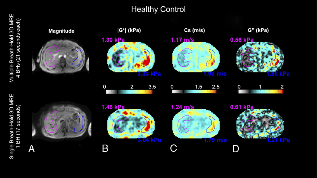
Methods: From November 2020 to December 2021, subjects were prospectively enrolled and divided into 2 groups. Group 1 included healthy volunteers (n = 10) who served as controls to compare the single breath-hold 3D MRE sequence with a multiple-breath-hold 3D MRE sequence. Group 2 included liver patients (n = 10) who served as participants to evaluate the clinical feasibility of the single breath-hold 3D MRE sequence in measuring liver fibrosis and inflammation. Controls and participants were scanned at 60 Hz mechanical excitation with the single breath-hold 3D MRE sequence to retrieve the magnitude of the complex-valued shear modulus (|G*| [kPa]), the shear wave speed (Cs [m/s]), and the loss modulus (G" [kPa]). The controls were also scanned with a multiple-breath-hold 3D MRE sequence for comparison, and the participants had histopathology (Ishak scores) for correlation with Cs and G".

Results: For the 10 controls, 5 were female, and the mean age and body mass index were 33.1 ± 9.5 years and 23.0 ± 2.1 kg/m 2 , respectively. For the 10 participants, 8 were female, and the mean age and body mass index were 45.1 ± 16.5 years and 33.1 ± 4.0 kg/m 2 (obese range), respectively. All participants were suspected of having nonalcoholic fatty liver disease. Bland-Altman analysis of the comparison in controls shows there are nonsignificant differences in |G*|, Cs, and G" below 6.5%, suggesting good consensus between the 2 sequences. For the participants, Cs and G" correlated significantly with Ishak fibrosis and inflammation grades, respectively ( ρ = 0.95, P < 0.001, and ρ = 0.84, P = 0.002).

Conclusion: The single breath-hold 3D MRE sequence may be effective in measuring liver fibrosis and inflammation in obese patients.


Conflict of interest statement

Conflicts of interest and sources of funding: Omar Darwish is a PhD candidate, and their university tuitions fees are covered by Siemens Healthineers. Dr Radhouene Neji is an employee of Siemens Healthineers United Kingdom, Dr Daniel Stäb is an employee of Siemens Healthcare Australia, and Dr Peter Speier is an employee of Siemens Healthcare Germany. For the remaining authors, none were declared. Sources of funding: CRUK City of London Centre Award (C7893/A26233), CRUK (EDDCPGM/100001), Siemens Healthineers, ITMO Aviesan 2020 (DESP/PB n°241), National Institutes of Health intramural fund, and by US Agency for International Development (USAID) and National Academy of Sciences through Subaward 2000012771 (any opinions, findings, conclusions, or recommendations expressed are those of the authors alone, and do not necessarily reflect the views of USAID or National Academy of Sciences).

Figures

FIGURE 1.
FIGURE 1.
Comparison of the different MRE acquisitions with respect to their timing: a previously published multiple–breath-hold 3D MRE sequence eXpresso (A), a previously published multiple–breath-hold 3D MRE sequence Ristretto (B), and the proposed single breath-hold 3D MRE sequence Intenso (C), where each square depicts a fractional GRE-MRE imaging shot acquiring 1 (single-band excitation) or 2 (SMS excitation) slices, respectively. The red blocks indicate time delays to shift from 1 wave offset, φi, to another wave offset, φk. In case of eXpresso, successive wave offsets are acquired, that is, k=i+1. For Ristretto, the shift is determined via Nd, that is, k=i+Nd. The proposed single breath-hold 3D MRE sequence Intenso is using optimal conditions, that is, Nd=1 and Nw=2. (D) Overview of the in-plane GRAPPA SMS formulation. Multiband radiofrequency (RF) excitation (1), with Nsms oversampling (2), standard in-plane acceleration scheme (3), and FOV-splitting for slice separation (4). In our implementation, R=5, Nsms=2, and Ri=2.5. (E) The proposed single breath-hold 3D MRE sequence Intenso consists of a spoiled GRE sequence (black) with dual-band RF pulses for SMS excitation (blue) and a Hadamard scheme for motion encoding (red).
FIGURE 2.
FIGURE 2.
Bland-Altman plots for the comparison of the proposed single breath-hold 3D MRE sequence (Intenso) with the multiple–breath-hold 3D MRE sequence (eXpresso) for the derived viscoelastic parameters (A–C), and repeatability analysis of the single breath-hold 3D MRE sequence (Intenso) (D–F).
FIGURE 3.
FIGURE 3.
MRE for a representative control using a multiple–breath-hold 3D MRE sequence (top row) and the single breath-hold 3D MRE sequence (bottom row). The outlines of the liver and spleen are highlighted in purple and blue, respectively. (A) Magnitude images, (B) the magnitude of the complex shear modulus |G*| (kPa), (C) the shear wave speed Cs (m/s), and (D) the loss modulus G″ (kPa). All maps show average values over the 4 innermost slices. The magnitude image of the multiple–breath-hold 3D MRE sequence (top row) is the magnitude of the fourth acquisition step where no motion encoding gradients are applied. On the other hand, the magnitude image of the single breath-hold 3D MRE sequence (bottom row) is the average of the magnitude images of the 4 motion encoding steps as the Hadamard motion encoding scheme applies motion encoding gradients during all 4 acquisition steps. However, visual inspection of the magnitude images shows very similar anatomical features in both sequences. Mean values of |G*|, Cs, and G″ in the liver were 1.30 ± 0.35 kPa, 1.17 ± 0.15 m/s, and 0.56 ± 0.24 kPa, respectively, for the multiple–breath-hold 3D MRE sequence and 1.46 ± 0.52 kPa, 1.24 ± 0.2 m/s, and 0.61 ± 0.35 kPa, respectively, for the single breath-hold 3D MRE sequence, reflecting a good agreement between the 2 sequences.
FIGURE 4.
FIGURE 4.
Dependence of the viscoelastic parameters measured with the single breath-hold 3D MRE sequence (Intenso) on the Ishak scores of the participants. (A) Dependence of the shear wave speed Cs (m/s) on Ishak liver fibrosis score. (B) Dependence of the loss modulus G″ (kPa) on Ishak liver inflammation grade.
FIGURE 5.
FIGURE 5.
Single breath-hold 3D MRE (Intenso) for a patient with advanced fibrosis and intermediate inflammation: (A) magnitude image, (B) wave image (curl), demonstrating efficient wave penetration within the liver, (C, D, E) reconstructed images of |G*|, Cs, and G″. All viscoelastic maps are averaged in the 4 innermost slices and the reported mean values are measured in the magenta-colored ROI to avoid large vessels and liver edges.

References

    1. Singh S, Venkatesh SK, Wang Z, et al. Diagnostic performance of magnetic resonance elastography in staging liver fibrosis: a systematic review and meta-analysis of individual participant data. Clin Gastroenterol Hepatol. 2015;13:440–51.e6. - PMC - PubMed
    1. Sinkus R, Lambert S, Abd-Elmoniem KZ, et al. Rheological determinants for simultaneous staging of hepatic fibrosis and inflammation in patients with chronic liver disease. NMR Biomed. 2018;31:e3956. - PMC - PubMed
    1. Shi Y, Qi YF, Lan GY, et al. Three-dimensional MR elastography depicts liver inflammation, fibrosis, and portal hypertension in chronic hepatitis B or C. Radiology. 2021;301:154–162. - PMC - PubMed
    1. Allen AM, Shah VH, Therneau TM, et al. The role of three-dimensional magnetic resonance elastography in the diagnosis of nonalcoholic steatohepatitis in obese patients undergoing bariatric surgery. Hepatology. 2020;71:510–521. - PMC - PubMed
    1. Garteiser P, Sahebjavaher RS, Ter Beek LC, et al. Rapid acquisition of multifrequency, multislice and multidirectional MR elastography data with a fractionally encoded gradient echo sequence. NMR Biomed. 2013;26:1326–1335. - PubMed

Publication types