

doi: 10.1055/a-1996-0604.
Epub 2023 Jan 31.
Introducer-assisted endoscopic transpapillary gallbladder biopsy for indeterminate gallbladder fundal wall thickness
Affiliations
- PMID: 36720267
- PMCID: PMC9889165
- DOI: 10.1055/a-1996-0604
Item in Clipboard
Introducer-assisted endoscopic transpapillary gallbladder biopsy for indeterminate gallbladder fundal wall thickness
Endoscopy.
2023 Dec.
No abstract available
Conflict of interest statement
The authors declare that they have no conflict of interest.
Figures
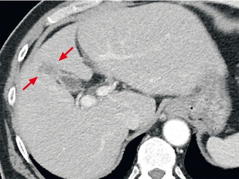
Contrast-enhanced abdominal computed tomography performed at admission revealed contrast-enhanced gallbladder fundal wall thickness with an obscure demarcation line of the liver parenchyma and gallbladder fossa (red arrows).

Contrast-enhanced abdominal ultrasonography.
a
Contrast-enhanced gallbladder fundal wall thickness with liver parenchyma in the early phase (yellow arrows). The image on the left is the reference image.
b
Direct liver invasion was suggested by the contrast-attenuated liver parenchyma on the gallbladder fossa in the delayed phase (red arrows). The image on the left is the reference image.

Endoscopic transpapillary gallbladder biopsy using biopsy forceps under fluoroscopic guidance. Biopsy forceps were passed through the outer sheath of the endoscopic introducer to the gallbladder fundus.

Microscopic findings of a specimen obtained from the gallbladder fundus. Severe atypical columnar cells with loss of polarity, nuclear enlargement, and stratification indicated a tubulo-papillary growth pattern. Histologically, the diagnosis was adenocarcinoma. Hematoxylin and eosin staining.
References
-
- Nagino M, Hirano S, Yoshitomi H et al. Clinical practice guidelines for the management of biliary tract cancers 2019: The 3rd English edition. J Hepatobiliary Pancreat Sci. 2021;28:26–54. - PubMed
-
- Pouw R E, Barret M, Biermann K et al. Endoscopic tissue sampling – Part 1: Upper gastrointestinal and hepatopancreatobiliary tracts. European Society of Gastrointestinal Endoscopy (ESGE) Guideline. Endoscopy. 2021;53:1174–1188. - PubMed
-
- Kito Y, Kato A, Yoshida M et al. Facile and secure deployment of plastic stent through an endoscopic tapered sheath for endoscopic ultrasound-guided drainage. Endoscopy. 2022;54:E674–E675. - PubMed
-
- Kamada H, Kobara H, Yamana H et al. Endoscopic direct visualization of gallbladder polypoid lesion using peroral digital single-operator cholangioscopy. Endoscopy. 2021;53:E263–E264. - PubMed
MeSH terms
LinkOut - more resources
Full Text Sources