Sex-based differences of antioxidant enzyme nanoparticle effects following traumatic brain injury
- PMID: 36720285
- PMCID: PMC10006352
- DOI: 10.1016/j.jconrel.2023.01.065
Sex-based differences of antioxidant enzyme nanoparticle effects following traumatic brain injury
Abstract
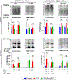
Following traumatic brain injury (TBI), reactive oxygen species (ROS) are released in excess, causing oxidative stress, carbonyl stress, and cell death, which induce the additional release of ROS. The limited accumulation and retention of small molecule antioxidants commonly used in clinical trials likely limit the target engagement and therapeutic effect in reducing secondary injury. Small molecule drugs also need to be administered every several hours to maintain bioavailability in the brain. Therefore, there is a need for a burst and sustained release system with high accumulation and retention in the injured brain. Here, we utilized Pro-NP™ with a size of 200 nm, which was designed to have a burst and sustained release of encapsulated antioxidants, Cu/Zn superoxide dismutase (SOD1) and catalase (CAT), to scavenge ROS for >24 h post-injection. Here, we utilized a controlled cortical impact (CCI) mouse model of TBI and found the accumulation of Pro-NP™ in the brain lesion was highest when injected immediately after injury, with a reduction in the accumulation with delayed administration of 1 h or more post-injury. Pro-NP™ treatment with 9000 U/kg SOD1 and 9800 U/kg CAT gave the highest reduction in ROS in both male and female mice. We found that Pro-NP™ treatment was effective in reducing carbonyl stress and necrosis at 1 d post-injury in the contralateral hemisphere in male mice, which showed a similar trend to untreated female mice. Although we found that male and female mice similarly benefit from Pro-NP™ treatment in reducing ROS levels 4 h post-injury, Pro-NP™ treatment did not significantly affect markers of post-traumatic oxidative stress in female CCI mice as compared to male CCI mice. These findings of protection by Pro-NP™ in male mice did not extend to 7 d post-injury, which suggests subsequent treatments with Pro-NP™ may be needed to afford protection into the chronic phase of injury. Overall, these different treatment effects of Pro-NP™ between male and female mice suggest important sex-based differences in response to antioxidant nanoparticle delivery and that there may exist a maximal benefit from local antioxidant activity in injured brain.
Keywords: Carbonyl stress; Lipid peroxidation products; PLGA nanoparticles; Reactive oxygen species; Traumatic brain injury; Treatment window.
Copyright © 2023 Elsevier B.V. All rights reserved.
Conflict of interest statement
Declaration of Competing Interest The study was in collaboration with ProTransit Nanotherapy, LLC, Omaha, NE, a start-up company based on patented work done at the University of Nebraska Medical Center. G.M. is founder of ProTransit Nanotherapy and has an equity interest in the company. ProTransit can make Pro-NP™ available to potential investigators under material transfer agreement.
Figures


References
-
- Aleman M, Prange T, Neurocranium and Brain, in: Equine Surgery, 2019, pp. 895–900.
-
- Leconte C, Benedetto C, Lentini F, Simon K, Ouaazizi C, Taib T, Cho AH, Plotkine M, Mongeau R, Marchand-Leroux C, Besson VC, Histological and behavioral evaluation after traumatic brain injury in mice: a ten months follow-up study, J Neurotrauma, (2019). - PubMed
-
- Mao X, Terpolilli NA, Wehn A, Chen S, Hellal F, Liu B, Seker B, Plesnila N, Progressive histopathological damage occurring up to one year after experimental traumatic brain injury is associated with cognitive decline and depression-like behavior, J Neurotrauma, (2019). - PubMed
-
- Janatpour ZC, Korotcov A, Bosomtwi A, Dardzinski BJ, Symes AJ, Subcutaneous Administration of Angiotensin-(1–7) Improves Recovery after Traumatic Brain Injury in Mice, Journal of Neurotrauma, 36 (2019) 3115–3131. - PubMed
Publication types
MeSH terms
Substances
Grants and funding
LinkOut - more resources
Full Text Sources
Other Literature Sources
Medical
Miscellaneous

