. 2023 Jul;152(1):167-181.e6.
doi: 10.1016/j.jaci.2023.01.016. Epub 2023 Jan 28.

Lung-resident CD69+ST2+ TH2 cells mediate long-term type 2 memory to inhaled antigen in mice

Affiliations

Lung-resident CD69+ST2+ TH2 cells mediate long-term type 2 memory to inhaled antigen in mice

Takao Kobayashi et al. J Allergy Clin Immunol. 2023 Jul.

Abstract

Background: Chronic airway diseases such as asthma are characterized by persistent type 2 immunity in the airways. We know little about the mechanisms that explain why type 2 inflammation continues in these diseases.

Objective: We used mouse models to investigate the mechanisms involved in long-lasting immune memory.

Methods: Naive mice were exposed intranasally to ovalbumin (OVA) antigen with Alternaria extract as an adjuvant. Type 2 memory was analyzed by parabiosis model, flow cytometry with in vivo antibody labeling, and intranasal OVA recall challenge. Gene-deficient mice were used to analyze the mechanisms.

Results: In the parabiosis model, mice previously exposed intranasally to OVA with Alternaria showed more robust antigen-specific immune responses and airway inflammation than mice with circulating OVA-specific T cells. After a single airway exposure to OVA with Alternaria, CD69+ST2+ TH2-type T cells, which highly express type 2 cytokine messenger RNA and lack CD62L expression, appeared in lung tissue within 5 days and persisted for at least 84 days. When exposed again to OVA in vivo, these cells produced type 2 cytokines quickly without involving circulating T cells. Development of tissue-resident CD69+ST2+ TH2 cells and long-term memory to an inhaled antigen were abrogated in mice deficient in ST2 or IL-33, but not TSLP receptor.

Conclusion: CD69+ST2+ TH2 memory cells develop quickly in lung tissue after initial allergen exposure and persist for a prolonged period. The ST2/IL-33 pathway may play a role in the development of immune memory in lung to certain allergens.

Keywords: IL-33; IL-5; T(H)2 cells; allergens; allergy; tissue-resident memory T cells.


Conflict of interest statement

All authors acknowledge no conflict of interest related to this manuscript.

Figures
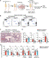
Figure 1.
Figure 1.
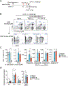
Tissue lymphocytes are necessary to produce strong memory responses to inhaled antigens. (A) Schematic overview of the experimental protocol. (B) Representative scattergrams of CD45.1 and CD45.2 expression on the CD3+CD4+ lymphocyte population from peripheral blood analysis by FACS, 21 days after parabiosis surgery. (C) Representative photomicrographs of lungs collected and examined by H&E stain 24 hours after the last i.n. OVA challenge. (D) Total number of cells in BAL fluids and (E) lung levels of cytokines and chemokines. Data are pooled from three experiments and presented as mean ± SEM (n=6 pairs). Each dot represents one mouse, and the paired mice are connected by lines. *P<0.05 between the groups indicated.
Figure 2.
Figure 2.
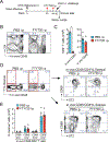
Antigen-specific Th2 cells accumulate in the lung tissues within 5 days of allergen exposure in naïve mice. (A) Schematic overview of the experimental protocol. (B-E) Lungs were collected on days 0, 3, or 5 immediately after i.v. injection with anti-CD45 Ab, and single-cell suspensions were analyzed by FACS. (B) Representative scattergrams showing CD4 expression and in-vivo CD45 labeling of lymphocytes. (C) Numbers of each cell population. Data are presented as mean ± SEM (n=4) and are representative of two experiments. (D) Representative scattergrams showing CD69 and ST2 expression on the in-vivo CD45-positive (intravenous) and -negative (tissue) CD4+ lymphocyte populations. (E) Numbers of each cell population. Data are presented as mean ± SEM (n=4) and are representative of two experiments. (F) Schematic overview of the experimental protocol for Panel G. (G) Lung levels of cytokines. *P<0.05 and **P<0.01 between the groups indicated. Data are presented as mean ± SEM (n=4–5) and are representative of two experiments. Alt, Alternaria.
Figure 3.
Figure 3.
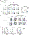
Th2-type memory cells persist in the lung tissues for more than 6 weeks after the initial inhaled allergen exposure. (A) Schematic overview of the experimental protocol. (B–D) Six hours after i.n. OVA challenge, lungs were collected and analyzed by FACS and ELISA. (B) Representative scattergrams showing expression of CD4 and in-vivo CD45 labeling (upper panels), and expression of CD69 and ST2 on the in-vivo CD45-negative (tissue) CD4+ cell population (lower panels). (C) Numbers of each cell population. (D) Lung levels of cytokines and CCL17. Data are presented as mean ± SEM (n=4–5 in each group). Each dot represents one mouse. *P<0.05 and **P<0.01 between the groups indicated.
Figure 4.
Figure 4.
ST2+CD69+CD4+ memory T cells in the lung tissues quickly produced IL-5 in response to OVA challenge in vivo. (A) Schematic overview of the experimental protocol. Six hours after i.n. OVA challenge, lungs were collected and analyzed by FACS. (B) Representative scattergrams showing expression of CD4 and in-vivo CD45 labeling. (C) Numbers of in-vivo CD45-positive (intravenous) and -negative (tissue) CD4+ T cells. (D) Representative scattergrams showing expression of IL-5-venus and in-vivo CD45 labeling. The IL-5-venus-positive and -negative cell populations were further analyzed for their expression of ST2 and CD69. (E) Numbers of each cell population. Data are presented as mean ± SEM (n=4 in each group). Each dot represents one mouse. *P<0.05 between the groups indicated.
Figure 5.
Figure 5.
The IL-33/ST2 pathway is indispensable for memory type 2 responses in the lungs. (A) Schematic overview of the experimental protocol for Panel B. (B) Total number of cells in BAL fluids, and lung levels of cytokines. Data are presented as mean ± SEM (n=4–5 in each group) and are representative of two experiments. (C) Schematic overview of the experimental protocols for Panels C and D. (D) Representative scattergrams showing expression of IL-4eGFP in the CXCR5CD4+ cell population in mLN and lung lymphocytes. (E) Numbers of IL-4eGFP+ CXCR5CD4+ T cells. Data are presented as mean ± SEM (n=3–6 in each group) and are representative of two experiments. N.D.; not determined. (F) Schematic overview of the experimental protocol for Panel G. (G) CD4+ T cells were isolated from mLNs and lungs and cultured with splenocytes from naïve mice with or without OVA antigen. The levels of cytokines in the supernatants were measured by ELISA. Data are presented as means ± SEM (n=3). *P<0.05 and **P<0.01 between the groups indicated.
Figure 6.
Figure 6.
Development and long-term presence of CD69+ST2+CD4+ memory T cells in the lung tissues in response to Alternaria exposure are dependent on IL-33. (A) Schematic overview of the experimental protocol. (B–E) Lungs were collected immediately after i.v. injection with anti-CD45 Ab, and single-cell suspensions were analyzed by FACS. (B) Representative scattergrams showing CD4 expression and in-vivo CD45 labeling of lymphocytes. (C) Kinetic change in the numbers of in-vivo CD45-negative (tissue) CD4+ T cells. Data are presented as mean ± SEM (n=3–5). (D) Representative scattergrams showing CD69 and ST2 expression on the in-vivo CD45-negative (tissue) CD4+ lymphocyte population. (E) Kinetic changes in the numbers of each cell population. Data are presented as mean ± SEM (n=3–5) and are representative of two experiments. **P<0.05 and **P<0.01 compared with WT BALB/c mice at the same time point.
Figure 7.
Figure 7.
Analyses of gene expression in several ST2+CD4+ T cell populations. (A) Schematic overview of the experimental protocol. (B) Representative scattergrams showing CD4 expression and in-vivo CD45 labeling of lymphocytes. Expression of ST2 and CD69 was examined by gating the in-vivo CD45-negative CD4+ cell populations in the lungs and mLNs. The cell populations marked by the red squares are those sorted for gene expression analyses. (C and D) mRNA expression in sorted lymphocyte populations was analyzed by nCounter Gene Expression Assay (NanoString). (C) Heat map showing gene expression from the cells sorted and pooled from two separate experiments. Representative genes in each cluster are indicated on the right. (D) Scattergrams showing the differences in gene expression between ST2+CD4+ cells in mLN versus CD69ST2+ CD4+ cells in the lungs (left panel) and between CD69ST2+ CD4+ cells and CD69+ST2+ CD4+ cells in the lungs (right panel). Dotted lines indicate 2-fold differences between the populations. (E and F) Lungs and mLNs were collected as described in Panel A, and single-cell suspensions from the specimens were analyzed by FACS by gating indicated cell populations. (E) Representative histograms. (F) Mean fluorescent intensity of each protein. Data are presented as mean ± SEM (n=4 in each group). Each dot represents one mouse. **P<0.05 and **P<0.01 between the groups indicated.

Similar articles

Cited by

References

    1. Cohn L, Elias JA, Chupp GL. Asthma: mechanisms of disease persistence and progression. Annu Rev Immunol. 2004;22:789–815. Epub 2004/03/23. doi: 10.1146/annurev.immunol.22.012703.104716. - DOI - PubMed
    1. Corris PA, Dark JH. Aetiology of asthma: lessons from lung transplantation. Lancet. 1993;341(8857):1369–71. Epub 1993/05/29. doi: 10.1016/0140-6736(93)90941-9. - DOI - PubMed
    1. Park CO, Kupper TS. The emerging role of resident memory T cells in protective immunity and inflammatory disease. Nat Med. 2015;21(7):688–97. Epub 2015/06/30. doi: 10.1038/nm.3883. - DOI - PMC - PubMed
    1. Jameson SC, Masopust D. Understanding Subset Diversity in T Cell Memory. Immunity. 2018;48(2):214–26. Epub 2018/02/22. doi: 10.1016/j.immuni.2018.02.010. - DOI - PMC - PubMed
    1. Masopust D, Soerens AG. Tissue-Resident T Cells and Other Resident Leukocytes. Annu Rev Immunol. 2019;37:521–46. Epub 2019/02/07. doi: 10.1146/annurev-immunol-042617-053214. - DOI - PMC - PubMed

Publication types