. 2023 Jan 31;6(4):e202201607.
doi: 10.26508/lsa.202201607. Print 2023 Apr.

Linear ubiquitination induces NEMO phase separation to activate NF-κB signaling

Affiliations

Linear ubiquitination induces NEMO phase separation to activate NF-κB signaling

Simran Goel et al. Life Sci Alliance. .

Abstract

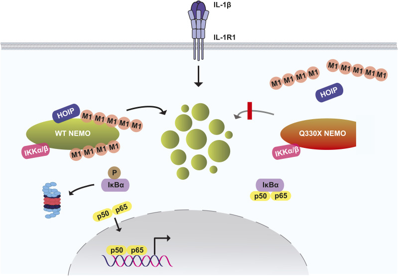
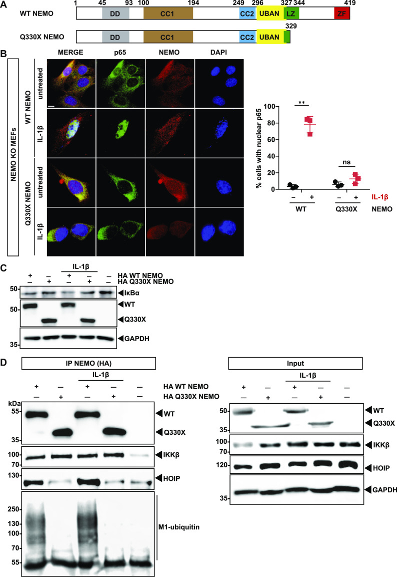
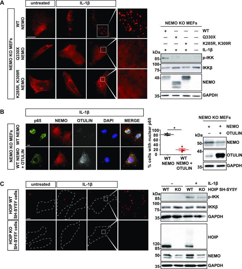
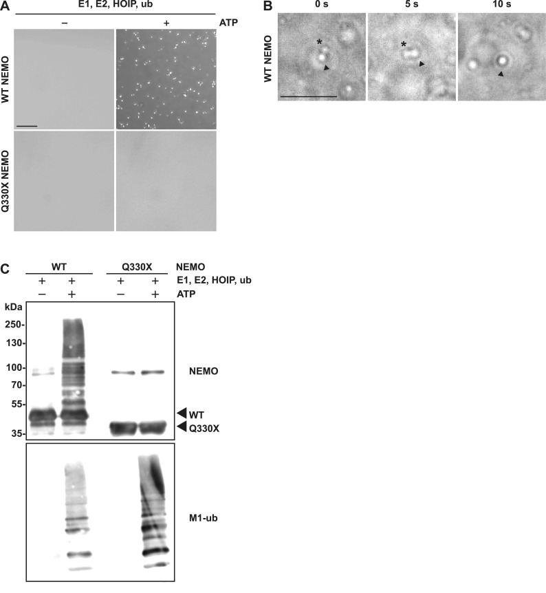
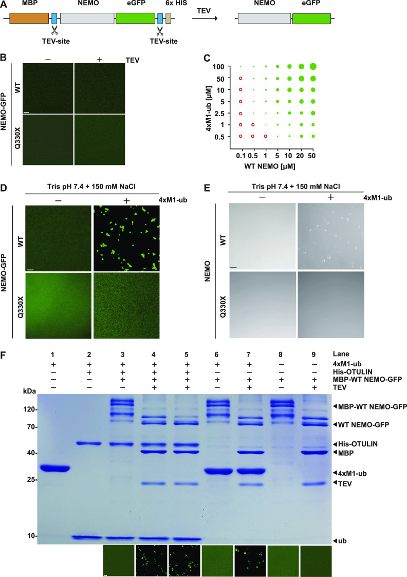
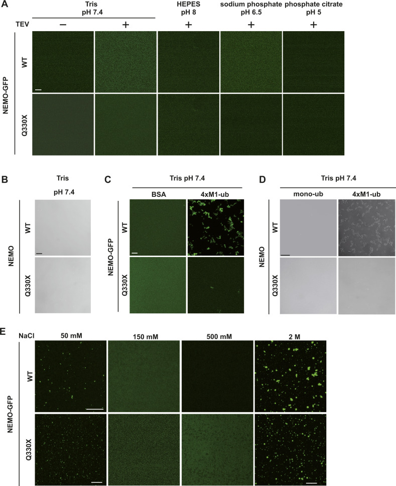
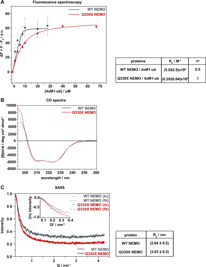
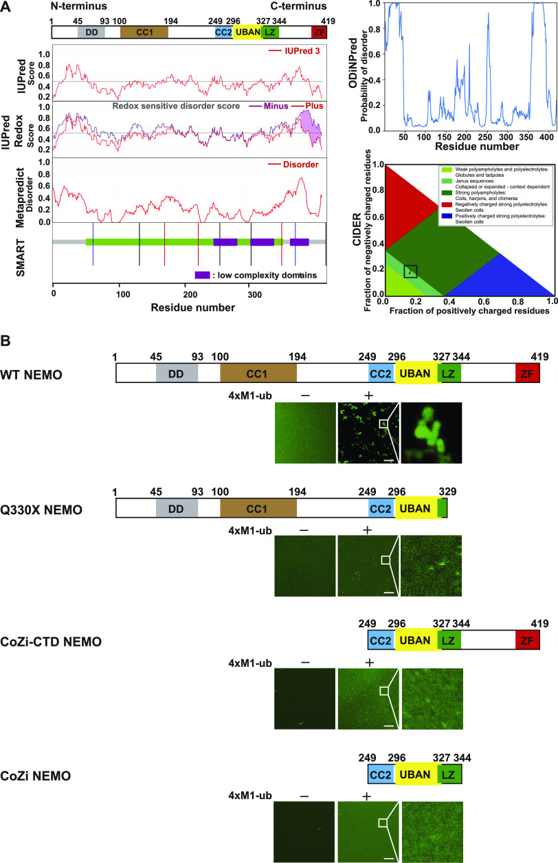
The NF-κB essential modulator NEMO is the core regulatory component of the inhibitor of κB kinase complex, which is a critical checkpoint in canonical NF-κB signaling downstream of innate and adaptive immune receptors. In response to various stimuli, such as TNF or IL-1β, NEMO binds to linear or M1-linked ubiquitin chains generated by LUBAC, promoting its oligomerization and subsequent activation of the associated kinases. Here we show that M1-ubiquitin chains induce phase separation of NEMO and the formation of NEMO assemblies in cells after exposure to IL-1β. Phase separation is promoted by both binding of NEMO to linear ubiquitin chains and covalent linkage of M1-ubiquitin to NEMO and is essential but not sufficient for its phase separation. Supporting the functional relevance of NEMO phase separation in signaling, a pathogenic NEMO mutant, which is impaired in both binding and linkage to linear ubiquitin chains, does not undergo phase separation and is defective in mediating IL-1β-induced NF-κB activation.


Conflict of interest statement

The authors declare that they have no conflict of interest.

Figures

None
Graphical abstract
Figure 1.
Figure 1.. The pathogenic Q330X NEMO mutant is defective in NF-κB signaling.
(A) Domain structure of WT human NEMO and of the pathogenic mutant Q330X NEMO. DD, dimerization domain (aa 45–93); CC1, coiled-coil 1 domain (aa 100–194); CC2, coiled-coil 2 domain (aa 249–292); UBAN, ubiquitin binding in ABIN and NEMO (aa 296–327); LZ, leucine zipper (aa 322–344); ZF, zinc finger (aa 389–419). (B) Q330X NEMO does not promote p65 translocation upon IL-1β stimulation. NEMO KO MEFs were reconstituted with HA-tagged WT NEMO or Q330X NEMO. 24 h later, the cells were stimulated with murine IL-1β (20 ng/ml) for 15 min or left untreated and then analyzed by immunocytochemistry and SR-SIM using antibodies against NEMO and p65 (scale bar, 10 µm). Data are displayed as mean ± SD and were analyzed by Kruskal-Wallis test followed by Dunn’s Multiple Comparison Test, n = 3, **P ≤ 0.01. (C). WT NEMO but not Q330X NEMO induces IκBα degradation upon IL-1β stimulation. HEK293T cells were transiently transfected with HA-tagged WT NEMO or HA-Q330X NEMO, as indicated. 1 d after transfection, the cells were stimulated with human IL-1β (25 ng/ml) for 15 min or left untreated and analyzed by immunoblotting using antibodies against IκBα, NEMO, and GAPDH. (D) The Q330X mutation disrupts binding of NEMO to HOIP and M1-linked ubiquitin. HEK293T cells were transiently transfected with HA-tagged WT NEMO or Q330X NEMO, as indicated. 1 d after transfection, the cells were stimulated with human IL-1β (25 ng/ml) for 15 min or left untreated and then lysed. HA-tagged NEMO was immunoprecipitated using anti-HA-beads, followed by immunoblotting using antibodies against HA, IKKβ, HOIP, and M1-ubiquitin. The input was immunoblotted for NEMO, IKKβ, HOIP, and GAPDH. Source data are available for this figure.
Figure 2.
Figure 2.. Linear ubiquitination promotes the formation of NEMO assemblies and IKK complex activation upon IL-1β receptor activation.
(A) In contrast to WT NEMO, Q330X and K285/309R NEMO do not form foci upon IL-1β treatment. NEMO KO MEFs were reconstituted with HA-tagged WT NEMO, Q330X NEMO or K285/309R NEMO. 24 h later, the cells were stimulated with murine IL-1β (20 ng/ml) for 15 min or left untreated and then analyzed after saponin extraction by immunocytochemistry using an antibody against NEMO (scale bar, 10 µm). Right Panel: IKK complex activation was analyzed by immunoblotting using an antibody against phospho-IKKα/β. Antibodies against IKKβ and NEMO were used to control expression levels. GAPDH was immunoblotted as input control. (B) IL-1β–induced nuclear translocation of p65 is impaired by OTULIN. The cells were treated as described in (A) and analyzed by immunocytochemistry using antibodies against NEMO, OTULIN, and p65 (scale bar, 10 µm). Right panel: Data are displayed as mean ± SD and were analyzed by Mann–Whitney test, n = 4, *P ≤ 0.05. Expression levels of HA-tagged WT NEMO and OTULIN were analyzed by immunoblotting using antibodies against NEMO and OTULIN. GAPDH was immunoblotted as input control. (C) IL-1β–induced formation of NEMO assemblies and IKK complex activation are compromised in HOIP-deficient cells. Control SH-SY5Y cells and HOIP KO SH-SY5Y cells were stimulated with human IL-1β (20 ng/ml) for 10 min and then analyzed by immunocytochemistry after saponin extraction using an antibody against NEMO (scale bar, 10 µm). Right Panel: IKK complex activation was analyzed by immunoblotting using an antibody against phospho-IKKα/β. Antibodies against IKKβ, HOIP, and NEMO were used to control expression. GAPDH was immunoblotted as input control. Source data are available for this figure.
Figure 3.
Figure 3.. WT NEMO undergoes phase separation in an M1-ubiquitin-dependent manner.
(A) In contrast to Q330X NEMO, WT NEMO undergoes LLPS upon linear ubiquitination by HOIP. The samples from the in vitro ubiquitination assay described in (C) were analyzed by bright-field microscopy. Representative images are shown (scale bar, 20 µm). (B) Liquid droplets formed by ubiquitinated NEMO are dynamic. WT NEMO ubiquitinated by HOIP as described in (C) were analyzed by bright-field microscopy to follow up fusion events. The images were taken every 5 s over a period of 10 s (scale bar, 20 µm). (C) WT but not Q330X NEMO is modified by M1-linked ubiquitin in vitro. Untagged WT NEMO and Q330X NEMO were subjected to an in vitro linear (M1-linked) ubiquitination assay by incubating the recombinant proteins (5 µM) with 50 µM mono-ubiquitin (ub), 1.5 µM ubiquitin-activating enzyme UBe1, 4 µM E2 ubiquitin-conjugating enzyme UbcH5c, 4 µM C-terminal HOIP comprising the RBR and LDD domains, ATP, and MgCl2 in 50 mM HEPES buffer (pH 7.4) containing 0.5 mM TCEP (tris(2-carboxyethyl)phosphine) for 2 h at RT. As a control, ATP was omitted (−ATP). The samples were analyzed by immunoblotting using antibodies against NEMO and M1-ubiquitin. Source data are available for this figure.
Figure S1.
Figure S1.. Recombinant WT NEMO and Q330X NEMO represent similar oligomerization profiles.
(A) Coomassie gels of recombinant fusion proteins composed of maltose-binding protein and WT NEMO-GFP or Q330X NEMO-GFP or CoZi NEMO-GFP purified from bacterial cells. (B) Gels and size exclusion chromatography profiles of recombinant untagged WT NEMO or Q330X NEMO purified from insect cells.
Figure 4.
Figure 4.. M1-linked tetra-ubiquitin induces phase transition of NEMO in vitro.
(A) Domain structure of recombinant WT NEMO-GFP or Q330X NEMO-GFP. MBP (maltose-binding protein), TEV (Tobacco Etch Virus protease). (B) WT NEMO-GFP and Q330X NEMO-GFP do not phase-separate. Fusion proteins were incubated in the presence of TEV protease (1 h at RT) to cleave off the N-terminal MBP and the C-terminal His6 tag and then WT NEMO-GFP and Q330X NEMO-GFP were analyzed by fluorescent microscopy using a laser scanning microscope. (C) Concentration-dependent phase separation of WT NEMO and M1-linked tetra-ubiquitin (4×M1-ub). WT NEMO-GFP was incubated in presence of recombinant M1-linked tetra-ubiquitin (4×M1-ub) at the concentrations indicated and analyzed by fluorescence microscopy. Red empty circles: no phase separation; green solid circles: phase separation; sizes of the circles illustrate the sizes of liquid droplets. (D, E) WT NEMO but not Q330X NEMO undergoes LLPS in the presence of M1-linked tetra-ubiquitin. GFP-tagged (D, E) or untagged (E) WT NEMO and Q330X NEMO (5 µM in 10 mM Tris, pH 7.4, and 150 mM sodium chloride [NaCl]) were incubated in the presence or absence of 10 µM 4×M1-ub and analyzed by fluorescence microscopy (D) or bright-field microscopy (E). Shown are representative images; scale bar, 10 µm. (F) Preformed NEMO assemblies persist upon OTULIN-mediated hydrolysis of M1-linked tetra-ubiquitin. MBP-WT NEMO-GFP was incubated with the components indicated (lanes 3–9). In the reaction corresponding to lane 4, recombinant OTULIN was added together with TEV protease, whereas in the reaction corresponding to lane 5 OTULIN was added 1 h after TEV protease. Lane 1: M1-linked tetra-ubiquitin only. Lane 2: M1-linked tetra-ubiquitin incubated with OTULIN for 1 h. The samples were analyzed by SDS–PAGE and Coomassie blue staining (upper panel) and laser scanning microscopy (lower panel). Source data are available for this figure.
Figure S2.
Figure S2.. Binding to M1-linked tetra-ubiquitin but not mono-ubiquitin induces LLPS of both WT untagged and NEMO-GFP fusion proteins.
(A) Recombinant WT NEMO-GFP and Q330X NEMO-GFP are soluble in different buffers at various pH values. Fusion proteins (5 µM) composed of MBP and WT NEMO-GFP or Q330X NEMO-GFP were incubated in different buffers as indicated in the absence (TEV−) or presence (TEV+) of TEV protease to cleave off the MBP tag for 1 h at RT and then analyzed by fluorescent microscopy using a laser scanning microscope (scale bar, 10 µm). (B) Recombinant untagged WT NEMO and Q330X NEMO (5 µM) are also soluble in Tris buffer at pH 7.4. Representative bright-field images are shown (scale bar, 10 µm). (C) WT NEMO-GFP undergoes LLPS when mixed with linear tetra-ubiquitin. Fusion proteins composed of MBP and WT NEMO-GFP or Q330X NEMO-GFP (5 µM in 10 mM Tris, pH 7.4) were incubated in presence of TEV protease for 1 h at RT plus 10 µM recombinant 4×M1-ub and then analyzed by fluorescence microscopy. BSA (10 µM) was used as a control (scale bar, 10 µm). (D) Mono-ubiquitin does not induce LLPS of WT NEMO. Untagged WT NEMO or Q330X NEMO was mixed with 10 µM 4×M1-ub or 40 µM mono-ubiquitin (mono-ub). Representative bright-field images are shown (scale bar, 20 µm). (E) NEMO displays reentrant phase transition behavior. Fusion proteins composed of MBP and WT NEMO-GFP or Q330X NEMO-GFP (5 µM in 10 mM Tris, pH 7.4) were incubated in presence of TEV protease for 1 h at RT with indicated salt concentrations and analyzed by fluorescence microscopy (scale bar, 10 µm).
Figure 5.
Figure 5.. Binding of Q330X NEMO to M1-linked ubiquitin is impaired.
(A) Q330X NEMO shows a reduced binding affinity for 4×M1-ub. Binding isotherms obtained by steady-state fluorescence spectroscopy for the complex formation between untagged WT NEMO (black squares) or Q330X NEMO (red circles) and recombinant 4×M1-ub at 25°C in 10 mM Tris–HCl buffer, pH 7.4. The solid lines represent the best fit of experimental data according to an equivalent and independent binding site model. an is the stoichiometry defined as mol of linear tetra-ubiquitin per mol of WT NEMO or Q330X NEMO. (B) WT NEMO and Q330X NEMO show similar, predominantly alpha-helical secondary structures. Circular dichroism spectra were recorded in the Far-UV region (below 260 nm) for WT NEMO and Q330X NEMO at 1 mg/ml concentration. (C) WT NEMO and Q330X NEMO show similar conformations in solution reflected by comparable radius of gyration values obtained by small-angle X-ray scattering. The small-angle X-ray scattering measurements of WT NEMO and mutant Q330X NEMO were made at a concentration of 5 mg/ml in 10 mM Tris, pH 7.4. The data were obtained by the program “PRIMUS” and are shown as a function of Guinier linear plot for intensity versus Q.
Figure 6.
Figure 6.. N- and C-terminal domains contribute to M1-ubiquitin–induced phase separation of NEMO.
(A) Bioinformatic analyses predicts IDRs in the N- and C-terminus of WT NEMO along with spanning low complexity domains. Schematic representation of the human NEMO domain structure is shown on top. DD, dimerization domain; CC, coiled coil; UBAN, ubiquitin binding in ABIN and NEMO; LZ, leucine zipper; ZF, zinc finger. The following bioinformatic tools were used: IUPred, prediction of intrinsically unstructured proteins; SMART, simple modular architecture research tool; ODiNPred, prediction of order and disorder by evaluation of NMR data. Classification of NEMO based on the fraction of charged residues by CIDER: classification of intrinsically disordered ensemble regions. (B) Both N- and C-terminal domains are essential for phase separation of NEMO. Fusion proteins (5 µM) composed of MBP and WT NEMO-GFP, Q330X NEMO-GFP or CoZi-CTD NEMO-GFP (aa 249–419), or CoZi NEMO-GFP (aa 249–344) were incubated in presence of TEV protease to cleave off the MBP tag for 1 h at RT in buffer containing 10 mM Tris pH 7.4 ± 10 µM 4×M1-ub and then analyzed by fluorescent microscopy using a laser scanning microscope (scale bar, 10 µm).

Similar articles

Cited by

References

    1. Abbott DW, Wilkins A, Asara JM, Cantley LC (2004) The Crohn[R8S2Q1M7]s disease protein, NOD2, requires RIP2 in order to induce ubiquitinylation of a novel site on NEMO. Curr Biol 14: 2217–2227. 10.1016/j.cub.2004.12.032 - DOI - PubMed
    1. Alberti S, Dormann D (2019) Liquid-liquid phase separation in disease. Annu Rev Genet 53: 171–194. 10.1146/annurev-genet-112618-043527 - DOI - PubMed
    1. Annibaldi A, Meier P (2018) Checkpoints in TNF-induced cell death: Implications in inflammation and cancer. Trends Mol Med 24: 49–65. 10.1016/j.molmed.2017.11.002 - DOI - PubMed
    1. Baker HA, Bernardini JP (2021) It[R8S2Q1M7]s not just a phase; ubiquitination in cytosolic protein quality control. Biochem Soc Trans 49: 365–377. 10.1042/bst20200694 - DOI - PMC - PubMed
    1. Berrow NS, Alderton D, Sainsbury S, Nettleship J, Assenberg R, Rahman N, Stuart DI, Owens RJ (2007) A versatile ligation-independent cloning method suitable for high-throughput expression screening applications. Nucleic Acids Res 35: e45. 10.1093/nar/gkm047 - DOI - PMC - PubMed

Publication types

LinkOut - more resources